
1. Introduction
A fetal disorder resulting from malpresentation during delivery represents an acute obstetrical crisis occurring when a fetus enters the final, expulsive phase of childbirth in any orientation other than a tucked, head-first position. Malpresentations include breech (buttocks or feet first), face, brow, and transverse (shoulder first) presentations. Unlike malposition, which involves a head-first fetus facing the wrong direction, malpresentation means a fundamentally incorrect anatomical part of the fetus is attempting to navigate the narrow maternal pelvis.
During the delivery phase, the massive mechanical forces of maternal pushing and uterine contractions are meant to expel a uniform, wedged-shaped fetal head. When a malpresentation occurs, these immense forces are misdirected against fragile fetal structures such as the cervical spine, facial bones, or the umbilical cord. This structural mismatch frequently results in a catastrophic mechanical arrest, subjecting the fetus to severe physical trauma and acute asphyxiation.
Modern obstetrical protocols mandate rigorous prenatal screening to identify malpresentations well before the onset of labor, allowing for planned surgical deliveries. However, unexpected malpresentations can emerge rapidly during the delivery phase. Managing these emergent crises requires advanced obstetrical extraction techniques, a profound understanding of fetal biomechanics, and the immediate availability of a surgical team to rescue the infant and prevent permanent structural and neurological injuries.
2. The Biomechanics of Malpresentation
The human birth canal is an unyielding bony passage perfectly contoured for the flexed fetal head, which possesses the ability to mold and compress. When a fetus presents in a malpresentation, this biomechanical harmony is destroyed. Each type of malpresentation introduces unique, formidable physical obstacles during the final descent.
In a face presentation, the fetal neck is sharply extended backward. The chin and facial bones act as the leading wedge. The facial bones cannot mold or compress like the cranial bones. As the face is forced against the maternal pelvis, the delicate facial tissues, eyes, and airway are subjected to severe crushing forces, often preventing the fetus from passing under the maternal pubic bone.
A brow presentation is the most mechanically disadvantageous cephalic orientation. The head is partially extended, presenting the absolute widest cranial diameter (the occipitomental diameter) to the birth canal. This diameter is universally too large to pass through a standard maternal pelvis, resulting in an immediate and absolute mechanical arrest during the pushing phase, making vaginal delivery physically impossible.
3. Risks of Breech Delivery and Head Entrapment
Breech presentations pose the most notorious risks during the expulsive phase of delivery. In a breech birth, the softer, smaller lower body of the fetus delivers first. The critical physiological danger arises because the largest and least compressible part of the fetus—the head—is delivered last.
As the lower body emerges, the fetal chest and shoulders must rotate precisely. If the arms become extended above the head, the descent is abruptly halted. The physician must perform complex, rapid internal maneuvers to sweep the fragile arms downward, carrying a high risk of fracturing the humerus or tearing the delicate nerves of the brachial plexus.
The most catastrophic complication is head entrapment. The softer fetal body may slip through an incompletely dilated cervix or a narrow pelvic outlet, but the rigid skull becomes firmly locked behind the maternal bones. The umbilical cord is instantly compressed between the trapped skull and the pelvis, completely cutting off the oxygen supply. The physician has only minutes to execute complex, forceful rotational maneuvers to extract the head before the infant suffers lethal anoxia.
4. Transverse Lie and Shoulder Presentation
A transverse lie occurs when the fetus rests horizontally across the maternal uterus, with the shoulder or arm resting directly over the pelvic inlet. During the delivery phase, if the amniotic sac ruptures and powerful contractions force the fetus downward, the shoulder becomes deeply impacted into the maternal pelvis, and the fetal arm frequently prolapses out of the vagina.
Vaginal delivery of a persistent transverse lie is physically impossible. The immense force of the uterine contractions attempting to push a horizontally oriented fetus through the vertical birth canal causes the uterus to contract violently around the fetus. This creates an extreme risk of a catastrophic maternal uterine rupture, a life-threatening surgical emergency resulting in massive internal hemorrhage.
For the fetus, a neglected transverse lie leads to rapid, severe hypoxia as the impacted shoulder compresses the surrounding blood vessels and the umbilical cord. A crash emergency cesarean section is the only absolute medical intervention capable of resolving this insurmountable mechanical obstruction and saving both the mother and the infant.
5. Fetal Cranial and Spinal Trauma
The mechanical forces required to extract a malpresenting fetus, particularly during an emergency breech extraction, frequently result in severe structural trauma. The delicate cervical spine of the fetus is uniquely vulnerable. Applying downward traction to the fetal body to deliver an entrapped head can stretch the spinal cord beyond its elastic limits.
This intense traction can lead to severe spinal cord contusions, hemorrhages, or complete transections (tearing) of the spinal cord in the cervical region. Infants suffering from severe spinal trauma frequently present at birth with profound respiratory failure, flaccid paralysis of the limbs, and an absence of deep tendon reflexes, resulting in permanent, devastating neurological disability or death.
Cranial trauma is also prevalent. The rapid, forceful extraction of the fetal head from behind the pelvic bones can cause depressed skull fractures or severe intracranial hemorrhages. The shearing forces tear the bridging veins within the fetal brain, leading to localized bleeding that compresses the fragile brain tissue and triggers acute neonatal seizures.
6. Facial and Airway Trauma in Face Presentations
When a fetus delivers in a face presentation, the soft tissues of the face absorb the massive compressive forces of the maternal pelvis and the pushing efforts. The infant is typically born with profound, disfiguring facial edema (swelling) and extensive bruising. The eyes may be severely swollen shut, and the delicate conjunctiva frequently exhibit bright red hemorrhages.
The most critical concern is trauma to the fetal airway. The crushing pressure against the neck and lower jaw can cause significant swelling of the fetal larynx and trachea. This internal swelling rapidly compromises the airway immediately after birth. The neonate may exhibit severe respiratory distress, marked by audible stridor (a high-pitched breathing sound) and deep chest retractions.
The neonatal resuscitation team must evaluate the airway instantly upon delivery. Severe laryngeal edema may prevent the infant from breathing independently and can make inserting a life-saving endotracheal breathing tube technically complex, requiring advanced pediatric airway management skills to secure oxygenation.
7. Umbilical Cord Prolapse During Extraction
Umbilical cord prolapse is a sudden, terrifying obstetrical emergency intricately linked with malpresentations, specifically footling breech and transverse presentations. Because the presenting parts in these orientations do not form a snug, uniform plug against the cervix, empty space remains in the lower pelvis.
During the delivery phase, as the amniotic fluid rushes out or as the physician manipulates the fetus to aid extraction, the umbilical cord can easily wash down past the presenting part and into the vaginal canal. Once the cord is exposed, the descending fetal body compresses it forcefully against the rigid maternal pelvic bones.
This complete occlusion of the cord halts the flow of oxygenated blood to the fetus instantly. The fetal heart rate monitor will display profound, sustained decelerations. The delivering physician must immediately elevate the presenting part of the fetus with their hand inside the vagina to relieve the pressure on the cord while the mother is rushed to the operating room for a surgical delivery.
8. Identifying Malpresentation During the Expulsive Phase
While prenatal ultrasounds aim to identify malpresentations weeks before labor, fetuses can occasionally change positions unpredictably, or a mother may present to the hospital fully dilated with no prior prenatal care. In these scenarios, diagnosing a malpresentation during the final delivery phase relies on precise physical examination.
During a sterile vaginal exam, the physician palpates the presenting fetal anatomy. Identifying a breech presentation requires feeling the soft buttocks, the cleft between them, or the distinct small bones of the fetal feet. A face presentation is diagnosed by palpating the distinct facial features: the nose, the orbital ridges of the eyes, and the mouth.
Differentiating a face presentation from a breech presentation can be exceptionally difficult when severe swelling is present; the swollen fetal mouth can easily be mistaken for the fetal anus. Accurate, rapid bedside ultrasound is frequently deployed in the delivery room to definitively confirm the anatomy and determine if immediate surgical intervention is mandated.
9. Structured Data: Trauma Risks by Presentation
Understanding the specific mechanical risks associated with each malpresentation guides the emergency response.
| Malpresentation Type | Primary Anatomical Obstacle | Specific Fetal Trauma Risk |
|---|---|---|
| Breech (Head Last) | Large head trapped behind cervix | Cervical spine injury, severe hypoxia, brachial plexus tear |
| Face Presentation | Face acting as the dilating wedge | Airway edema, severe facial bruising, eye trauma |
| Brow Presentation | Widest cranial diameter presenting | Absolute mechanical arrest, skull fractures from impaction |
| Transverse Lie | Shoulder impacted in the pelvis | Umbilical cord prolapse, crushed torso, anoxic brain injury |
10. Abandoning the Vaginal Delivery
The primary obstetrical mandate when managing a severe malpresentation during the delivery phase is recognizing the absolute limit of safe vaginal extraction. Attempting to force a physically incompatible fetus through the birth canal ensures catastrophic maternal and fetal trauma.
For brow and transverse presentations, vaginal delivery is unequivocally contraindicated. The physician must declare an immediate obstetrical emergency. For face presentations, a vaginal delivery is only possible if the fetal chin is pointing toward the maternal front (mentum anterior). If the chin points toward the maternal spine (mentum posterior), the head cannot extend under the pubic bone, and the delivery will arrest completely.
If a breech extraction arrests, or if any malpresentation causes sustained, profound drops in the fetal heart rate, the physician must cease all vaginal extraction efforts. Applying excessive manual traction or using instruments like forceps in these desperate situations frequently results in lethal fetal spinal and cranial injuries.
11. Emergency Cesarean Section
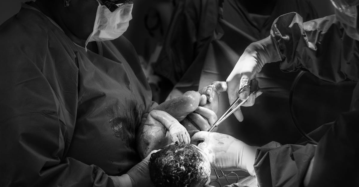
The definitive, life-saving intervention for an arrested, malpresenting fetus is a crash emergency cesarean section. This major abdominal surgery completely bypasses the obstructed bony birth canal, instantly removing the mechanical stress on the trapped fetal structures and permanently halting the hypoxic cascade.
The urgency is extreme. The mother is rushed to the operating room, frequently placed under immediate general anesthesia to save critical minutes, and the surgeon performs a rapid incision to extract the fetus. However, extracting a malpresenting fetus during surgery presents unique challenges.
If a transverse fetus has a prolapsed arm, or if a breech fetus has a deeply impacted head wedged down in the pelvis, the surgeon must execute complex, delicate internal maneuvers to safely dislodge and elevate the fragile fetal structures back into the abdominal cavity before they can be extracted, carrying a risk of causing minor fractures even during the surgical rescue.
12. Neonatal Resuscitation Readiness
Because infants delivered following an emergency extraction for a malpresentation are at immense risk for severe trauma and profound asphyxia, a fully equipped neonatal resuscitation team must be present in the operating room. The pediatric team anticipates a severely compromised infant and prepares for advanced, immediate interventions.
Upon birth, the infant is swiftly transferred to a radiant warmer. If the infant is apneic, limp, or bradycardic (dangerously slow heart rate), the team clears the airway and initiates positive pressure ventilation. If the infant sustained severe facial trauma from a face presentation, the team may need to perform immediate, complex endotracheal intubation to secure a swollen airway.
The resuscitation must be deliberate, recognizing that the infant may have sustained hidden spinal trauma during the extraction. The neck and spine must be stabilized and handled with extreme care during ventilation and chest compressions until a thorough physical and radiological examination can definitively rule out vertebral fractures or spinal cord injury.
13. Immediate Postnatal Assessment
Once the infant is stabilized and breathing adequately, the pediatrician performs a meticulous, structured trauma assessment. The physician carefully evaluates the symmetry of movement in all four limbs. An arm hanging limply at the side is a glaring clinical sign of a brachial plexus nerve tear or a fractured clavicle resulting from a difficult breech extraction.
The skull is palpated for distinct, depressed fractures, and the facial structures are examined for severe bruising and nerve palsies. A thorough neurological evaluation assesses muscle tone, pupil reactivity, and normal neonatal reflexes. Any indication of profound lethargy, abnormal eye movements, or generalized stiffness prompts an immediate transfer to the neonatal intensive care unit.
For infants born in a breech presentation, the pediatrician pays specific attention to the hip joints. The abnormal intrauterine positioning and the forceful extraction maneuvers significantly elevate the risk of developmental dysplasia of the hip, requiring early ultrasound screening to ensure the hip joints are stable and properly formed.
14. Orthopedic and Neurological Rehabilitation
The management of birth trauma injuries sustained during a malpositioned extraction continues long after the delivery. Simple skeletal injuries, such as a fractured clavicle or humerus, heal remarkably fast in a newborn. The pediatric orthopedic team typically applies a soft splint or pins the sleeve to immobilize the limb, allowing the bone to remodel perfectly within a few weeks.
Nerve injuries, specifically brachial plexus trauma, require dedicated, long-term rehabilitation. Parents are instructed on specific, gentle range-of-motion exercises to prevent the affected joints from developing permanent, rigid contractures while the stretched nerves slowly heal. Severe nerve avulsions require evaluation by a specialized pediatric microsurgeon to restore limited functionality.
Infants who suffered intracranial hemorrhages require serial brain imaging and vigilant neurological monitoring. The pediatric team watches closely for the development of post-hemorrhagic hydrocephalus or the onset of neonatal seizures, ensuring rapid medical intervention to protect developing brain tissue.
15. The Psychological Impact on the Mother
Enduring an unexpected, terrifying obstetrical emergency culminating in a crash surgical extraction is profoundly traumatizing for the expectant mother. The sudden influx of a massive medical team, the frantic rush to the operating room, and the potential transfer of the newborn to the intensive care unit replace the anticipated joy of childbirth with severe psychological shock.
Comprehensive obstetrical care must address this psychological burden directly. The delivering physician should provide a clear, empathetic debriefing once the mother is stable, explicitly explaining that the malpresentation was a mechanical anomaly beyond her control and detailing the medical necessity of the rapid interventions.
Facilitating early involvement of the parents in the neonatal care, providing specialized lactation support, and offering access to perinatal mental health counselors are essential components of holistic, trauma-informed postpartum care, significantly reducing the risk of severe postpartum depression and post-traumatic stress disorder.
16. Long-Term Prognosis
The long-term prognosis for an infant who has navigated a traumatic extraction for a malpresentation depends entirely on the specific anatomical structures damaged and the duration of any associated hypoxia. Infants sustaining soft tissue injuries, facial bruising, or simple clavicular fractures generally recover completely with no residual physical or developmental deficits.
Infants diagnosed with moderate to severe hypoxic-ischemic encephalopathy due to prolonged cord compression face a guarded prognosis. To mitigate permanent brain damage, intensive care units frequently utilize therapeutic hypothermia, deliberately lowering the body temperature for seventy-two hours to halt cellular death in the brain.
Despite advanced therapies, severe cases of spinal cord trauma or profound hypoxia carry a substantial risk of permanent neurodevelopmental disorders, including cerebral palsy, lifelong paralysis, and severe intellectual disabilities. These children require comprehensive, lifelong pediatric care and intensive physical and occupational therapy.
17. When to Seek Urgent Pediatric Care
Following a difficult instrumental or surgical delivery for a malpresentation, parents must be highly vigilant for signs of delayed complications once they bring the newborn home. If the infant develops a high-pitched, abnormal cry, refuses to feed, or becomes exceptionally difficult to awaken, parents must proceed immediately to a pediatric emergency department, as these indicate delayed brain swelling or bleeding.
If a large, firm lump on the head begins to grow rapidly, or if the infant skin and eyes develop a deep, distinct yellow color (jaundice) within the first few days of life, urgent medical evaluation is required. The breakdown of trapped blood from severe bruising can cause bilirubin levels to spike to dangerous levels, requiring prompt treatment with phototherapy lights.
Additionally, if parents notice that the infant is completely avoiding using one arm or leg, or if the infant cries out in severe pain during routine diaper changes or when being dressed, they should contact their pediatrician immediately to rule out an undiagnosed fracture or joint dislocation that may have been missed during the initial hospital evaluation.
18. Frequently Asked Questions (FAQ)
1. How does a doctor know if the baby is presenting face first?
During a vaginal exam in labor, the doctor feels for the distinct features of the face—the nose, mouth, and eye ridges—instead of the smooth, hard top of the skull. An ultrasound is quickly used in the delivery room to confirm the exact position.
2. Why is a breech baby head getting stuck so dangerous?
The baby lower body is small and can slip out, but the head is large and hard. If the head gets stuck behind the pelvic bone, the umbilical cord is instantly crushed, cutting off the oxygen supply. The doctor has only minutes to extract the head safely.
3. Will the severe bruising on my baby face cause permanent damage?
Generally, no. The severe bruising and swelling from a face presentation look alarming but usually heal completely within a week or two without leaving any permanent scars or structural damage to the facial bones.
4. Why did they do a C-section so fast when my baby was sideways?
A sideways (transverse) baby cannot physically pass through the birth canal. When your water breaks, the umbilical cord or the baby arm can fall out, causing an immediate, life-threatening emergency that requires surgery in minutes to save the baby life.
5. How long does a broken collarbone take to heal in a newborn?
Newborn bones possess an incredible regenerative capacity. A fractured collarbone (clavicle) typically heals completely within a few weeks without any specific casting, leaving no permanent structural deformity as the child grows.
19. Bibliography
Disclaimer: The content is for informational purposes only and does not replace medical advice. Always consult your doctor for personalized treatment.
