1. Introduction to Iliotibial Band Friction Syndrome
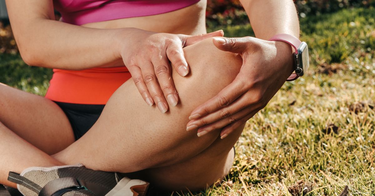
Iliotibial band friction syndrome is a highly prevalent overuse injury, recognized as one of the leading causes of lateral knee pain among endurance athletes, particularly long-distance runners and cyclists. The condition is characterized by a sharp, localized inflammatory pain on the outer aspect of the knee joint, which reliably intensifies during repetitive flexion and extension of the leg.
The syndrome derives its name from the iliotibial band, a thick, tough tract of fascial connective tissue that runs down the outside of the thigh, connecting the hip musculature to the tibia below the knee. The pathology occurs when biomechanical flaws, muscle imbalances, or sheer repetitive volume cause this dense band to compress and irritate the highly sensitive structures located at the side of the knee.
Managing this frustrating condition requires a detailed assessment of the athlete’s kinematics. Because it is fundamentally an injury of faulty mechanics rather than structural trauma, treatment relies heavily on active physical therapy, targeted strengthening, and precise alterations to training volume, rather than surgical intervention, to restore pain-free athletic function.
2. Anatomy and Biomechanics of the IT Band
The iliotibial band is not a muscle; it is a dense, fibrous thickening of the fascia lata that envelops the thigh. It originates at the pelvis, specifically receiving tendinous fibers from two major hip muscles: the tensor fasciae latae and the gluteus maximus. The band courses straight down the lateral aspect of the thigh and anchors firmly onto a small bump on the outer shin bone known as Gerdy’s tubercle.
Functionally, the iliotibial band acts as a critical stabilizer for the lateral knee during running. When the knee is fully straight, the band sits slightly in front of the lateral femoral epicondyle, a bony prominence on the outer side of the thigh bone. As the knee bends past 30 degrees, the band shifts backward, sliding directly over this bony prominence.
This continuous back-and-forth movement across the epicondyle during the running or cycling stride creates a zone of potential mechanical conflict. In a healthy, mechanically sound athlete, this transition is smooth and frictionless. However, when specific biomechanical flaws alter the tension on the band, this sliding motion becomes pathological.
3. Pathophysiology of the Friction Zone
Historically, sports medicine literature attributed the pain strictly to the physical friction of the dense iliotibial band repeatedly snapping back and forth directly over the rigid bone of the lateral femoral epicondyle. This mechanical rubbing was believed to cause inflammation in a small, fluid-filled sac called a bursa, located between the bone and the band.
However, modern anatomical and radiological studies have refined this understanding. It is now widely accepted that the primary source of pain is the severe compression of a highly innervated and highly vascularized layer of fat and connective tissue that resides deep to the iliotibial band in this specific area.
When the tension on the band is abnormally high, it acts like a tight rubber band being pulled across a curved surface. As the knee bends through the critical 30-degree angle—the exact angle of the knee when the foot strikes the ground during running—the band forcefully compresses this sensitive fat pad against the bone, triggering acute localized inflammation and sharp nerve pain.
4. Common Triggers in Endurance Athletes
This syndrome is definitively classified as an overuse injury, meaning it rarely results from a single traumatic event. It develops progressively due to the continuous repetition of a flawed movement pattern. Long-distance runners are exceptionally susceptible because a single long run involves thousands of consecutive knee flexions through the exact 30-degree impingement zone.
Training errors are the most frequent immediate triggers. A sudden, aggressive increase in weekly running mileage, incorporating excessive downhill running (which keeps the knee longer in the impingement zone), or running exclusively on cambered (sloped) roads can rapidly provoke the syndrome.
Cyclists are also highly prone to this injury if their bicycle is improperly fitted. Cleats that force the toes to point too far inward, or a saddle that is positioned too high, force the cyclist to overextend the knee through the friction zone with every pedal stroke, rapidly inducing lateral knee pain.
5. Anatomic Risk Factors and Muscle Imbalance
Underlying the training errors are almost universally present biomechanical flaws and muscle imbalances. The most critical factor driving increased tension in the iliotibial band is severe weakness in the hip abductor muscles, specifically the gluteus medius.
The gluteus medius is responsible for stabilizing the pelvis when the athlete is standing on one leg during the running stride. If this muscle is weak, the pelvis tilts downward on the opposite side. This pelvic drop forces the femur (thigh bone) to rotate inward and adduct (move toward the midline).
This inward collapse of the leg places a massive, unnatural stretch on the iliotibial band attached to the outside of the hip, pulling it violently tight against the side of the knee. Therefore, the pain felt at the knee is merely the symptom of a mechanical failure originating higher up at the hip.
6. Clinical Symptoms and Pain Patterns
The clinical presentation is highly distinct. Patients report a sharp, burning, or stinging pain precisely localized to the lateral aspect of the knee, typically one to two centimeters above the joint line. The pain is rarely present while walking normally or resting.
Instead, the pain reliably initiates after a specific, predictable distance or time into a run or bike ride. Initially, the athlete may be able to run through the discomfort, but as the inflammation mounts, the pain becomes sharp and debilitating, forcing the athlete to stop entirely. The pain typically worsens noticeably when running downhill or descending stairs.
Identifying these specific mechanical triggers requires precise evaluation. The sharp nature of this activity-dependent pain differentiates it from internal joint issues requiring different assessments, similar to the protocols utilized when investigating generalized knee pain caused by internal meniscal tears or cartilage wear.
7. Orthopedic Physical Examination
The diagnosis of iliotibial band friction syndrome is primarily clinical, relying on a focused physical examination to reproduce the patient’s specific pain. The physician begins by palpating the lateral femoral epicondyle, checking for localized tenderness and subtle swelling.
To clarify the diagnostic process, the table below outlines the two hallmark orthopedic provocation tests.
| Orthopedic Test | Maneuver Description | Clinical Interpretation |
|---|---|---|
| Noble Compression Test | Physician presses on the lateral epicondyle while passively extending the knee from a bent position. | A sharp pain elicited exactly at 30 degrees of flexion confirms compression of the fat pad beneath the IT band. |
| Ober Test | Patient lies on healthy side; physician pulls the affected leg backward and attempts to drop the knee toward the table. | Inability of the knee to drop indicates severe tightness and lack of flexibility in the tensor fasciae latae and IT band. |
8. Diagnostic Imaging Considerations
Because the diagnosis is highly reliant on the clinical history and physical provocation tests, diagnostic imaging is rarely necessary for the initial confirmation of iliotibial band friction syndrome. Standard X-rays of the knee will invariably appear perfectly normal, as the condition involves soft tissue and mechanics, not bone pathology.
However, if the pain fails to respond to months of dedicated physical therapy, or if the clinical presentation is atypical (e.g., locking, clicking, or severe joint swelling), a magnetic resonance imaging scan is mandated.
An MRI is utilized specifically to rule out other severe internal derangements of the knee, such as a lateral meniscus tear or underlying cartilage damage. In severe cases of friction syndrome, the MRI may reveal a localized bright signal of edema (fluid and inflammation) situated deep to the iliotibial band near the epicondyle.
9. Acute Phase Conservative Management
The immediate goal of treatment in the acute phase is to rapidly extinguish the localized inflammation and break the pain cycle. The absolute cornerstone of this phase is relative rest. The athlete must temporarily cease all activities that provoke the pain, specifically avoiding running and cycling.
During this rest period, cross-training activities that do not involve repetitive knee flexion past 30 degrees, such as deep-water running or upper-body ergometry, can be utilized to maintain cardiovascular fitness without aggravating the injury.
Aggressive application of ice to the lateral knee immediately following any activity, combined with a short course of oral non-steroidal anti-inflammatory drugs, is highly effective at reducing the acute swelling and chemical irritation within the compressed fat pad.
10. Physical Therapy and Kinematic Correction
Once the acute, sharp pain subsides, the treatment focus shifts to the definitive, long-term solution: rigorous physical therapy to correct the underlying biomechanical flaws. The primary objective is to fundamentally strengthen the weak hip abductors and external rotators, particularly the gluteus medius.
Therapists employ targeted exercises such as lateral band walks, clam shells, and single-leg squats, ensuring the patient learns to fire the gluteal muscles correctly to stabilize the pelvis and prevent the inward collapse of the knee during movement.
Furthermore, physical therapists utilize deep tissue mobilization techniques, such as foam rolling and manual massage, targeting the muscular bellies of the tensor fasciae latae and gluteus maximus to reduce tension feeding into the fibrous band. Notably, aggressive foam rolling directly over the inflamed lateral knee bone is discouraged, as it simply crushes the already irritated fat pad further.
11. Pharmacological and Injection Therapies
If the patient remains severely limited by pain despite several weeks of dedicated rest and physical therapy, more aggressive medical interventions are considered.
A targeted corticosteroid injection is a potent option. The sports medicine physician injects a localized, powerful anti-inflammatory steroid directly into the space between the iliotibial band and the lateral epicondyle. This intervention is often highly successful at entirely eradicating the localized swelling and sharp pain.
However, a steroid injection is not a cure. It acts as a powerful window of pain relief that allows the athlete to engage fully and pain-free in the rigorous physical therapy required to fix the underlying muscular imbalances. Relying on injections without correcting the mechanics will invariably lead to a rapid relapse once the medication wears off.
12. Gait Analysis and Biomechanical Adjustments
For endurance athletes who suffer from recurrent episodes, a formal biomechanical gait analysis is an invaluable clinical tool. The athlete is filmed while running on a treadmill, allowing clinicians to analyze their specific movement patterns frame by frame.
The analysis frequently reveals subtle, correctable errors, such as a “crossover gait.” In this flawed pattern, the runner’s foot strikes the ground too close to, or completely crossing over, the midline of their body. This narrow stride drastically increases the stretch and tension on the lateral aspect of the hip and knee.
Clinicians implement gait retraining strategies, providing cues to slightly widen the running stance or increase the step rate (cadence). These minor, deliberate alterations in form significantly reduce the mechanical load placed on the iliotibial band with each stride. Cyclists undergo a professional bike fit to adjust cleat alignment and saddle height to eliminate the mechanical impingement.
13. Surgical Intervention Criteria
Surgical intervention for iliotibial band friction syndrome is exceptionally rare and is considered only as an absolute last resort. It is reserved exclusively for athletes who have failed a minimum of six to twelve months of rigorous, documented conservative therapy, including steroid injections, and remain completely disabled from their sport.
The surgical procedure involves a localized release or lengthening of the iliotibial band. Through a small incision on the outside of the knee, the surgeon isolates the dense fibrous band and cuts a small triangular or Z-shaped piece out of the posterior aspect of the band specifically where it crosses the lateral epicondyle.
This minor structural alteration relieves the extreme tension, preventing the band from compressing the underlying tissues when the knee bends. Postoperative recovery involves immediate mobilization and an aggressive return to physical therapy to ensure scar tissue does not restrict the newly released tissues.
14. Return to Sport Guidelines
Returning to endurance sports following this injury requires profound patience and strict adherence to a structured, graduated protocol. Returning too quickly to high mileage guarantees a severe relapse.
Athletes must begin with a walk-run interval program, carefully monitoring their knee for any sensation of tightness or stinging during and immediately after the session. Mileage is increased by no more than ten percent per week. The athlete is instructed to immediately stop the run at the very first indication of sharp lateral knee pain, rather than attempting to push through it.
Maintaining the hip strengthening exercises acquired during physical therapy is a lifelong requirement for these athletes. The gluteal strength must be maintained at a high level to ensure the pelvis remains stable and the knee tracking remains optimal during periods of heavy training volume.
15. When to Consult a Sports Medicine Specialist
Athletes experiencing lateral knee pain should seek evaluation from a sports medicine specialist or a specialized physical therapist if the pain forces them to alter their running form, if it causes them to stop their workouts prematurely, or if the pain persists for more than two weeks despite resting and icing.
Immediate medical evaluation is required if the knee becomes visibly swollen, feels hot to the touch, or if the athlete experiences sudden locking or instability in the joint. These symptoms are not characteristic of simple friction syndrome and strongly suggest an acute structural injury inside the knee joint requiring advanced imaging.
Proactive engagement with a specialist ensures that the biomechanical root of the problem is identified and corrected early, preventing a temporary inflammatory issue from transitioning into a chronic, season-ending disability.
16. Frequently Asked Questions (FAQ)
1. Is the iliotibial band a muscle that I can stretch out?
No. The IT band is a very thick, rigid piece of connective tissue (fascia), similar to a tendon. You cannot physically stretch the band itself. Physical therapy focuses on stretching and relaxing the actual muscles at the hip that pull on the band, thereby reducing the tension.
2. Should I foam roll directly on the side of my knee where it hurts?
Absolutely not. The pain on the side of your knee is caused by the band crushing an inflamed pad of tissue against the bone. Pressing a hard foam roller directly into this tender spot will only crush it further and worsen the inflammation. Foam roll the muscles high up on your hip and thigh instead.
3. Will changing my running shoes cure the pain?
Shoes alone will not cure the syndrome. While worn-out shoes or those lacking proper support can contribute to poor mechanics, the root cause is almost always weak hip muscles. You must strengthen your glutes to permanently solve the problem.
4. How long does it usually take to recover from this injury?
With dedicated physical therapy, absolute avoidance of running during the acute phase, and gradual return to activity, most athletes recover fully in four to eight weeks. Attempting to run through the pain will stretch the timeline to several months.
5. Can I cycle instead of running while I recover?
It depends entirely on your bike fit. If cycling does not trigger the specific sharp pain on the outside of your knee, it is an excellent way to maintain fitness. However, if your saddle is too high, cycling will irritate the exact same spot as running.
17. Bibliography
Disclaimer: The content is for informational purposes only and does not replace medical advice. Always consult your doctor for personalized treatment.