
1. Introduction to Impacted Third Molar Teeth
An impacted third molar, universally known as a wisdom tooth, is a highly common developmental dental condition. It occurs when a tooth fails to fully erupt into its functional, proper position within the dental arch due to a physical obstruction, a lack of adequate anatomical space in the jawbone, or an abnormal angle of development.
Positioned at the very back of the mouth, third molars are the final teeth to develop, typically attempting to erupt during late adolescence or early adulthood. In modern human anatomy, the evolutionary shortening of the jaw frequently leaves insufficient room to accommodate these large, late-arriving grinding teeth. Consequently, they become trapped entirely within the jawbone or partially emerge through the gum tissue, establishing a permanent state of impaction.
Managing an impacted third molar requires precise clinical and radiographic evaluation. While some impacted wisdom teeth remain completely asymptomatic and pathology-free throughout life, the vast majority eventually provoke significant clinical complications. Prompt surgical extraction is frequently indicated to prevent severe localized infections, structural damage to adjacent healthy teeth, and the development of destructive pathological cysts.
2. Dental Anatomy and the Eruption Timeline
To understand the mechanics of impaction, the normal chronology of dental development must be reviewed. The human dentition consists of primary (baby) teeth and permanent adult teeth. The molars are large, multi-rooted teeth engineered for crushing and grinding food. Adults typically develop three molars in each quadrant of the mouth.
The first molars usually erupt around age six, followed by the second molars near age twelve. The third molars, forming the extreme posterior boundary of the dental arch, develop deep within the bone during childhood but do not begin their upward migration until the late teenage years, earning them the moniker “wisdom teeth.”
For a tooth to erupt successfully, it requires an unimpeded vertical pathway through the bone and soft tissue, and sufficient horizontal space on the alveolar ridge. When the skeletal growth of the mandible (lower jaw) or maxilla (upper jaw) ceases before adequate space is created behind the second molars, the stage is set for unavoidable impaction.
3. Mechanisms and Etiology of Impaction
The primary driver of third molar impaction is a fundamental discrepancy between the size of the teeth and the overall length of the jawbone. This physical mismatch physically blocks the tooth from achieving a normal, upright position. The dense bone of the ascending ramus in the lower jaw frequently acts as an unyielding barrier to the erupting tooth.
Evolutionary biology plays a significant role. Over millennia, changes in human dietary habits—transitioning from tough, coarse foods requiring massive grinding power to softer, processed diets—have led to a gradual evolutionary decrease in overall jaw size. However, the genetic coding for tooth size and the number of teeth has not decreased proportionately.
Additionally, the orientation of the tooth bud during early embryonic development dictates the final angle of eruption. If the tooth bud develops at a severe angle, the tooth will grow horizontally or diagonally, driving itself directly into the adjacent second molar or deep into the surrounding jawbone rather than upward into the oral cavity.
4. Classifications of Third Molar Impactions
Dental professionals meticulously classify impacted third molars based on their anatomical position and the specific angle of the tooth’s crown relative to the adjacent second molar. This precise classification dictates the complexity of the required surgical extraction.
To clarify the structural nature of these impactions, the table below outlines the primary angulation categories.
| Angulation Type | Anatomical Description | Clinical Complication Risk |
|---|---|---|
| Mesioangular | The tooth is tilted forward, aggressively pushing against the adjacent second molar. | High risk of causing deep cavities and root damage to the healthy second molar. |
| Vertical | The tooth points straight up but is trapped beneath the bone or tight gum tissue. | High risk of recurrent soft tissue infections (pericoronitis). |
| Horizontal | The tooth is lying completely flat, growing sideways at a 90-degree angle. | Requires complex surgical bone removal; high risk of nerve proximity issues. |
| Distoangular | The tooth is tilted backward, away from the second molar, facing the back of the jaw. | Surgically challenging to extract due to dense bone coverage and posterior position. |
5. The Danger of Pericoronitis
The most frequent and acutely painful complication associated with partially impacted wisdom teeth is pericoronitis. When a third molar only partially breaches the gum line, a flap of thick gingival tissue, known as an operculum, remains draped over the crown of the tooth.
This tissue flap creates a deep, dark, and highly inaccessible pocket between the gum and the partially erupted tooth. Food debris, plaque, and aggressive oral bacteria rapidly accumulate within this space. Because this area cannot be effectively cleaned with a standard toothbrush or dental floss, a fierce, localized bacterial infection inevitably ensues.
Pericoronitis causes the operculum to become massively swollen, bright red, and exquisitely tender. The inflammation frequently spreads to the surrounding cheek and throat muscles. If left untreated, the localized infection can rapidly escalate into a life-threatening, deep fascial space infection in the neck, requiring emergency hospitalization and intravenous antibiotics.
6. Structural Damage to Adjacent Teeth
An impacted third molar, particularly one locked in a mesioangular or horizontal position, poses a profound physical threat to the adjacent, perfectly healthy second molar. The impacted crown forcefully presses against the root or the back surface of the second molar.
Because the area between the two teeth is impossible to keep clean, highly destructive bacteria thrive. This environment rapidly fosters severe dental caries (cavities) on the distal surface of the second molar. These cavities occur deep below the gum line, making them notoriously difficult, and often impossible, to repair with standard dental fillings.
Furthermore, the continuous physical pressure exerted by the impacted wisdom tooth can trigger root resorption. The body essentially begins to dissolve the root of the healthy second molar due to the constant mechanical stress. If the damage is extensive, the patient risks losing not just the useless wisdom tooth, but also the vital, functional second molar.
7. Cyst and Tumor Formation
A less common but anatomically devastating complication involves the formation of pathology surrounding a completely submerged, asymptomatic wisdom tooth. Before a tooth erupts, its crown is enveloped in a small, fluid-filled sac called a dental follicle.
If the tooth remains impacted deep within the jawbone, this follicle can degenerate and slowly fill with fluid, expanding to form a dentigerous cyst. As the cyst enlarges, it exerts immense hydraulic pressure on the surrounding bone. This pressure silently and painlessly dissolves the jawbone, creating a massive, hollow void.
An expanding cyst can severely hollow out the mandible, leaving the bone dangerously thin and highly susceptible to spontaneous, pathological fractures resulting from minor trauma. In exceptionally rare instances, the cellular lining of these cysts can mutate into aggressive, benign jaw tumors, such as ameloblastomas, requiring major reconstructive facial surgery to eradicate.
8. Clinical Symptoms of Impaction
While completely impacted teeth deep in the bone may remain clinically silent for decades, partially impacted teeth reliably generate distinct symptoms. The primary complaint is an insidious, throbbing ache located at the very back of the jaw, which frequently radiates upward to the ear or forward along the jawline.
This specific pain referral pattern parallels the diagnostic evaluation utilized for severe jaw pain, ensuring that issues like temporomandibular joint dysfunction are clinically differentiated from acute dental infections.
Patients experiencing active pericoronitis report severe localized swelling, a foul taste in the mouth resulting from purulent drainage, and significant halitosis (bad breath). A critical warning sign is trismus—the sudden, severe inability to open the mouth fully. Trismus indicates that the acute bacterial infection has spread deeply into the muscles of mastication, requiring urgent surgical evaluation.
9. Diagnostic Radiography and Imaging
Clinical examination alone is entirely insufficient to evaluate an impacted wisdom tooth. Comprehensive dental radiography is an absolute mandate to map the specific anatomy, determine the angle of impaction, and assess the proximity of the roots to vital facial structures.
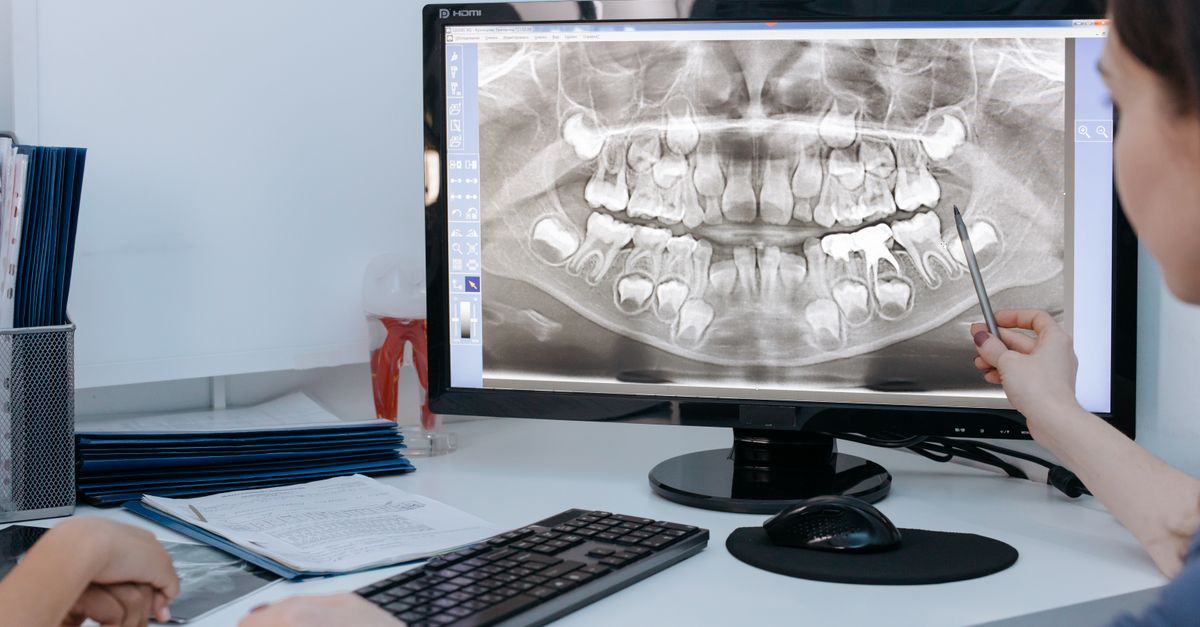
A panoramic radiograph is the standard initial imaging modality. This single, wide-angle X-ray captures the entire upper and lower jaw, visualizing all four third molars simultaneously. It reveals the exact position of the impacted teeth in relation to the second molars, the sinuses in the upper jaw, and the inferior alveolar nerve canal in the lower jaw.
When the panoramic X-ray indicates that the roots of a lower wisdom tooth are dangerously entwined with the major sensory nerve of the jaw, a Cone Beam Computed Tomography (CBCT) scan is ordered. This advanced 3D imaging provides unparalleled, millimeter-accurate visualization, allowing the oral surgeon to plan a safe extraction trajectory that avoids catastrophic nerve damage.
10. Indications for Surgical Extraction
The decision to surgically extract impacted third molars is based on a careful assessment of current pathology and the probability of future complications. Preventative extraction during late adolescence is frequently recommended, as the roots are not yet fully formed, the surrounding bone is softer, and the patient’s biological healing capacity is optimal.
Definitive clinical indications for immediate extraction include recurrent episodes of pericoronitis, the presence of non-restorable cavities on the wisdom tooth or the adjacent second molar, or radiographic evidence indicating the formation of a cyst or tumor.
Conversely, if a wisdom tooth is completely embedded deep within the bone, is covered by healthy tissue, exhibits no signs of disease, and poses a high surgical risk of nerve damage, the surgeon may elect a strategy of active surveillance. This involves leaving the tooth in place and monitoring it with panoramic X-rays every few years to ensure no silent pathology develops.
11. The Surgical Extraction Procedure
The surgical removal of an impacted third molar is an intricate oral surgery procedure. Depending on the complexity and the patient’s anxiety level, the surgery is performed under local anesthesia, intravenous conscious sedation, or full general anesthesia.
The oral and maxillofacial surgeon begins by making a precise incision in the gum tissue to create a surgical flap, exposing the hidden bone and the impacted tooth. Often, the tooth is entirely encased in dense bone. The surgeon utilizes a specialized, high-speed surgical drill to carefully remove the overlying bone, liberating the crown of the tooth.
Because impacted teeth are frequently locked at severe angles, attempting to pull the entire tooth out in one piece would cause massive bone trauma. Instead, the surgeon meticulously sections the tooth, cutting it into smaller, manageable fragments. These individual fragments are then gently elevated and removed from the socket, minimizing trauma to the surrounding jawbone.
12. Postoperative Care and Healing
Following the successful extraction, the surgical site is thoroughly irrigated with sterile saline, and the gum flap is sutured securely back into place. Proper postoperative care is absolutely critical to ensure rapid healing and prevent painful complications.
Patients will experience significant localized swelling, mild bruising, and moderate discomfort peaking approximately 48 hours post-surgery. Aggressive application of ice packs to the face during the first 24 hours drastically reduces this inflammatory swelling. Pain is managed through a strict regimen of scheduled non-steroidal anti-inflammatory drugs, occasionally supplemented with stronger analgesics for the first few days.
Strict adherence to a soft-food diet is mandatory to prevent mechanical trauma to the surgical sites. Patients are explicitly instructed not to use straws, spit forcefully, or smoke for several days. The negative pressure generated in the mouth by these actions can violently dislodge the vital blood clot forming in the socket, precipitating the most dreaded postoperative complication: a dry socket.
13. The Dry Socket Complication
Alveolar osteitis, commonly referred to as a dry socket, is an intensely painful complication occurring in approximately five percent of lower wisdom tooth extractions. It occurs when the biological blood clot that naturally forms within the surgical socket to protect the exposed bone is prematurely dissolved or physically dislodged.
Without this protective clot, the raw jawbone and highly sensitive nerve endings within the socket are directly exposed to air, food debris, and oral fluids. The resulting pain is excruciating, typically beginning three to five days after the surgery, and frequently radiating deeply into the ear or eye.
Managing a dry socket does not require antibiotics, as it is an inflammatory process, not an infection. The patient must return to the oral surgeon, who will gently flush the socket and pack it with a specialized, medicated dressing. This dressing contains powerful soothing agents, such as eugenol, which provide almost instantaneous, profound pain relief while the body heals the exposed bone via secondary intention.
14. Nerve Injury Risks
The most significant anatomical risk associated with extracting lower impacted wisdom teeth is injury to the inferior alveolar nerve. This major nerve travels through a canal within the lower jawbone, providing feeling to the lower lip, chin, and teeth. The roots of fully developed lower wisdom teeth frequently wrap around or compress this nerve canal.
During complex extractions, the physical manipulation of the tooth fragments can bruise, stretch, or rarely, severe this nerve. A nerve injury manifests as profound numbness, persistent tingling sensations, or altered feeling in the lower lip and chin that persists long after the local anesthesia has worn off.
In the vast majority of cases, this nerve injury is a temporary neuropraxia caused by swelling or bruising. Sensation typically returns fully within a few weeks to several months as the nerve heals. Permanent nerve damage is a severe, albeit exceptionally rare, complication that underscores the necessity of having complex impactions managed by board-certified oral and maxillofacial surgeons utilizing advanced 3D imaging.
15. When to Seek an Oral Surgery Consultation
Any individual experiencing recurrent pain in the back of the jaw, sudden difficulty opening their mouth, or swelling of the gums behind their last visible tooth should seek an immediate evaluation from a dental professional.
Late adolescence—typically between the ages of 16 and 19—is the optimal time for a proactive, comprehensive evaluation of the third molars. During this precise biological window, the roots of the wisdom teeth are only partially formed, making them structurally easier to remove, and pulling them before they become impacted prevents decades of potential dental complications.
By engaging proactively with an oral and maxillofacial surgeon, patients can secure a definitive diagnosis through advanced 3D imaging, allowing for a highly controlled, planned surgical intervention rather than suffering through an acute, painful emergency infection years later.
16. Frequently Asked Questions (FAQ)
1. Does everyone have to get their wisdom teeth removed?
No. If your jaw is large enough to accommodate the teeth, they grow in completely straight, and you can easily reach them with a toothbrush to keep them perfectly clean, they do not need to be removed. However, this scenario is quite rare in modern anatomy.
2. Why do dentists recommend removing them even if they do not hurt?
Impacted teeth are a ticking time bomb. They often remain silent while silently dissolving the bone around them, creating large cysts, or rotting the healthy tooth next to them. Waiting until they cause severe pain means the damage to your jaw or adjacent teeth has already occurred.
3. Will I be awake when the surgeon cuts the teeth out?
It depends on your preference and the complexity of the surgery. For severe impactions, oral surgeons usually provide deep intravenous sedation. You will be completely asleep, feel absolutely no pain during the procedure, and wake up with no memory of the surgery.
4. What happens if I use a straw after surgery?
Using a straw creates a powerful vacuum in your mouth. This suction can violently rip the healing blood clot out of the hole in your jawbone. This leaves the raw bone and nerves exposed to the air, causing an agonizing condition called a dry socket.
5. How long will my face be swollen after the surgery?
Swelling is a normal part of the healing process. It usually peaks roughly 48 hours after the surgery and then slowly goes down. By the end of the first week, the majority of the visible swelling should be completely resolved.
17. Bibliography
Disclaimer: The content is for informational purposes only and does not replace medical advice. Always consult your doctor for personalized treatment.
