1. Introduction
A Cytomegalovirus (CMV) infection involves a widespread DNA virus that establishes lifelong latency within the human host, typically causing mild or unnoticeable symptoms in healthy individuals but posing severe, potentially fatal risks to fetuses and immunocompromised patients. The bottom line up front is that while the majority of the population carries this virus silently, clinical intervention—primarily involving specialized antiviral medications—is strictly required when the virus reactivates in patients with weakened immune systems, causing end-organ damage, or when it is transmitted during pregnancy.
CMV belongs to the Herpesviridae family, sharing the biological hallmark of establishing a permanent, dormant presence in specific cells after the initial acute infection resolves. Managing this virus in a clinical setting requires understanding the delicate balance between the host’s cellular immunity and viral replication.
2. Virology and Cellular Mechanics
Cytomegalovirus is an enveloped, double-stranded DNA virus. The name “cytomegalo” translates to “large cell,” reflecting the characteristic cytopathic effect the virus induces. When observed under a microscope, cells actively infected with CMV become significantly enlarged and contain distinct viral inclusion bodies, often described as having an “owl’s eye” appearance.
The virus exhibits a slow replication cycle compared to other viruses. It primarily targets endothelial cells, epithelial cells, and macrophages. By infecting these widespread cell types, the virus facilitates its dissemination throughout various organ systems before the immune system can mount a comprehensive neutralizing response.
3. Mechanisms of Latency and Reactivation
Following a primary infection, a robust human immune response forces the virus into a state of latency. CMV establishes this dormant state predominantly within hematopoietic stem cells residing in the bone marrow and circulating monocytes in the peripheral blood.
During latency, the virus does not actively produce infectious viral particles. Its DNA remains quietly integrated or episomal within the host cell nucleus. However, if the host’s immune surveillance wanes—due to stress, aging, immunosuppressive medications, or acquired immunodeficiencies—the virus can reactivate, initiating a new cycle of replication and potentially causing symptomatic tissue-invasive disease.
4. Transmission Pathways
CMV is transmitted through direct contact with infected bodily fluids. Because the virus is shed intermittently in saliva, urine, blood, tears, semen, and breast milk, transmission requires close, intimate, or prolonged physical contact.
In children, transmission frequently occurs in daycare settings through the sharing of toys contaminated with saliva or urine. Adults commonly acquire the virus through sexual contact or by caring for infected children. Additionally, the virus can be transmitted iatrogenically via blood transfusions or solid organ transplants if the donor tissue harbors latent virus.
5. Primary Infection in Healthy Individuals
In an individual with a competent immune system, a primary CMV infection is usually entirely asymptomatic. The immune system efficiently controls the viral replication without the host ever realizing they were infected.
When symptoms do occur in healthy adults or adolescents, they typically manifest as a mononucleosis-like syndrome. The patient may present with prolonged fatigue, fever, myalgia, and enlarged lymph nodes (lymphadenopathy). Unlike the classic infectious mononucleosis caused by the Epstein-Barr virus, CMV mononucleosis is usually characterized by the absence of severe pharyngitis (sore throat) and negative heterophile antibody testing.
6. Congenital CMV Infection
Congenital CMV is a primary clinical concern, representing the most common infectious cause of birth defects globally. It occurs when a pregnant person experiences a primary infection, or a significant reactivation, and the virus crosses the placenta to infect the developing fetus.
The risk of severe fetal damage is statistically highest when the primary maternal infection occurs during the first trimester of pregnancy. The virus disrupts normal cellular proliferation and differentiation, particularly within the developing fetal brain, leading to significant neurological and structural abnormalities.
7. Neonatal Complications and Long-Term Effects
While the majority of infants born with congenital CMV are asymptomatic at birth, approximately ten percent present with severe clinical signs. These include a petechial rash (blueberry muffin appearance), jaundice, an enlarged liver and spleen (hepatosplenomegaly), microcephaly (abnormally small head), and intrauterine growth restriction.
Even infants who appear asymptomatic at birth face significant long-term neurological risks. Congenital CMV is a leading non-genetic cause of progressive sensorineural hearing loss. Affected children may also experience developmental delays, vision impairment, and intellectual disability, underscoring the need for vigilant pediatric follow-up.
8. CMV in Solid Organ Transplant Recipients
Solid organ transplant recipients are highly vulnerable to CMV due to the heavy immunosuppressive medications required to prevent organ rejection. In this population, CMV is considered a major opportunist and a significant cause of morbidity.
The highest risk occurs when an organ from a CMV-positive donor is transplanted into a CMV-negative recipient. The recipient receives a massive viral load within the transplanted tissue without possessing any pre-existing neutralizing antibodies. This can lead to severe CMV syndrome, characterized by persistent fever, profound bone marrow suppression (leukopenia and thrombocytopenia), and direct viral damage to the transplanted organ.
9. Tissue-Invasive Disease
When CMV replication is entirely unchecked by the immune system, the virus can cause severe tissue-invasive disease, attacking specific organ systems. CMV pneumonia (pneumonitis) is a rapidly progressive and highly lethal complication, particularly in hematopoietic stem cell transplant patients.
Gastrointestinal CMV disease can manifest as severe esophagitis or colitis, causing ulcerations, debilitating pain, and severe diarrhea. CMV hepatitis and encephalitis are other life-threatening manifestations that highlight the virus’s ability to destroy tissues universally when cellular immunity is compromised. Recognizing these systemic issues often complements viral hepatitis overview protocols.
10. Diagnostic Laboratory Testing
Diagnosing a CMV infection requires precision, as the goal is to differentiate between asymptomatic latent infection, active viral shedding, and actual tissue-invasive disease. The specific test utilized depends entirely on the patient’s immune status and presenting symptoms.
| Diagnostic Modality | Clinical Application | Interpretation |
|---|---|---|
| CMV Serology (IgG / IgM) | Healthy patients, pregnant individuals | Determines past exposure (IgG) or suggests recent primary infection (IgM). Not useful in immunocompromised states. |
| Quantitative PCR (Viral Load) | Transplant and immunocompromised patients | Measures the exact amount of viral DNA in the blood; essential for monitoring reactivation and treatment response. |
| Tissue Biopsy and Histopathology | Suspected tissue-invasive disease (e.g., colitis) | Gold standard for diagnosing end-organ damage; reveals classic “owl’s eye” viral inclusions in cells. |
11. Monitoring Viral Loads
For highly vulnerable patients, such as stem cell transplant recipients, clinicians do not wait for symptoms to appear. Instead, they employ a strategy known as preemptive therapy.
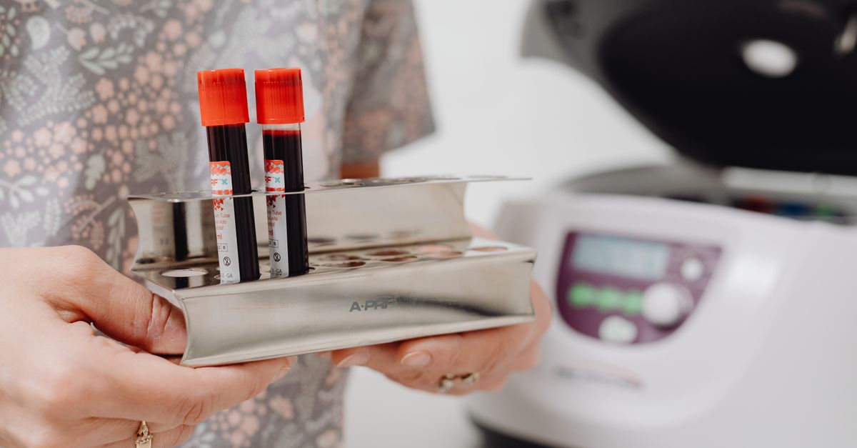
The patient’s blood is drawn weekly, and a quantitative Polymerase Chain Reaction (PCR) test is performed to measure the CMV viral load. If the PCR detects rising levels of viral DNA—indicating that the virus is reactivating and multiplying—the clinician immediately initiates antiviral medication to suppress the virus before it can progress to tissue-invasive disease.
12. Pharmacological Antiviral Therapy
Unlike bacterial infections, CMV cannot be treated with standard antibiotics. The pharmacological management of active, symptomatic CMV requires specialized antiviral medications that interrupt the viral DNA replication process.
Intravenous ganciclovir and its oral prodrug, valganciclovir, are the first-line therapies. These medications inhibit the viral DNA polymerase enzyme. While highly effective, they carry significant clinical toxicities, most notably severe bone marrow suppression, which can cause dangerous drops in white blood cell counts (neutropenia), necessitating careful clinical monitoring during the treatment course.
13. Managing Drug Toxicity and Resistance
If a patient cannot tolerate the bone marrow suppression caused by ganciclovir, or if the virus develops a genetic mutation rendering the drug ineffective (resistance), alternative antiviral agents must be utilized.
Foscarnet is a second-line medication that also blocks viral DNA synthesis but binds to a different site on the enzyme. However, foscarnet is notoriously nephrotoxic and can cause severe kidney damage and electrolyte imbalances. Cidofovir is another option, though it also carries a profound risk of renal toxicity, restricting its use to specialized clinical scenarios under the guidance of infectious disease experts.
14. Prevention and Prophylaxis
In solid organ transplantation, prophylactic therapy is the standard of care for high-risk donor-recipient pairs. The recipient is placed on oral valganciclovir for several months immediately following the transplant surgery. This continuous antiviral pressure suppresses the virus during the period of maximum immunosuppression.
For pregnant individuals, prevention relies on rigorous hygiene. Because a vaccine is not yet commercially available, pregnant people are counseled to avoid sharing food, utensils, or pacifiers with young children and to wash their hands thoroughly after changing diapers, as toddler urine and saliva are primary vectors for transmission.
15. Long-term Outlook and Prognosis
For the vast majority of the global population with a healthy immune system, CMV remains a benign, silent lifelong companion that does not alter life expectancy or general health.
For immunocompromised patients and congenitally infected infants, the prognosis relies entirely on rapid diagnostic testing, proactive monitoring, and the judicious use of antiviral therapies. Modern medical protocols, including preemptive therapy and robust antiviral agents, have substantially reduced the mortality associated with CMV, transforming a once-fatal complication into a highly manageable condition.
16. Frequently Asked Questions (FAQ)
1. Is Cytomegalovirus a type of herpes?
Yes, it belongs to the herpesvirus family. Like other herpes viruses (such as the ones that cause cold sores or chickenpox), once you catch it, the virus goes to sleep (becomes latent) and stays in your body for the rest of your life.
2. How do you know if you have a CMV infection?
Most healthy people never know they have it because it causes no symptoms. If symptoms do occur, they usually feel like a mild case of the flu or mononucleosis, with fatigue and a low fever.
3. Why is this virus dangerous for pregnant women?
If a pregnant woman catches the virus for the first time, it can cross the placenta and infect the developing baby. This congenital infection can cause severe developmental issues, including permanent hearing loss and neurological delays.
4. Can antibiotics cure a CMV infection?
No. Antibiotics only kill bacteria, not viruses. CMV requires specialized antiviral medications, such as valganciclovir, which are usually only prescribed for patients with weakened immune systems who cannot fight off the virus naturally.
5. How can I avoid catching CMV?
Because the virus is shed in bodily fluids like saliva and urine, careful hygiene is the best prevention. Thorough handwashing, especially after changing diapers or wiping a child’s nose, and avoiding sharing cups or utensils are key preventative measures.
17. Bibliography
Disclaimer: The content is for informational purposes only and does not replace medical advice. Always consult your doctor for personalized treatment.