1. Introduction
An inflammatory disease of the mucous membrane, medically referred to as mucositis or mucosal inflammation, involves the swelling, irritation, and potential ulceration of the protective linings within the body. These linings cover various internal cavities, including the gastrointestinal tract, respiratory system, and urogenital pathways. The condition arises when the epithelial cells forming these barriers are damaged by infection, immune system dysregulation, or toxic exposures, prompting an acute or chronic cellular immune response.
This article provides a comprehensive clinical overview of mucosal inflammatory conditions. It details the underlying pathophysiology, common triggers, diagnostic protocols, and established therapeutic strategies. Understanding the mechanisms of mucosal injury is essential for achieving proper symptom relief and preventing long-term structural changes to the affected tissues.
2. Anatomy and Function of Mucous Membranes
Mucous membranes are specialized epithelial tissues that line organs and body cavities exposed to the external environment. They are structurally composed of one or more layers of epithelial cells resting on a layer of connective tissue known as the lamina propria. These tissues are highly vascularized, allowing for rapid cellular turnover and a constant supply of immune cells.
The primary function of these membranes is protective. They secrete mucus, a viscous fluid containing mucins, antimicrobial peptides, and immunoglobulins, primarily immunoglobulin A. This secretion forms a physical and chemical barrier against pathogens, abrasive substances, and dehydration. Additionally, mucous membranes facilitate vital physiological processes such as nutrient absorption in the intestines and gas exchange in the lungs.
When these structural and functional barriers are compromised, the underlying tissues become vulnerable. The loss of mucosal integrity allows antigens and pathogens to penetrate deeper layers, setting the stage for a localized or widespread inflammatory response.
3. Pathophysiology of Mucosal Inflammation
The pathophysiological mechanism of mucosal inflammation begins with an initial injury to the epithelial layer. This insult triggers the release of pro-inflammatory cytokines, including tumor necrosis factor-alpha and interleukins, from damaged epithelial cells and resident macrophages. These chemical signals cause local vasodilation and increased capillary permeability.
As blood vessels dilate, fluid and circulating immune cells, such as neutrophils and lymphocytes, migrate into the lamina propria. This influx of cells and fluid manifests clinically as swelling, redness, and heat in the affected tissue. The recruited immune cells release further chemical mediators, reactive oxygen species, and enzymes aimed at neutralizing the offending agent but often resulting in collateral damage to the surrounding healthy tissue.
In cases where the inflammatory stimulus persists, the condition can transition from an acute response to a chronic state. Chronic mucosal inflammation involves a shift in the cellular infiltrate toward macrophages, plasma cells, and T-lymphocytes. Prolonged inflammation can lead to tissue remodeling, characterized by fibrosis, atrophy, or metaplasia, which fundamentally alters the normal function of the affected organ.
4. Common Types of Mucosal Inflammatory Diseases
Mucosal inflammation can manifest in nearly any organ system lined by these protective tissues. Stomatitis involves the inflammation of the oral mucosa, often presenting as painful ulcers or widespread redness. This condition can severely impact a patient’s ability to eat and speak. Similarly, esophagitis affects the lining of the esophagus and is frequently associated with acid reflux or specific infections.
In the respiratory tract, rhinitis and bronchitis represent common forms of mucosal inflammation. Rhinitis involves the nasal passages, typically reacting to airborne allergens or viral pathogens, leading to increased mucus production and tissue swelling. Bronchitis affects the lower respiratory airways, causing a persistent cough and impaired airflow due to bronchial mucosal thickening.
The gastrointestinal tract is another frequent site for these conditions. Enteritis and colitis describe inflammation of the small intestine and colon, respectively. These conditions can range from acute infectious episodes to chronic autoimmune conditions like inflammatory bowel disease. Each specific manifestation requires a tailored diagnostic and therapeutic approach based on its anatomical location.
5. The Role of Immune System Dysregulation
Immune system dysregulation plays a central role in many non-infectious inflammatory diseases of the mucous membranes. In conditions such as mucous membrane pemphigoid, the immune system inappropriately produces autoantibodies directed against structural proteins in the basement membrane zone. This targeted attack causes the epithelial layer to detach from the underlying connective tissue, leading to blister formation and subsequent ulceration.
Similarly, conditions like lichen planus involve a cell-mediated immune response where cytotoxic T-cells mistakenly target the basal keratinocytes of the oral or genital mucosa. The exact trigger for this autoimmune reaction remains poorly understood, but it results in a chronic, painful desquamation of the mucosal surface.
Managing these immune-mediated forms of mucosal inflammation requires complex interventions aimed at modulating or suppressing the overactive immune response. Unlike infectious causes, these conditions are often chronic and require long-term monitoring to prevent severe complications, including irreversible scarring.
6. Infectious Causes and Pathogens
Bacterial, viral, and fungal infections are prevalent triggers for acute mucosal inflammation. Viral pathogens, such as the herpes simplex virus, frequently infect the oral and genital mucosa. The virus replicates within the epithelial cells, causing them to lyse and form painful vesicular lesions that eventually ulcerate.
Fungal infections, particularly those caused by Candida species, commonly affect the mucous membranes of the mouth, esophagus, and vagina. Candidiasis typically occurs when the normal microbial flora is disrupted, often following systemic antibiotic use or in individuals with compromised immune function. The fungal overgrowth induces a localized inflammatory response characterized by white plaques and underlying erythema.
Bacterial infections can also precipitate severe mucosal inflammation. For instance, Helicobacter pylori specifically colonizes the gastric mucosa, inducing chronic inflammation that can lead to peptic ulcer disease or gastric atrophy. Identifying the specific pathogen is critical for administering targeted antimicrobial therapy.
7. Environmental and Chemical Triggers
Exposure to noxious environmental agents or harsh chemicals can directly damage the mucosal epithelium and initiate an inflammatory cascade. Tobacco smoke is a well-documented irritant that severely affects the respiratory and oral mucosa. The toxic compounds in smoke impair ciliary function and induce a state of chronic inflammation, predisposing individuals to recurrent infections and cellular dysplasia.
Therapeutic interventions can also act as significant chemical triggers. Cytotoxic chemotherapy and localized radiation therapy frequently cause severe oral and gastrointestinal mucositis. These treatments target rapidly dividing cells, inadvertently destroying the highly proliferative epithelial cells of the mucous membranes. This leads to profound mucosal breakdown, severe pain, and a high risk of secondary infections.
Dietary factors and occupational exposures also contribute to mucosal irritation. Frequent consumption of highly acidic or spicy foods can exacerbate pre-existing oral or esophageal inflammation. Similarly, inhaling industrial chemicals or particulate matter can induce chronic rhinitis or bronchitis in susceptible individuals.
8. Clinical Manifestations and Symptoms
The clinical presentation of mucosal inflammation varies widely depending on the anatomical site involved. However, certain hallmark signs are consistently observed across different tissues. Erythema, or redness, is typically the first visible sign, resulting from increased local blood flow. This is frequently accompanied by edema, causing the tissue to appear swollen and boggy.
Pain is a prominent and often debilitating symptom. Because mucous membranes are rich in sensory nerve endings, any breach in the epithelial barrier exposes these nerves to chemical irritants and temperature fluctuations. This can manifest as a burning sensation in the mouth, sharp pain during swallowing in esophagitis, or severe abdominal cramping in colitis.
In advanced or severe cases, the inflammation progresses to ulceration. Ulcers represent areas where the entire epithelial layer has been lost, exposing the underlying vascular connective tissue. These lesions are highly prone to bleeding and represent a significant portal of entry for secondary bacterial or fungal infections.
9. Diagnostic Approaches and Biopsy
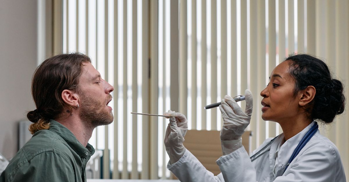
Accurate diagnosis of mucosal inflammatory diseases requires a systematic clinical evaluation. The initial assessment involves a detailed patient history, focusing on symptom onset, potential exposures, dietary habits, and current medications. A thorough physical examination of accessible mucous membranes, such as the oral cavity or nasal passages, provides immediate clinical clues.
For deeper mucosal surfaces, endoscopic evaluation is frequently necessary. Procedures such as esophagogastroduodenoscopy or colonoscopy allow clinicians to visualize the mucosal lining directly. Endoscopy can reveal subtle signs of inflammation, such as friability, loss of vascular pattern, or the presence of microscopic ulcerations.
When the etiology remains unclear or when autoimmune conditions are suspected, a tissue biopsy is paramount. Histopathological examination of the mucosal tissue can identify specific inflammatory cell infiltrates, structural abnormalities, or the presence of specific pathogens. Direct immunofluorescence testing on biopsy specimens is particularly crucial for diagnosing blistering diseases like mucous membrane pemphigoid.
10. Data Summary: Common Causes and Symptoms
To better understand the multifaceted nature of these conditions, the following table summarizes the relationship between common causes and their associated clinical presentations.
| Category of Cause | Common Triggers | Typical Symptoms |
|---|---|---|
| Infectious | Herpes Simplex, Candida, H. pylori | Vesicles, white plaques, localized pain, mucosal bleeding |
| Immune-Mediated | Pemphigoid, Lichen Planus, IBD | Chronic ulceration, desquamation, recurrent inflammation, systemic symptoms |
| Chemical / Toxic | Chemotherapy, Radiation, Tobacco | Widespread erythema, severe burning sensation, tissue sloughing |
| Allergic | Pollen, Food Allergens, Medications | Edema, increased mucus production, pruritus, sudden onset |
11. Differential Diagnosis
Distinguishing between various causes of mucosal inflammation is a critical step in formulating an effective treatment plan. A primary challenge in the differential diagnosis is separating benign inflammatory processes from pre-malignant or malignant lesions. Chronic mucosal ulcers that do not heal within a standard timeframe must be evaluated for squamous cell carcinoma or other malignancies.
Clinicians must also differentiate between localized infections and systemic autoimmune conditions. For example, oral ulcers might be a localized response to trauma or a manifestation of systemic lupus erythematosus or Behcet disease. This distinction often relies on a comprehensive review of systems and targeted serological testing.
Furthermore, nutritional deficiencies must be ruled out. Deficiencies in vitamin B12, iron, or folate can cause atrophic glossitis and generalized mucosal erythema that closely mimics primary inflammatory diseases. A complete blood count and specific vitamin assays are standard components of the diagnostic workup.
12. Treatment Strategies: Topical and Systemic
The therapeutic approach to mucosal inflammation is dictated by the underlying cause and the severity of the symptoms. For localized, mild inflammation, topical treatments are frequently sufficient. These may include anesthetic rinses containing viscous lidocaine to manage pain, or topical corticosteroids to suppress local immune responses and reduce edema.
In cases driven by infectious agents, targeted antimicrobial therapy is required. Antiviral medications, antifungal troches, or specific antibiotic regimens are deployed based on microbiological findings. Eradicating the pathogen allows the mucosal barrier to regenerate and restore its protective function.
For severe or systemic immune-mediated conditions, systemic therapy is necessary. Oral corticosteroids are often used to gain rapid control over severe inflammation. For long-term management, corticosteroid-sparing agents such as azathioprine, mycophenolate mofetil, or biologic therapies targeting specific inflammatory pathways may be prescribed. These treatments require careful monitoring for potential adverse effects, including systemic immunosuppression.
13. Nutritional Support and Lifestyle Modifications
Maintaining adequate nutrition is a significant challenge for patients with severe mucosal inflammation, particularly when the oral cavity or gastrointestinal tract is involved. Pain associated with eating can lead to reduced caloric intake, resulting in unintended weight loss and delayed tissue healing. Dietary modifications, such as consuming soft, bland foods and avoiding acidic, spicy, or thermally hot meals, can help minimize irritation.
Hydration is equally critical. The mucous membranes rely on adequate systemic hydration to produce the protective mucus layer. Patients are encouraged to consume frequent, small sips of water or use synthetic saliva substitutes if normal salivary gland function is impaired.
Lifestyle modifications play a crucial role in preventing recurrent episodes. Smoking cessation is imperative, as tobacco smoke continuously degrades mucosal integrity and impedes cellular repair. Additionally, practicing excellent oral hygiene and using soft-bristled toothbrushes can prevent mechanical trauma to delicate oral tissues, which is a common trigger for localized inflammatory flares.
14. Complications of Untreated Mucosal Inflammation
Failing to adequately manage mucosal inflammation can result in severe and potentially irreversible complications. Chronic inflammation leads to continuous tissue remodeling, which can result in stricture formation. In the esophagus or intestines, strictures cause a narrowing of the lumen, obstructing the passage of food or waste and often necessitating surgical intervention.
Another significant complication is mucosal scarring, medically known as cicatrization. In conditions like mucous membrane pemphigoid, scarring of the ocular conjunctiva can lead to symblepharon formation, where the eyelid fuses to the eyeball, potentially resulting in blindness. Scarring in the upper airway can similarly compromise respiratory function.
Furthermore, chronic inflammation is a known risk factor for dysplastic cellular changes. Tissues subjected to constant cellular turnover and oxidative stress are at an increased risk of malignant transformation. Regular clinical surveillance is necessary for patients with long-standing mucosal inflammatory conditions to detect any neoplastic changes early.
15. When to See a Healthcare Professional
Patients should seek prompt medical evaluation when mucosal inflammation interferes with fundamental daily activities such as eating, drinking, or breathing. The inability to maintain adequate hydration due to oral or esophageal pain is a medical emergency that requires immediate intervention and potential intravenous fluid support.
Additionally, the appearance of systemic symptoms accompanying mucosal changes warrants immediate clinical attention. High fever, unexplained weight loss, extreme fatigue, or the development of cutaneous rashes alongside mucosal lesions may indicate a widespread infectious or autoimmune process that requires comprehensive diagnostic testing.
Persistent mucosal ulcers that fail to heal after two weeks, regardless of whether they are painful or painless, must be evaluated by a healthcare professional. Such non-healing lesions necessitate a biopsy to definitively rule out underlying malignancy or chronic granulomatous diseases.
16. Current Research and Future Therapies
The landscape of treatment for mucosal inflammatory diseases is rapidly evolving due to continuous scientific research. Current studies are heavily focused on understanding the precise molecular pathways that drive chronic mucosal inflammation. By mapping these pathways, researchers are developing targeted biologic therapies that can intercept specific pro-inflammatory cytokines without causing generalized immunosuppression.
Another promising area of research involves the microbiome and its interaction with mucosal immunity. There is growing evidence that dysbiosis, or an imbalance in the normal bacterial flora residing on mucous membranes, significantly contributes to inflammatory states. Interventions aimed at restoring microbial balance, such as advanced probiotics or targeted microbiota transplants, are undergoing clinical trials.
Advancements in tissue engineering also offer hope for patients with severe mucosal damage. Researchers are exploring methods to cultivate autologous epithelial cells in the laboratory, which could theoretically be grafted onto areas of extensive ulceration or scarring to restore normal anatomical structure and function.
17. Frequently Asked Questions (FAQ)
1. Is mucosal inflammation contagious?
Mucosal inflammation itself is not contagious. However, if the inflammation is caused by an infectious agent, such as a virus or bacteria, that underlying pathogen can be transmitted to others through direct contact or respiratory droplets.
2. Can stress cause inflammatory diseases of the mucous membrane?
While stress does not directly cause these diseases, it can significantly alter immune function. High levels of physiological or psychological stress can trigger flare-ups in patients with underlying autoimmune or chronic inflammatory conditions.
3. How long does it take for a mucosal ulcer to heal?
Minor, acute mucosal ulcers typically heal within ten to fourteen days with proper care. Ulcers that persist beyond this timeframe require clinical evaluation to determine the underlying cause and to rule out more serious conditions.
4. Are there specific diets that help reduce mucosal inflammation?
Patients often benefit from an anti-inflammatory diet that avoids spicy, highly acidic, and rough-textured foods. Ensuring adequate intake of vitamins and maintaining hydration are essential for supporting mucosal repair.
5. What is the role of biologic medications in treating these conditions?
Biologic medications target specific parts of the immune system that drive inflammation. They are generally reserved for severe, chronic cases of immune-mediated mucosal diseases that do not respond to conventional corticosteroid therapies.
18. Bibliography
Disclaimer: The content is for informational purposes only and does not replace medical advice. Always consult your doctor for personalized treatment.