
1. Introduction
An injury of the external ear encompasses any structural damage to the auricle, also known as the pinna, and the external auditory canal. These injuries are typically the direct result of blunt force trauma, sharp lacerations, thermal insults, or the introduction of foreign bodies into the canal. Due to the exposed anatomical position of the auricle on the lateral aspect of the skull, it is remarkably susceptible to mechanical disruption during physical altercations, contact sports, and motor vehicle collisions.
The external ear serves a critical physiological function in the collection and directional funneling of acoustic waves toward the tympanic membrane. The structural framework relies on a delicate, complex architecture of elastic cartilage tightly bound by a thin layer of skin. When trauma compromises this framework, the resulting inflammatory cascade and fluid accumulation can rapidly compromise the restricted vascular supply of the cartilage.
Timely and precise clinical intervention is imperative to prevent permanent aesthetic deformity and preserve auditory function. Understanding the localized vascular anatomy and the specific pathophysiological mechanisms driving tissue necrosis allows healthcare providers to implement evidence-based drainage, repair, and stabilization protocols to optimize healing.
2. Anatomy of the Auricle and External Canal
The auricle is uniquely constructed entirely of flexible, elastic cartilage, with the exception of the fatty lobule at the inferior pole. This cartilaginous skeleton is sculpted into intricate ridges and depressions, including the helix, antihelix, tragus, and concha. The overlying skin is exceptionally thin and adheres directly to the perichondrium, the dense connective tissue layer encasing the cartilage.
The external auditory canal extends inward from the concha to the tympanic membrane. The outer third of this canal is cartilaginous and contains specialized apocrine glands responsible for cerumen production. The inner two-thirds consist of a bony tunnel lined with a fragile, highly sensitive layer of squamous epithelium that lacks a protective subcutaneous fat layer.
Crucially, the auricular cartilage lacks its own internal blood supply. It depends entirely on the diffusion of oxygen and nutrients from the vascular network within the tightly adherent perichondrium. Any traumatic event that separates the perichondrium from the underlying cartilage immediately halts this vital diffusion process, placing the structural framework at significant risk for ischemic necrosis.
3. Biomechanics of Blunt Force Trauma
Blunt trauma to the lateral head frequently transfers significant kinetic energy directly to the auricle. This is commonly observed in contact sports such as wrestling, rugby, and martial arts, where the ear is subjected to recurrent compressive and shearing forces against hard surfaces or opponents.
When a shearing force is applied to the auricle, the skin and perichondrium are forcibly slid across the underlying cartilage. This mechanical stress tears the delicate perforating blood vessels that bridge the tissue layers. The resulting hemorrhage pools rapidly within the newly created potential space between the perichondrium and the cartilage matrix.
Because the skin is tightly bound to the cartilage architecture, the pooling blood faces notable resistance. This restricted space causes the localized pressure to rise quickly, resulting in a tense, firm swelling that fundamentally alters the normal anatomical contours of the ear.
4. Auricular Hematoma Formation
The accumulation of blood between the perichondrium and the cartilage is clinically defined as an auricular hematoma. This condition presents as a distinct, tender, and fluctuant swelling, most commonly located in the anterior upper half of the auricle, specifically within the scaphoid fossa or the conchal bowl.
The formation of an auricular hematoma constitutes a localized vascular emergency. The pooling blood creates a physical barrier that prevents the diffusion of arterial oxygen from the perichondrial vascular bed to the avascular cartilage. Prolonged separation rapidly induces ischemic hypoxia within the cartilaginous cells.
If the hematoma is not evacuated promptly, typically within hours of the injury, the deprived chondrocytes undergo necrosis. The localized death of these structural cells initiates a chaotic biological repair process that replaces the delicate elastic cartilage with dense, disorganized fibrous tissue, permanently altering the ear shape.
5. The Pathophysiology of Cauliflower Ear
The long-term sequela of an untreated or inadequately managed auricular hematoma is a permanent cosmetic and structural deformity known as cauliflower ear. This term describes the thick, nodular, and asymmetrical appearance of the auricle resulting from the aberrant healing of necrotic cartilage.
Following the ischemic death of the native chondrocytes, the body attempts to repair the structural defect. Fibroblasts migrate into the hematoma cavity and begin synthesizing an unorganized matrix of dense collagen. Concurrently, new, irregular neocartilage is generated in a haphazard manner, lacking the refined anatomical ridges of the original structure.
This fibrotic and cartilaginous mass thickens the auricle, effectively obliterating the normal anatomical contours. Once this firm, fibrous tissue is fully established, it is entirely irreversible through standard conservative measures and requires complex plastic reconstructive surgery to improve the cosmetic appearance.
6. Penetrating Trauma and Lacerations
Sharp trauma and penetrating injuries to the external ear occur through various mechanisms, including human or animal bites, lacerations from shattered glass, or tearing injuries related to earrings and body modifications. These injuries range from simple superficial skin cuts to complex, full-thickness lacerations that bisect the cartilaginous skeleton.
Lacerations involving the cartilage present specific clinical challenges. The cartilage edges lack the intrinsic vascularity to mount a standard cellular repair response, making the tissue notoriously slow to heal and exceptionally prone to secondary bacterial infection.
Traction injuries to the earlobe are a frequent presentation in the clinical setting. A sudden downward pull on an earring can easily split the soft, fatty tissue of the lobule. While structurally less complicated than cartilage lacerations, these injuries require meticulous, layered surgical closure to restore the natural rounded contour of the earlobe edge.
7. Thermal Injuries and Frostbite
The auricle is remarkably susceptible to thermal insults due to its prominent anatomical position, high surface area-to-volume ratio, and relative lack of insulating subcutaneous fat. Exposure to profound cold environments frequently results in frostbite, a condition characterized by localized tissue freezing and subsequent ischemic damage.
During severe cold exposure, the body induces localized peripheral vasoconstriction to preserve central core temperature. This drastically reduces arterial blood flow to the auricle. As tissue temperatures drop, ice crystals form within the extracellular spaces, drawing water out of the cells and causing profound cellular dehydration and structural collapse.
Upon rewarming, the damaged endothelial cells lining the local capillaries leak substantial amounts of fluid, causing prominent edema and blister formation. The sluggish blood flow frequently coagulates, forming microthrombi that permanently occlude the vascular supply, leading to delayed tissue necrosis and demarcation of the affected auricular margins.
8. Foreign Body Obstructions and Canal Abrasions
Trauma to the external auditory canal is frequently self-inflicted through the insertion of foreign objects, such as cotton swabs, hairpins, or matchsticks, typically in an attempt to remove cerumen or relieve localized pruritus. The skin lining the medial portion of the canal is extraordinarily thin and fragile.
Mechanical abrasion from these objects easily lacerates the delicate epithelium, causing localized bleeding and sharp, acute ear pain. This physical breach of the cutaneous barrier provides a direct portal of entry for opportunistic pathogens residing in the canal, significantly elevating the risk of developing a secondary bacterial infection known as acute otitis externa.
In pediatric populations, the insertion of small toys or beads into the canal is common. Attempting to remove spherical or tightly wedged objects without specialized otolaryngology instruments often pushes the object deeper, risking severe abrasion of the canal walls or traumatic perforation of the tympanic membrane.
9. Differentiating Superficial from Structural Damage
A critical step in evaluating an external ear injury is determining whether the trauma is confined to the superficial soft tissues or involves the underlying cartilaginous framework. This distinction dictates the complexity of the required medical intervention.
| Clinical Feature | Superficial Soft Tissue Injury | Structural Cartilage Injury |
|---|---|---|
| Visual Inspection | Localized redness, minor superficial abrasions. | Loss of normal anatomical ridges, full-thickness cuts. |
| Palpation Findings | Soft, pliable tissue with mild tenderness. | Fluctuant fluid pockets (hematoma) or palpable cartilage gaps. |
| Bleeding Pattern | Minor oozing from skin edges. | Deep, persistent bleeding or dark blood pooling under the skin. |
| Healing Prognosis | Excellent with basic wound care. | Risk of permanent deformity and serious infection. |
Injuries limited to the skin generally heal predictably with standard cleansing and topical antibiotics. Any evidence of cartilage exposure, hematoma formation, or structural deformity necessitates advanced surgical wound management to ensure the viability of the auricular skeleton.
10. Clinical Evaluation and Otoscopic Examination
The comprehensive assessment of ear trauma begins with a meticulous visual inspection of the entire auricle and the surrounding periauricular tissue. The clinician evaluates the symmetry of the ears, noting any blunting of the normal cartilaginous landmarks, which is the hallmark sign of an accumulating hematoma.
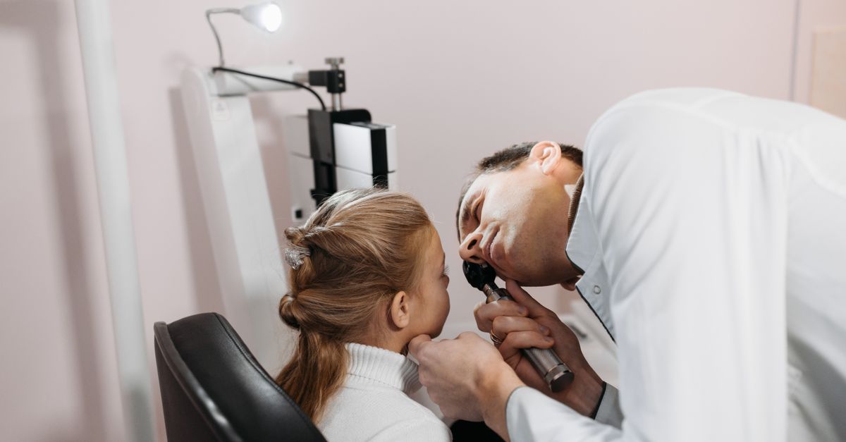
An otoscopic examination is a mandatory component of the clinical evaluation to assess the integrity of the external auditory canal and the tympanic membrane. The clinician carefully inserts the otoscope to search for canal lacerations, pooling blood, or obstructive foreign bodies.
Visualizing the tympanic membrane is essential to rule out a traumatic perforation or the presence of a hemotympanum, which refers to blood accumulating in the middle ear space behind the eardrum. These findings suggest a more profound transfer of kinetic energy and mandate a thorough assessment of auditory and vestibular function.
11. Diagnostic Imaging Modalities
While the majority of external ear injuries are diagnosed through direct clinical examination, specific traumatic scenarios warrant diagnostic imaging. Plain radiographs offer minimal value due to the cartilaginous nature of the auricle.
If the patient sustained substantial blunt force trauma to the lateral skull and presents with bleeding from the external canal, severe localized swelling, or signs of a skull base fracture, a high-resolution Computed Tomography scan of the temporal bone is indicated.
The Computed Tomography scan provides detailed cross-sectional images of the bony architecture. It can definitively identify fractures of the anterior canal wall resulting from mandibular condyle impaction, evaluate the status of the middle ear ossicles, and rule out deep structural damage that extends beyond the external cartilaginous framework.
12. Non-Surgical Management and Infection Control
For simple lacerations and superficial abrasions of the auricle, conservative management focuses on meticulous wound hygiene and infection prevention. The injury site is irrigated gently with sterile saline to remove particulate debris and coagulated blood.
Because the blood supply to the ear cartilage is tenuous, preventing secondary bacterial infection is paramount. Pathogens such as Pseudomonas aeruginosa and Staphylococcus aureus are common culprits in auricular infections. Prophylactic oral antibiotics covering these specific organisms are frequently prescribed for injuries involving exposed cartilage or wounds resulting from animal or human bites.
Topical antibiotic ointments are applied to skin lacerations to maintain a moist wound-healing environment. Patients are advised to protect the ear from further mechanical trauma and to avoid sleeping directly on the injured side to prevent sustained pressure from compromising the recovering microvascular circulation.
13. Surgical Drainage and Cartilage Repair
The definitive treatment for an auricular hematoma is immediate surgical evacuation. Using a sterile technique, a clinician makes a small incision along the natural contours of the ear, typically within the scaphoid fossa, to access the fluid cavity. The pooled blood and any developing clots are thoroughly suctioned and irrigated away.
Once the hematoma is evacuated, simply closing the incision is insufficient. The potential space between the skin and cartilage remains, and blood will reliably reaccumulate. To prevent recurrence, the clinician must apply a compressive bolster dressing.
This specialized dressing involves suturing sterile gauze rolls, silicone splints, or dental rolls tightly against both the anterior and posterior surfaces of the auricle. The sutures pass directly through the cartilage, physically clamping the skin back down onto the perichondrium. This sustained mechanical pressure eliminates the dead space, restoring the vital diffusion of oxygen to the chondrocytes.
14. Reconstructive Interventions for the Pinna
Complex, full-thickness lacerations or avulsions of the auricle require meticulous reconstructive techniques to restore cosmetic symmetry. The primary surgical objective is to perfectly align the severed edges of the cartilaginous skeleton.
Surgeons utilize fine, absorbable sutures to approximate the cartilage, ensuring that the stitches only pass through the perichondrium to avoid tearing the fragile cartilage matrix. The overlying skin is then closed carefully to minimize tension across the wound edges.
In cases involving significant tissue loss, such as severe animal bites, local tissue flaps or cartilage grafts harvested from the contralateral ear or the rib cage may be necessary to rebuild the structural framework. These complex reconstructive procedures are typically staged over several months to ensure adequate vascularization and optimal aesthetic outcomes.
15. Frequently Asked Questions (FAQ)
1. Why does my ear look swollen and lose its shape after wrestling?
The repetitive rubbing and impact during wrestling can tear the blood vessels between the skin and the ear cartilage. Blood pools in this space, creating a swollen, firm pocket called an auricular hematoma, which obscures the normal folds of the ear.
2. What exactly is cauliflower ear?
If a blood pocket on the ear is not drained by a doctor, the underlying cartilage dies from a lack of oxygen. The body tries to heal it by forming thick, hard scar tissue and irregular new cartilage, resulting in a permanently bumpy, deformed ear.
3. Should I try to drain a swollen ear with a needle at home?
No. Draining an ear hematoma requires strict sterile conditions. Doing it at home introduces a profound risk of causing a severe bacterial infection in the cartilage, which can destroy the entire structure of the ear.
4. How long does a pressure bandage need to stay on after draining the ear?
A doctor will typically leave a specialized compressive bolster dressing securely stitched to the ear for approximately one week. This constant pressure is necessary to ensure the skin heals tightly back against the cartilage and prevents blood from refilling the space.
5. Can a cut on the cartilage heal on its own?
Cartilage has a very poor blood supply and heals remarkably slowly. While a minor skin scratch will heal, a deep cut that goes into the cartilage usually requires careful stitches and prescription antibiotics to heal properly without becoming infected.
16. Bibliography
Disclaimer: The content is for informational purposes only and does not replace medical advice. Always consult your doctor for personalized treatment.
