
1. Introduction to Skin Fissures
A skin fissure is a sharply defined, linear cleavage or crack in the skin that extends through the superficial epidermis and penetrates deeply into the underlying dermis. Unlike superficial abrasions or minor cuts resulting from acute trauma, fissures develop slowly as a consequence of severe structural compromise within the skin barrier. These painful lesions frequently occur on areas of the body subject to constant mechanical stress and movement, particularly the heels of the feet, the tips of the fingers, and the web spaces between toes.
The development of a fissure signifies a complete failure of the skin’s natural elasticity and moisture retention capabilities. When the epidermis becomes excessively dry, thickened, and inelastic, even minor physical movements can cause the rigid tissue to fracture. Because the dermal layer contains a dense network of sensory nerves and blood capillaries, fissures are characteristically painful and prone to mild bleeding.
Clinical management of skin fissures requires a multifaceted dermatological approach. Treatment must focus not only on healing the acute wound but also on correcting the underlying dryness or inflammatory condition that led to the skin’s structural failure. Proper hydration, mechanical offloading, and addressing any underlying dermatoses are essential for achieving complete resolution and preventing recurrent fissuring.
2. Anatomy of the Epidermal Barrier
To understand why skin fractures, it is necessary to examine the microscopic structure of the epidermis. The outermost layer of the skin is the stratum corneum, composed of flattened, dead keratinocytes tightly bound together by a complex mixture of specialized lipids. This lipid matrix functions similarly to mortar between bricks, creating an impermeable barrier that traps internal moisture and blocks external environmental irritants.
Beneath the epidermis lies the dermis, a thicker layer containing collagen and elastin fibers that provide the skin with its structural strength and flexibility. The dermis also houses the vascular network and the intricate sensory nerve endings responsible for transmitting pain signals.
When the stratum corneum is healthy and well hydrated, it is highly pliable and easily stretches to accommodate joint movement and pressure. However, when the lipid barrier is depleted by environmental factors, harsh chemicals, or underlying skin diseases, the stratum corneum loses its water content rapidly. The dehydrated skin becomes brittle, hyperkeratotic, and fundamentally incapable of stretching, setting the stage for mechanical fracture.
3. Pathophysiology of Fissuring
The transition from dry skin to a full thickness fissure is a progressive biomechanical failure. As the stratum corneum dehydrates, the body often attempts to reinforce the weakened barrier by accelerating the production of keratinocytes. This compensatory mechanism results in hyperkeratosis, a thickening of the outer skin layer, commonly observed as calluses on the heels or palms.
While thicker skin might seem more protective, hyperkeratotic tissue is exceptionally rigid and lacks the elasticity of normal skin. When mechanical stress is applied to this thickened area, such as the weight of the body pressing down on a callused heel during walking, the inelastic skin cannot deform. Instead, the mechanical tension concentrates at a focal point, causing the tissue to split open.
The initial crack typically begins superficially but rapidly propagates downward under continued stress. Once the cleavage breaches the dermal-epidermal junction and enters the dermis, the highly sensitive nerve endings are exposed to air and physical friction. This exposure triggers the sharp, intense pain characteristic of a deep skin fissure.
4. Common Environmental and Lifestyle Causes
Environmental factors are primary drivers in the degradation of the epidermal barrier. Low ambient humidity, particularly during cold winter months, draws moisture directly out of the stratum corneum. The frequent transition between cold outdoor air and dry, centrally heated indoor environments severely taxes the skin’s ability to maintain hydration, leading to widespread xerosis.
Occupational exposures play a significant role, particularly for individuals engaged in wet work. Healthcare workers, culinary professionals, and cleaners who frequently wash their hands or use harsh alkaline soaps strip the natural protective lipids from their skin. This chronic depletion prevents the stratum corneum from recovering, making the fingertips and knuckles highly susceptible to cracking.
Footwear choices heavily influence the development of fissures on the feet. Wearing open-backed shoes or sandals allows the fat pad of the heel to expand laterally upon striking the ground. If the skin on the heel is already dry and hyperkeratotic, this lateral mechanical expansion forces the rigid callus to split, creating deep, painful heel fissures.
5. Underlying Dermatological Conditions
Beyond environmental dryness, several chronic inflammatory skin diseases directly impair epidermal function and strongly predispose patients to fissuring. Atopic dermatitis, or eczema, involves a genetic defect in the skin barrier proteins, leading to chronic moisture loss, severe inflammation, and recurrent cracking, particularly in the flexural folds of the skin.
Psoriasis is an autoimmune condition characterized by the rapid, chaotic turnover of skin cells, resulting in thick, scaly plaques. When these rigid psoriatic plaques develop on the palms or the soles of the feet, the inelastic tissue frequently fractures, causing severe pain and disability.
Contact dermatitis, whether irritant or allergic, triggers localized inflammation that damages the stratum corneum. Continuous exposure to an offending agent, such as specific industrial chemicals or even certain components of cosmetic products, leads to chronic skin thickening, drying, and eventual cleavage of the affected area.
6. Fungal Infections and Skin Cracking
Fungal infections, particularly tinea pedis, commonly known as athlete’s foot, are a frequent and often overlooked cause of skin fissures, especially in the web spaces between the toes. Dermatophyte fungi thrive in the warm, moist environment enclosed within modern footwear and actively digest the keratin of the stratum corneum.
As the fungus invades the superficial skin layers, it incites an inflammatory response that leads to scaling, maceration, and a breakdown of tissue integrity. The skin in the interdigital spaces becomes fragile and easily tears, creating painful superficial fissures.
In some variations of tinea pedis, the infection causes a dry, moccasin-like scaling across the entire sole of the foot. This widespread hyperkeratosis severely diminishes skin elasticity, culminating in deep fissures along the weight-bearing margins of the heel. Accurately diagnosing a fungal etiology is crucial, as the fissures will not resolve permanently without appropriate antifungal eradication.
7. Clinical Symptoms and Presentation
The clinical presentation of a skin fissure is distinct and readily identifiable. Patients typically present with a sharply defined, linear crack surrounded by thickened, dry, or scaly skin. The edges of the fissure are often hyperkeratotic and rigid. Depending on the depth of the cleavage, the base of the fissure may appear red and raw, indicating exposure of the dermal vascular bed.
Pain is the predominant symptom. The discomfort is usually described as a sharp, stinging, or burning sensation that is significantly exacerbated by movement, pressure, or exposure to water and irritating substances. For heel fissures, simply standing or walking can elicit severe pain, altering the patient’s gait.
If the fissure is deep enough to traumatize the dermal capillaries, minor bleeding will occur. While the blood loss is negligible, the presence of blood indicates a complete breach of the protective barrier, elevating the clinical concern for secondary complications.
8. Potential Complications: The Risk of Infection
A skin fissure represents an open portal directly into the sterile layers of the dermis, bypassing the body’s primary immunological defense mechanism. The most significant complication of an untreated fissure is the development of a secondary bacterial infection.
Pathogens naturally residing on the skin surface, such as Staphylococcus aureus, can easily colonize the nutrient-rich, moist environment at the base of a deep fissure. Clinical signs of a secondary bacterial infection include increased, localized warmth, spreading erythema extending beyond the edges of the crack, escalating pain, and the presence of purulent exudate or a yellow crust.
In patients with compromised immune systems, peripheral neuropathy, or poor peripheral circulation, such as those with poorly controlled diabetes mellitus, a minor infected fissure can rapidly progress to a severe localized infection like cellulitis or even lead to deep tissue ulceration requiring aggressive medical intervention.
9. Clinical Diagnosis and Evaluation
The diagnosis of a skin fissure is primarily clinical, based on a direct visual and physical examination by a healthcare provider. The physician will assess the depth, length, and location of the fissures, as well as the overall condition of the surrounding epidermis.
A comprehensive medical history is essential to identify the underlying etiology. The clinician will inquire about occupational exposures, daily skincare routines, footwear habits, and any personal or family history of inflammatory dermatoses like psoriasis or eczema.
If a fungal infection is suspected, particularly on the feet, the physician may perform a potassium hydroxide preparation. This involves scraping a small amount of scale from the edge of the fissure, treating it with a chemical solution, and examining it under a microscope to visually confirm the presence of fungal hyphae. This simple in-office test guides appropriate targeted therapy.
| Underlying Cause | Common Locations | Distinguishing Clinical Features |
|---|---|---|
| Severe Xerosis | Heels, knuckles, fingertips. | Thick, dry callus; worse in winter. |
| Tinea Pedis | Between toes, soles of feet. | Scaling, maceration, mild itching. |
| Psoriasis | Palms, soles, extensor surfaces. | Thick red plaques with silvery scale. |
| Contact Dermatitis | Hands, areas of chemical exposure. | Erythema, vesicles, distinct boundaries. |
10. Topical Emollients and Keratolytics
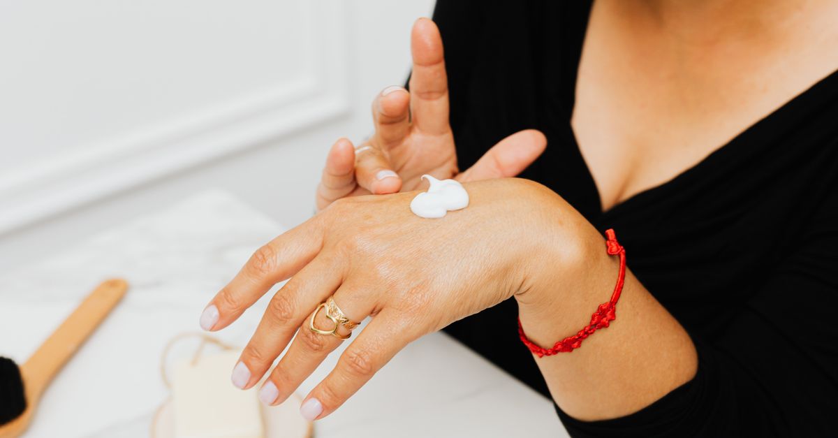
The foundation of treating non-infectious skin fissures is aggressive and consistent moisturization to restore the epidermal barrier. Standard lotions are often inadequate because their high water content evaporates quickly, providing only transient relief. Instead, thick, lipid-rich emollients, such as heavy creams or pure petroleum jelly, are strictly recommended.
These heavy emollients act as occlusive agents, physically trapping the body’s internal moisture within the stratum corneum. For optimal results, emollients must be applied immediately after bathing, while the skin is still damp, to lock in the maximum amount of water.
If the skin surrounding the fissure is highly hyperkeratotic, keratolytic agents are introduced. Creams containing urea, salicylic acid, or alpha-hydroxy acids actively break down the bonds between dead skin cells, helping to gently dissolve the rigid callus. Reducing the thickness of the callus restores flexibility to the skin margins, significantly reducing the mechanical tension that forces the fissure open.
11. Advanced Wound Care and Liquid Bandages
For deep, painful fissures, simply applying a moisturizer may not be sufficient, as the crack is constantly pulled apart by normal movement. In these instances, physically sealing the wound is necessary to allow the dermal tissue to heal from the base upward.
Medical grade liquid bandages containing cyanoacrylate are highly effective for treating fissures on the fingertips and knuckles. When painted over the crack, the liquid polymerizes instantly, forming a waterproof, flexible seal that holds the wound edges together. This immediately eliminates pain by shielding the exposed nerve endings and creates an optimal microenvironment for cellular repair.
For severe heel fissures, specialized hydrocolloid dressings may be applied. These dressings provide a moist wound healing environment while offering a significant degree of physical cushioning, reducing the mechanical impact of walking on the compromised tissue.
12. Pharmacological Interventions
When fissures are secondary to an underlying inflammatory skin disease, targeted pharmacological therapy is required to halt the pathological process. If a patient is diagnosed with eczema or contact dermatitis, a physician will typically prescribe a short course of a topical corticosteroid ointment. The steroid rapidly suppresses the localized immune response, reducing inflammation and allowing the epidermal barrier to regenerate.
For fissures caused by tinea pedis, topical antifungal creams, such as terbinafine or clotrimazole, are strictly necessary. The antifungal medication must be applied consistently, often for several weeks, to ensure complete eradication of the dermatophyte. Emollients should be used concurrently to repair the damaged skin barrier.
In cases where a secondary bacterial infection has developed within the fissure, topical or systemic antibiotics are prescribed based on the severity of the infection. Treating the infection is paramount before aggressive moisturizing therapies can safely resume.
13. Prevention Strategies and Skincare Routines
Preventing the recurrence of skin fissures requires a permanent alteration in daily skincare habits. The most critical preventative measure is avoiding the depletion of natural skin lipids. Patients should replace harsh, highly alkaline bar soaps with mild, synthetic liquid cleansers that do not disrupt the skin’s slightly acidic pH.
Bathing should be kept brief and utilize lukewarm water, as prolonged exposure to hot water aggressively strips oils from the epidermis. Routine, daily application of heavy emollients, particularly before bed, ensures continuous hydration of the stratum corneum.
For individuals working in wet environments or handling harsh chemicals, wearing protective barrier gloves is non-negotiable. Gloves prevent direct contact with irritants and preserve the skin’s structural integrity. During cold winter months, wearing gloves outdoors protects the hands from the dehydrating effects of freezing temperatures and low humidity.
14. Footwear Modifications for Heel Fissures
Managing and preventing heel fissures requires specific attention to footwear biomechanics. The primary goal is to prevent the lateral expansion of the heel’s fat pad during the strike phase of the walking cycle.
Patients prone to heel fissures must strictly avoid open-backed shoes, flip-flops, and thin-soled sandals. Instead, they should wear supportive shoes with a deep, closed heel cup. A firm heel counter physically contains the fat pad, directing the mechanical force of walking upward rather than outward, which significantly reduces the tensile stress placed on the skin of the heel.
Using a pumice stone gently on wet heels a few times a week can safely remove minor hyperkeratosis before it becomes a rigid, unyielding callus. This mechanical debridement, combined with daily urea-based creams, ensures the skin remains soft and pliable enough to withstand the mechanical forces of ambulation.
15. Frequently Asked Questions (FAQ)
1. Why do skin fissures hurt so much more than normal dry skin?
Normal dry skin only affects the outer dead layers. A fissure splits open deep enough to expose the living dermis, which is packed with highly sensitive nerve endings.
2. Can I use superglue to close a crack on my finger?
While similar in concept, household superglue is toxic and can cause skin irritation. You should only use medical-grade liquid bandages specifically formulated for skin application.
3. Why do fissures seem to happen mostly in the winter?
Cold air holds very little moisture, and indoor heating further dries the air. This environment aggressively pulls water out of your skin, leading to severe dryness and cracking.
4. Should I use a file to scrub away the hard skin around the crack?
Aggressive filing can worsen the injury. It is better to use chemical exfoliants like urea creams to soften the callus gradually, or have a medical professional safely debride the tissue.
5. Are skin fissures contagious?
A fissure caused by simple dry skin or eczema is not contagious. However, if the cracking is caused by a fungal infection like athlete’s foot, the underlying fungus can be spread to others.
6. How long does a deep fissure take to heal?
With aggressive moisturization and sealing the wound, a minor fissure can heal in a week. Deep or infected fissures may take several weeks and require prescription medications.
16. Bibliography
Disclaimer: The content is for informational purposes only and does not replace medical advice. Always consult your doctor for personalized treatment.
