
1. Introduction
An injury of the musculoskeletal system refers to structural damage affecting the complex network of bones, joints, ligaments, tendons, and muscles that provide the human body with form, stability, and movement. When any component of this biomechanical architecture is compromised through acute trauma or chronic physical stress, it leads to significant pain, localized inflammation, and a restriction in physical mobility. The clinical objective is to accurately identify the damaged tissue, facilitate biological healing, and restore the patient functional capacity.
The musculoskeletal system operates under a delicate balance of tension and compression. Every physical action, from walking to lifting heavy objects, distributes mechanical forces across this network. When these forces exceed the structural tolerance of a tissue, failure occurs. The resulting damage can range from microscopic muscle tears to catastrophic bone fractures, each demanding a specific medical approach.
Understanding the principles of orthopedic healing is essential for effective treatment. Unlike many other systems in the body, musculoskeletal tissues possess a remarkable ability to regenerate and repair themselves, provided they are stabilized and protected during the initial healing phases. Comprehensive care combines medical intervention, biomechanical support, and structured physical rehabilitation.
2. Components of the Musculoskeletal System
To comprehend how musculoskeletal injuries occur, it is necessary to understand the distinct tissues involved. Bones provide the rigid structural framework and protect vital internal organs. They are dynamic, living tissues that constantly remodel themselves in response to mechanical stress.
Joints are the anatomical locations where two or more bones intersect, allowing for fluid skeletal movement. These articulations are cushioned by articular cartilage, a smooth, frictionless tissue that prevents bones from grinding against one another. The joint capsule encloses the joint space and secretes synovial fluid to lubricate the moving surfaces.
Connecting these bones are ligaments, robust bands of fibrous connective tissue that dictate the normal range of motion and provide critical joint stability. Muscles supply the contractile force necessary for movement, and tendons are the thick, cord-like structures that anchor these muscles directly to the bone. Disruption of any single component cascades into dysfunction for the entire regional system.
3. Types of Musculoskeletal Injuries
Musculoskeletal injuries are clinically classified by the specific anatomical structure that has sustained the damage. Accurate classification directs the diagnostic pathway and treatment protocol.
| Injury Classification | Affected Structure | Pathological Description |
|---|---|---|
| Fracture | Bone | Structural break or crack in bone continuity due to excessive force |
| Dislocation | Joint | Complete displacement of bone ends from their normal articular alignment |
| Sprain | Ligament | Overstretching or tearing of stabilizing joint ligaments |
| Strain | Muscle or Tendon | Tearing of contractile muscle fibers or the tendon anchoring them |
| Tendinopathy | Tendon | Degenerative changes and inflammation due to chronic mechanical overload |
Patients frequently confuse sprains and strains. A clinical mnemonic is that a sprain affects a joint, while a strain affects a muscle. Both invoke an intense inflammatory response but require different rehabilitation timelines depending on their blood supply.
4. Bone Fractures: Classification and Healing
Bone fractures occur when mechanical stress surpasses the bone ultimate tensile strength. They are broadly categorized into closed fractures, where the skin remains intact, and open fractures, where a bone fragment pierces the skin, introducing a severe risk of deep infection. Fractures are further described by their pattern, such as transverse, oblique, or comminuted, indicating multiple bone fragments.
Bone healing is an extraordinary physiological process. Immediately following a fracture, a hematoma forms around the break. Within days, specialized cells form a soft fibrocartilage callus to temporarily stabilize the bone ends. Over subsequent weeks, osteoblasts deposit new bone matrix, converting the soft callus into a hard, rigid bony callus.
For this biological process to succeed, the fracture ends must be held in precise anatomical alignment and kept entirely motionless. If motion occurs at the fracture site, the bone may fail to fuse, resulting in a clinical complication known as a non-union, which requires surgical correction.
5. Sprains and Ligamentous Injuries
Ligaments are essential for maintaining the structural integrity of joints during movement. When a joint is forced beyond its physiological range of motion, the restraining ligaments are stretched or torn. Ankle sprains are the most common musculoskeletal injury worldwide, typically occurring when the foot forcibly rolls inward.
Orthopedic specialists grade ligament injuries on a scale of one to three. A grade one sprain involves microscopic stretching of fibers with mild pain and no joint instability. Grade two indicates a partial tear, characterized by significant swelling, bruising, and a feeling of joint looseness. A grade three sprain is a complete rupture of the ligament, causing severe pain and profound mechanical instability.
Because ligaments have a relatively poor blood supply compared to muscles, they heal at a slower rate. Complete ruptures of critical ligaments, such as the anterior cruciate ligament in the knee, often fail to heal correctly on their own and routinely require surgical reconstruction in active individuals.
6. Muscle Strains and Tendon Ruptures
Muscles and tendons act as a single functional unit to generate movement. Strains typically occur during explosive physical activities when a muscle is forced to contract powerfully while being stretched simultaneously. The hamstring and calf muscles are notorious for sustaining this type of eccentric load injury.
Similar to sprains, muscle strains are graded by severity. Mild strains cause localized cramping and tightness, while severe strains involve a complete tearing of the muscle belly, often resulting in a visible indentation under the skin and a massive hematoma.
Tendon ruptures are catastrophic failures of the connective cord attaching muscle to bone. The Achilles tendon is frequently ruptured during sudden acceleration sports. Patients describe a sensation akin to being kicked in the back of the ankle, accompanied by an immediate inability to push off the toes. Tendon ruptures generally require prolonged immobilization or surgical repair.
7. Joint Dislocations and Subluxations
A joint dislocation is a severe orthopedic trauma where the articulating bones are completely displaced from their normal anatomical positions. The shoulder and finger joints are highly susceptible due to their extensive range of motion. A dislocation causes intense, acute pain and visible physical deformity.
A subluxation is a partial or temporary dislocation where the joint surfaces briefly lose contact but spontaneously snap back into place. While seemingly less severe, subluxations still cause significant damage to the surrounding joint capsule and ligaments, leaving the joint prone to chronic instability.
Dislocations are considered medical emergencies. The displaced bones can severely stretch or compress adjacent nerves and blood vessels. A physician must perform a closed reduction, a specialized maneuver to safely realign the bones, restoring normal anatomy and preventing permanent neurovascular damage.
8. Mechanisms of Injury: Trauma and Biomechanics
The etiology of musculoskeletal injuries is divided into acute macrotrauma and chronic microtrauma. Macrotrauma involves a single, identifiable event with immense force. High-velocity impacts, such as motor vehicle collisions or falls from significant heights, routinely cause complex fractures and multi-ligament trauma.
Microtrauma, conversely, is the result of repetitive biomechanical stress over an extended period. Running long distances with improper foot biomechanics, lifting weights with poor form, or performing repetitive occupational tasks places continuous, low-grade stress on specific tissues.
When the rate of this microscopic tissue damage outpaces the body natural cellular repair mechanisms, chronic overuse injuries develop. Conditions like stress fractures, plantar fasciitis, and tennis elbow are classic examples of microtraumatic failure. Treatment focuses on identifying and correcting the underlying biomechanical fault rather than simply managing the pain.
9. Clinical Evaluation and Symptoms
The cardinal symptoms of a musculoskeletal injury are pain, localized swelling, erythema, and a notable decrease in functional capacity. During the clinical evaluation, the physician takes a detailed history to understand the precise vector of force that caused the injury.
Physical examination involves methodical palpation to identify the exact point of maximum tenderness, which correlates with the damaged structure. The clinician evaluates the active and passive range of motion, noting any mechanical blocks or apprehension from the patient.
Specific orthopedic provocation tests are utilized to stress individual ligaments and tendons. For example, the Lachman test is performed to assess the integrity of the knee cruciate ligaments. A comprehensive neurovascular examination is also mandatory to ensure that pulses are present and nerve sensation is intact distal to the injury site.
10. Diagnostic Imaging Modalities
Diagnostic imaging is crucial for confirming clinical suspicions and defining the extent of structural damage. Standard X-rays are the universal starting point. They provide rapid, high-contrast images of the skeletal architecture, easily identifying fractures, joint space narrowing, and bone alignment.
When X-rays are inconclusive or soft tissue damage is suspected, advanced imaging is employed. Magnetic Resonance Imaging is the gold standard for visualizing non-bony tissues. MRI provides exceptional detail of ligaments, menisci, muscle tears, and hidden bone bruising that standard X-rays cannot detect.
Computed Tomography scans offer three-dimensional cross-sectional images of bone. CT is particularly valuable for surgical planning in complex, multi-fragment fractures, especially in intricate areas like the pelvis or the articular surfaces of major joints. Ultrasound imaging is also increasingly used for real-time dynamic assessment of superficial tendon gliding and muscle tears.
11. The Role of Inflammation in Tissue Healing
Inflammation is often viewed negatively by patients, but it is a vital, non-negotiable phase of biological healing. Immediately after an injury, blood vessels dilate, flooding the area with immune cells and growth factors. This causes the characteristic redness, heat, and swelling.
The inflammatory cascade clears away dead cellular debris and sets the stage for tissue regeneration. Suppressing this phase entirely with excessive use of non-steroidal anti-inflammatory drugs in the first few days can actually delay the overall healing process.
However, prolonged or excessive inflammation leads to excessive scar tissue formation and persistent pain. The clinical goal is not to eliminate inflammation, but to control it. Utilizing ice, compression, and elevation optimizes the inflammatory environment, ensuring the tissue proceeds efficiently into the subsequent repair and remodeling phases.
12. Immobilization and Orthopedic Interventions
The fundamental principle of treating structural musculoskeletal failure is protection. Injured tissues must be shielded from mechanical loads to heal properly. Immobilization is achieved through various orthopedic devices depending on the injury severity.
Casts, made of fiberglass or plaster, provide rigid, circumferential support, ensuring absolute immobility for bone fractures. Orthopedic walking boots offer similar support but allow for staged weight-bearing and easy removal for hygiene. Braces and functional splints limit specific ranges of motion while allowing safe mobility, making them ideal for ligament sprains.
When tissues are torn entirely or bones are severely displaced, surgical intervention is required. Orthopedic surgeons utilize techniques such as open reduction and internal fixation, using metallic plates, screws, and intramedullary nails to physically rebuild the skeleton and secure the anatomy for healing.
13. Physiotherapy and Functional Restoration
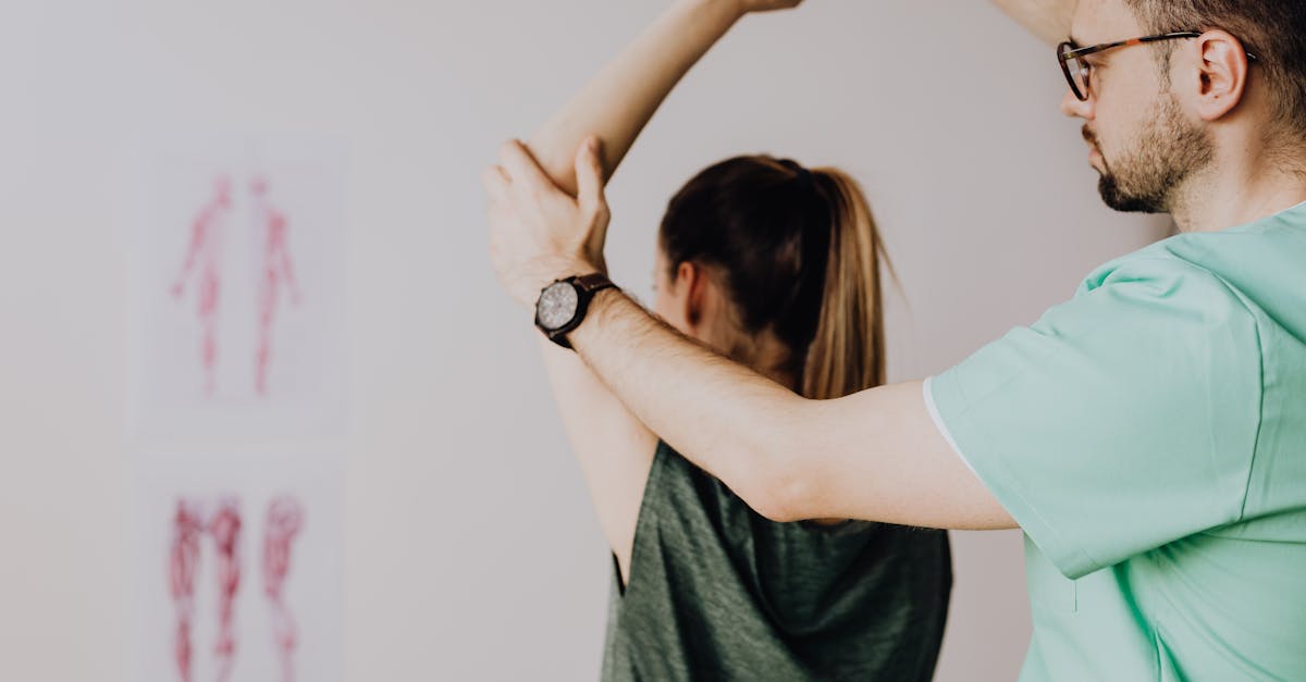
Healing the damaged tissue is only the first phase of recovery; restoring the biomechanical function of the limb is the ultimate goal. Following a period of immobilization, joints become stiff, and muscles experience rapid disuse atrophy. Physical therapy is mandatory to reverse these effects.
Rehabilitation begins with passive stretching to break up restrictive scar tissue and restore joint mobility. As the tissue matures, progressive resistance training is introduced. Strengthening the surrounding musculature provides dynamic stability to the previously injured joint, compensating for any residual ligamentous laxity.
The final stage of functional restoration focuses on proprioception, balance, and sport-specific biomechanics. Retraining the nervous system to coordinate complex movements safely is the most effective strategy for preventing future musculoskeletal injuries and ensuring a successful return to pre-injury activities.
14. Frequently Asked Questions (FAQ)
1. What is the difference between a bone fracture and a bone break?
Medically speaking, there is no difference. Both terms describe a disruption in the structural continuity of a bone. The severity depends on the pattern of the break, not the terminology used.
2. Should I use ice or heat on a muscle strain?
For acute injuries, ice should be applied for the first two to three days to control bleeding, limit excessive swelling, and reduce pain. Heat can be introduced later to relax muscle spasms and promote blood flow for healing.
3. Why do joints get stiff after wearing a cast?
Immobilization prevents the normal gliding of joint surfaces and allows surrounding connective tissues to tighten and shorten. Muscles also lose mass and flexibility rapidly when not in use, requiring physical therapy to stretch and rebuild.
4. Can a torn ligament heal without surgery?
Minor and partial ligament tears often heal well with bracing and physical therapy. However, complete ruptures of major stabilizing ligaments, like the ACL, generally lack the blood supply to heal naturally and require surgical repair in active people.
5. What is a stress fracture?
A stress fracture is a tiny crack in a bone caused by repetitive, cumulative mechanical overload rather than a single acute trauma. It is an overuse injury common in distance runners and military recruits.
15. Bibliography
Disclaimer: The content is for informational purposes only and does not replace medical advice. Always consult your doctor for personalized treatment.
