1. Introduction
A peripheral nerve injury involves damage to the intricate network of neural pathways that transmit information between the central nervous system and the rest of the body. These nerves serve as the vital communication lines allowing the brain to command muscle movements and receive sensory feedback from the skin, joints, and internal organs. When this communication is disrupted by physical trauma or mechanical compression, patients experience distinct motor and sensory deficits that require meticulous medical evaluation.
The peripheral nervous system is remarkably complex, comprising motor, sensory, and autonomic fibers bundled together in tough connective tissue sheaths. Unlike the central nervous system, peripheral nerves possess a unique biological capacity to regenerate following certain types of injury. This regenerative potential forms the foundation of clinical treatment and rehabilitation protocols.
Effective medical management depends upon determining the exact anatomical location and severity of the nerve damage. Physicians utilize detailed clinical examinations and advanced electrodiagnostic testing to map the injury. Treatment strategies span from conservative observation and physical therapy for minor stretch injuries to intricate microsurgical reconstruction for completely severed nerves.
2. Anatomy of the Peripheral Nervous System
Understanding peripheral nerve trauma requires familiarity with the cellular and structural anatomy of these tissues. The fundamental unit is the neuron, which consists of a cell body residing near the spinal cord and a long projection called an axon. The axon functions as a biological wire, conducting electrical impulses over long distances to target muscles or sensory receptors.
Many axons are encased in a protective myelin sheath produced by specialized Schwann cells. This myelin acts as insulation, significantly increasing the speed of electrical conduction. Axons are further bundled into fascicles, which are wrapped in distinct layers of connective tissue known as the endoneurium, perineurium, and epineurium.
The epineurium provides the outer mechanical strength of the nerve, allowing it to withstand a certain degree of stretching and compression during normal bodily movements. When traumatic forces exceed the tensile strength of these protective layers, the delicate internal axons are compromised, leading to immediate neurological dysfunction.
3. Mechanisms of Peripheral Nerve Damage
Peripheral nerves are subjected to various forms of physical stress. Clinicians categorize these injuries based on the mechanical force applied, as this directly influences the resulting tissue damage and the potential for spontaneous recovery.
| Mechanism of Injury | Common Examples | Clinical Pathophysiology |
|---|---|---|
| Compression | Carpal tunnel syndrome, tourniquet use | Localized ischemia and demyelination |
| Traction or Stretching | Joint dislocations, difficult childbirth | Elongation leading to axonal tearing |
| Laceration | Knife wounds, broken glass accidents | Complete anatomical transection of the nerve |
| Crush Injury | Industrial accidents, heavy object impacts | Disruption of internal fibers with an intact outer sheath |
Crush and stretch injuries are the most frequently encountered in orthopedic trauma. While the nerve may appear physically continuous to the naked eye, the internal axonal pathways are often severely damaged, necessitating careful observation to determine if surgical intervention will become necessary.
4. Classification Systems: Seddon and Sunderland
To standardize communication and predict outcomes, neurologists utilize specific classification systems. The Seddon classification divides nerve injuries into three progressive grades: neuropraxia, axonotmesis, and neurotmesis.
Neuropraxia represents the mildest form of injury, typically caused by brief compression. The nerve structurally remains completely intact, but there is a temporary block in electrical conduction due to localized damage to the myelin sheath. Recovery is generally spontaneous and complete within weeks to months.
Axonotmesis involves the tearing of the internal axons, while the outer connective tissue sheaths remain intact. This triggers a specific biological process of axonal degeneration. Recovery is possible but slow, as the nerve fibers must regenerate along the preserved sheaths.
Neurotmesis is the most severe grade, characterized by complete disruption of the nerve and all its supporting connective tissues. Spontaneous recovery is impossible in these cases, and surgical repair is absolutely mandatory to restore any degree of anatomical continuity.
5. Wallerian Degeneration and Regeneration
When an axon is physically severed or severely crushed, the portion of the nerve distal to the injury site undergoes a programmed cellular breakdown known as Wallerian degeneration. Within forty-eight hours of the injury, the disconnected axon and its myelin sheath disintegrate and are cleared away by immune system cells called macrophages.
This clearing process is essential, as it creates an empty physical tube within the surviving connective tissue sheaths. The surviving cell body near the spinal cord then increases its metabolic activity, preparing to synthesize the proteins required to build a new axon.
The regenerating axon sends out microscopic sprouts that attempt to find the empty connective tissue tubes. If successful, the nerve grows down the tube toward its original target at a rate of approximately one millimeter per day. This remarkably slow pace explains why recovery from peripheral nerve trauma requires substantial time and patience.
6. Clinical Presentation and Sensory Symptoms
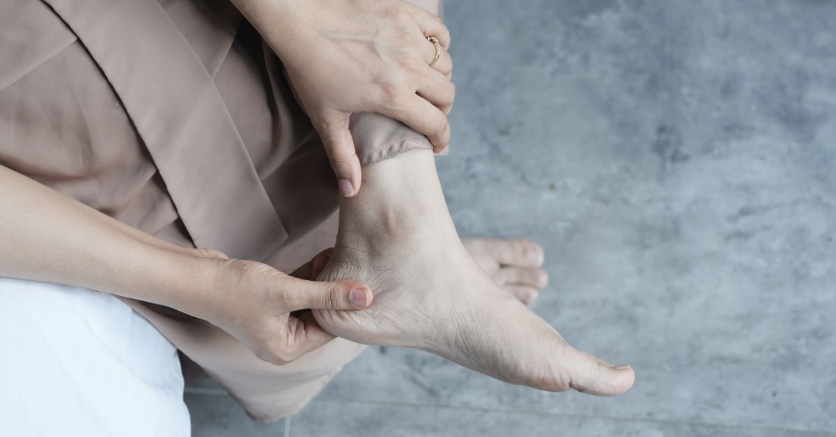
The symptomatology of a peripheral nerve injury corresponds directly to the specific nerve involved and the types of fibers it carries. Sensory symptoms are often the first clinical indicator of nerve pathology. Patients frequently describe altered sensation, known medically as paresthesia, which manifests as tingling, burning, or a prickling feeling in the affected area.
As the injury progresses, paresthesia can evolve into profound numbness, rendering the patient unable to distinguish between sharp and dull objects or hot and cold temperatures. This lack of sensory feedback significantly increases the risk of secondary injuries, such as accidental burns or unnoticed lacerations on the hands or feet.
Neuropathic pain is a distinct and often debilitating symptom. Unlike the aching pain of a muscle strain, neuropathic pain originates from the damaged nerve itself. It is frequently described as a severe, electrical shooting pain that travels along the anatomical path of the nerve, often worsening at night.
7. Motor Deficits and Muscle Atrophy
When motor nerve fibers are damaged, the muscles they supply lose their command signals, resulting in immediate weakness or complete flaccid paralysis. The distribution of this weakness provides clinicians with a clear anatomical map of the nerve injury.
If the nerve supply is not restored, the affected muscle tissue begins to shrink, a physiological process termed denervation atrophy. Without continuous electrical stimulation from the nerve, muscle fibers rapidly lose their mass and contractile proteins. Visible wasting of the muscle belly becomes apparent within a few weeks of the injury.
Preventing irreversible muscle atrophy is a primary focus of early clinical intervention. If a nerve takes too long to regenerate, the target muscle may become completely replaced by dense fibrous scar tissue, rendering it permanently non-functional even if the nerve eventually arrives.
8. Autonomic Nervous System Dysfunction
Many major peripheral nerves carry autonomic fibers, which regulate involuntary physiological processes such as blood flow and sweat production. Damage to these fibers produces subtle but clinically significant signs in the skin of the affected limb.
Following a severe nerve injury, the skin over the denervated area often becomes unusually dry and loses its ability to sweat, a condition known as anhidrosis. The skin may also appear flushed or unusually pale due to the loss of vascular control, which dictates how small blood vessels dilate or constrict in response to temperature changes.
Over time, these autonomic changes alter the texture of the skin, making it appear thin, shiny, and hairless. Recognizing these signs helps physicians confirm the extent of the nerve damage and differentiate a true peripheral nerve injury from other orthopedic conditions.
9. Diagnostic Assessment and Physical Examination
The diagnostic protocol begins with a comprehensive neurological examination. The physician systematically tests sensory perception using specialized monofilaments, pinpricks, and tuning forks to map the exact boundaries of numbness. These sensory maps correspond to specific nerve territories, known as dermatomes.
Motor function is assessed by evaluating the strength of individual muscles against resistance. The physician grades muscle strength on a standardized scale to monitor progression or deterioration over time. Deep tendon reflexes are also tested; a diminished or absent reflex is a classic indicator of peripheral nerve pathway disruption.
The Tinel sign is a valuable clinical test. The physician gently taps along the course of the injured nerve. A positive result elicits a sudden, tingling sensation radiating distally into the limb. The location of the positive Tinel sign helps track the physical progress of a regenerating nerve tip over several months.
10. Electromyography and Nerve Conduction Studies
Electrodiagnostic testing is the gold standard for confirming the diagnosis and quantifying the severity of a peripheral nerve injury. These tests are typically performed three to four weeks after the initial trauma, allowing sufficient time for Wallerian degeneration to become electrically measurable.
Nerve conduction studies involve applying small electrical pulses to the skin over the nerve and measuring the speed and amplitude of the resulting signal. Slowed conduction velocities indicate damage to the myelin sheath, while a reduction in the signal strength suggests damage to the internal axons.
Electromyography involves inserting a fine needle electrode directly into specific muscles to record their electrical activity. Abnormal spontaneous electrical discharges, such as fibrillations, confirm that the muscle has lost its nerve supply. This objective data is crucial for determining whether surgical exploration is warranted.
11. Advanced Diagnostic Imaging
While electrodiagnostic tests assess nerve function, advanced imaging provides detailed anatomical visualization of the nerve structure. High-resolution magnetic resonance imaging is increasingly utilized in peripheral nerve evaluation. MRI can identify physical abnormalities such as tumors, compressive bone fragments, or dense scar tissue entrapping the nerve.
Magnetic resonance neurography is a specialized technique that suppresses signals from surrounding fat and muscle, making the peripheral nerves stand out brightly on the scan. This allows radiologists to pinpoint the exact site of structural disruption or detect the presence of a neuroma, a painful tangle of disorganized nerve fibers attempting to regenerate.
High-frequency ultrasound is another valuable diagnostic tool. It offers real-time, dynamic imaging of the nerve as it glides through anatomical tunnels during joint movement, making it particularly useful for diagnosing compression neuropathies and identifying superficial nerve lacerations.
12. Conservative Management and Pharmacotherapy
For injuries classified as neuropraxia or mild axonotmesis, the initial treatment pathway is predominantly conservative. The primary objective is to protect the limb from further mechanical trauma while awaiting spontaneous biological recovery.
Bracing and splinting are employed to maintain affected joints in functional, neutral positions. This prevents the development of joint contractures and protects paralyzed muscles from being overstretched by the opposing healthy muscles. Physical therapy is initiated immediately to maintain joint mobility through passive range-of-motion exercises.
Managing neuropathic pain requires specific pharmacological interventions. Standard analgesic medications are generally ineffective against nerve-generated pain. Physicians utilize specialized medications such as gabapentin, pregabalin, or certain tricyclic antidepressants, which function by stabilizing the hyperactive electrical membranes of the damaged nerve fibers.
13. Principles of Surgical Reconstruction
Surgical intervention is indicated when a nerve is completely transected, when a compressive lesion must be removed, or when electrodiagnostic tests show no signs of spontaneous regeneration after a sufficient observation period. The procedures are performed using intricate microsurgical techniques.
For a clean, sharp nerve laceration, primary end-to-end repair is the preferred treatment. The surgeon meticulously aligns the nerve fascicles and sutures the outer epineurium together using microscopic threads, restoring a physical pathway for the axons to follow.
If there is a significant gap between the nerve ends due to severe trauma or retraction, a nerve graft is required. The surgeon harvests a less critical sensory nerve from another part of the body to bridge the defect. This graft acts as a biological scaffold, providing empty tubes to guide the regenerating axons toward the target muscle.
14. Rehabilitation and Sensory Re-education
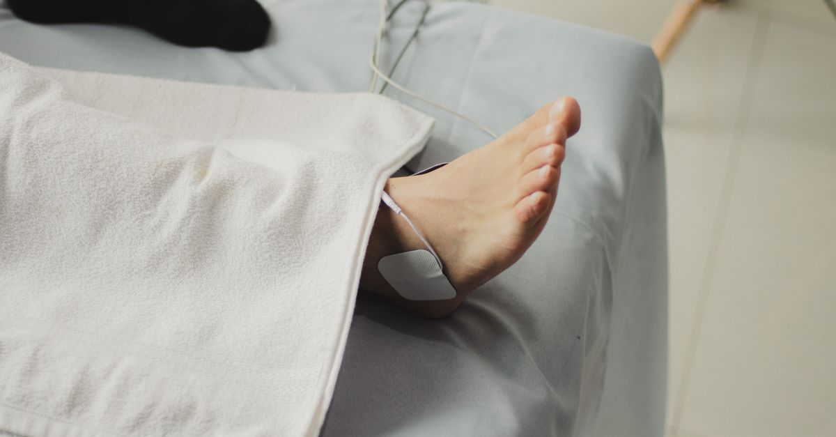
Rehabilitation is an arduous but essential phase of recovery, often extending for months or years. As the nerve slowly regenerates, the targeted muscles begin to receive faint electrical signals. Therapists use functional electrical stimulation and targeted biofeedback to help patients isolate and consciously contract these re-innervated muscles.
Sensory re-education is equally critical. When regenerating nerve fibers eventually reach the skin, the brain often misinterprets the new signals, perceiving light touch as painful or confusing the exact location of the stimulus. This can be closely associated with persistent numbness or tingling in the extremities.
Therapists guide patients through structured exercises using various textures, temperatures, and pressures to retrain the central nervous system. This process of neuroplasticity allows the brain to recalibrate its sensory mapping, ultimately restoring functional dexterity and protecting the limb from future harm.
15. Frequently Asked Questions (FAQ)
1. How long does a peripheral nerve take to heal?
Peripheral nerves regenerate very slowly, at an average rate of one millimeter per day or one inch per month. Recovery can take anywhere from a few weeks for minor compression injuries to well over a year for severe trauma.
2. Will I regain full feeling after a nerve repair?
Complete restoration of normal sensation is challenging after a severe injury. Most patients experience significant improvement, but minor deficits in fine touch or temperature perception often remain permanently.
3. Why is my injured nerve causing a burning pain?
This is known as neuropathic pain. When the protective outer layers of a nerve are damaged, the nerve fibers can spontaneously fire disorganized electrical signals to the brain, which are interpreted as a burning or electrical sensation.
4. What is a neuroma?
A neuroma is a disorganized mass of nerve tissue that forms when regenerating nerve fibers cannot find their correct anatomical pathway. They grow into a tangled scar that is typically very sensitive and painful to the touch.
5. Is physical therapy necessary if my nerve is cut?
Yes, physical therapy is absolutely essential. While waiting for the nerve to grow back, daily therapy prevents the joints from stiffening permanently and keeps the paralyzed muscles from shortening into severe contractures.
16. Bibliography
Disclaimer: The content is for informational purposes only and does not replace medical advice. Always consult your doctor for personalized treatment.