1. Introduction
An upper arm injury involves any structural damage to the humerus bone, the surrounding musculature, the tendinous attachments, or the traversing neural and vascular networks located between the shoulder and the elbow. The primary clinical objective in managing these injuries is to restore normal skeletal alignment, promote unhindered soft tissue healing, and preserve the mechanical function of the entire upper extremity. Prompt medical evaluation is required to identify fractures, relieve pain, and rule out any secondary neurological compromise that could permanently impair limb function.
The upper arm is anatomically designed to facilitate a wide range of motion while providing substantial leverage for lifting and pushing tasks. The central humerus acts as the structural pillar, surrounded by the biceps brachii anteriorly and the triceps brachii posteriorly. Because this region frequently absorbs the kinetic energy of falls and direct impacts, it is vulnerable to both blunt force trauma and significant mechanical overload.
Medical management depends entirely on identifying the exact anatomical structures involved. Treatment protocols vary significantly, ranging from simple immobilization for minor muscle strains to intricate surgical fixation for displaced bone fractures. A thorough understanding of the specific injury mechanics ensures that patients receive the appropriate therapeutic interventions to maximize their functional recovery.
2. Anatomy of the Upper Arm
The structural foundation of the upper arm is the humerus, the longest and largest bone of the upper extremity. The proximal end forms the ball of the shoulder joint, while the distal end articulates with the radius and ulna bones to form the elbow joint. The central portion is known as the humeral shaft.
Surrounding the humerus are the primary muscles responsible for elbow movement. The biceps brachii, located on the front of the arm, flexes the elbow and supinates the forearm. The triceps brachii, located on the back of the arm, is responsible for extending the elbow. These muscles are anchored to the bone by robust, fibrous tendons.
Crucial neurovascular structures traverse the upper arm. The brachial artery descends along the medial aspect, providing the main blood supply to the arm and hand. The radial nerve spirals tightly around the back of the humeral shaft in a shallow groove, making it exceptionally vulnerable to injury when the mid-shaft of the humerus fractures.
3. Mechanisms of Traumatic Injury
Upper arm injuries are categorized based on the direction and magnitude of the mechanical forces applied. The specific vector of force provides clinicians with critical diagnostic information regarding the likely pattern of tissue damage.
| Mechanism of Injury | Common Etiology | Typical Structural Damage |
|---|---|---|
| Direct Blunt Force | Automobile collisions, sports impacts | Transverse humeral shaft fractures, deep contusions |
| Indirect Torsional Force | Falling onto an outstretched hand, throwing | Spiral humeral fractures, proximal humerus fractures |
| Sudden Eccentric Loading | Catching a heavy falling object | Biceps or triceps tendon ruptures, muscle strains |
| Penetrating Trauma | Deep lacerations, puncture wounds | Vascular lacerations, peripheral nerve transection |
A fall onto an outstretched hand is a frequent presentation in emergency settings. The kinetic energy travels up the forearm and frequently localizes in the surgical neck of the humerus, causing a characteristic proximal fracture that is particularly common in older adults with decreased bone density.
4. Humeral Shaft Fractures
A fracture of the humeral shaft involves a break in the central portion of the bone. These fractures typically result from a direct blow or a severe twisting injury. Patients present with severe pain, significant localized swelling, and an obvious visible deformity where the arm appears bent or unnaturally angulated between the shoulder and elbow.
The bone can break transversely straight across, obliquely at an angle, or spiral down the shaft depending on the twisting forces involved. In severe impacts, the bone may comminute, meaning it shatters into multiple fragments.
Most humeral shaft fractures heal remarkably well without surgery because the surrounding thick muscle envelope acts as a natural biological splint. Physicians generally utilize a specialized functional brace that gently compresses the muscles around the bone, guiding it into correct alignment while it knits together over several weeks.
5. Proximal and Distal Humerus Fractures
Fractures occurring near the joints present unique clinical challenges. Proximal humerus fractures occur near the shoulder joint. These are frequently seen in elderly patients following minor falls. If the bone fragments remain aligned, treatment involves simple sling immobilization. However, if the segments are displaced, surgical reconstruction is required to restore shoulder mobility.
Distal humerus fractures occur just above the elbow joint. These injuries are notoriously difficult to treat because the fracture lines often extend directly into the articular surface of the joint cartilage.
Surgical intervention is almost universally required for displaced distal humerus fractures. The surgeon must meticulously reassemble the joint surface using thin titanium plates and screws to prevent the development of post-traumatic arthritis and ensure the elbow regains a functional range of motion.
6. Biceps and Triceps Muscle Strains
A strain occurs when the contractile muscle fibers are stretched beyond their physiological limits, leading to microscopic or macroscopic tearing. Biceps strains frequently occur during heavy lifting activities where the elbow is forcefully extended while the muscle is actively trying to contract.
Triceps strains are less common but occur when the muscle forcefully contracts to straighten the arm against a sudden, heavy resistance, such as a fall or a blocking motion in sports.
Patients with muscle strains experience sudden, sharp pain in the muscle belly, followed by localized swelling and bruising. Unlike complete ruptures, strains do not cause a structural deformity in the contour of the arm. Conservative management involving rest, cold therapy, and gradual stretching usually results in a complete functional recovery.
7. Tendon Ruptures in the Upper Arm
A tendon rupture represents a complete structural failure of the connective cord attaching a muscle to the bone. In the upper arm, a rupture of the long head of the biceps tendon at the shoulder is common. Patients often hear a distinct popping sound, followed by the appearance of a large bulge in the lower part of the upper arm, colloquially known as a “Popeye deformity.”
Ruptures of the distal biceps tendon at the elbow are distinct clinical entities. They usually occur in middle-aged individuals lifting surprisingly heavy loads. The patient loses a significant amount of forearm twisting strength and elbow bending power.
While a rupture of the proximal biceps tendon at the shoulder often heals well without surgery, a rupture of the distal biceps tendon at the elbow generally requires prompt surgical reattachment to the radius bone to restore normal arm strength and endurance.
8. Radial Nerve Palsy
The most significant acute complication of a humeral shaft fracture is an injury to the radial nerve. Because the radial nerve wraps tightly around the mid-shaft of the humerus, the sharp edges of a broken bone can easily stretch, bruise, or sever this delicate neural structure during the traumatic event.
If the radial nerve is injured, the patient develops a condition known as a wrist drop. The patient will be entirely unable to lift their wrist or straighten their fingers against gravity. Additionally, they will experience a patch of numbness on the back of the hand near the base of the thumb.
Fortunately, the vast majority of these nerve injuries are neuropraxias, meaning the nerve is only bruised or temporarily stretched. Most radial nerve palsies associated with closed humeral fractures resolve spontaneously over three to six months as the nerve biologically repairs itself.
9. Clinical Symptoms and Presentation
The cardinal symptom of any upper arm injury is acute pain that intensifies with movement. The specific location of the pain provides critical diagnostic clues. Pain localized near the shoulder suggests a proximal fracture or biceps tendon issue, while deep, aching pain in the mid-arm points toward a shaft fracture.
Swelling and ecchymosis develop rapidly. The bruising may initially appear at the injury site but will often track downward toward the elbow and forearm over several days due to the effects of gravity on the extravasated blood.
A noticeable structural deformity, an inability to raise the arm, or an audible grinding sound known as crepitus when the arm is moved are definitive signs of a bone fracture. Patients may also experience muscle spasms as the body attempts to rigidly splint the broken bone fragments.
10. When to Seek Urgent Medical Care
While minor muscle soreness can be monitored at home, specific signs mandate immediate emergency evaluation. An obvious visual deformity of the arm or a bone fragment visibly pushing against the skin requires urgent orthopedic alignment to prevent the bone from puncturing the skin and causing a severe infection.
Any sudden loss of sensation, profound numbness, or a tingling feeling extending down into the forearm or hand indicates that a nerve is being actively compressed. This symptom is frequently linked to broader arm weakness and requires a prompt neurological assessment.
If the hand turns pale, cold, or bluish, this indicates that the brachial artery has been compressed or severed, cutting off the blood supply to the limb. This constitutes an absolute surgical emergency where every minute is critical for salvaging the arm.
11. Diagnostic Imaging Protocols
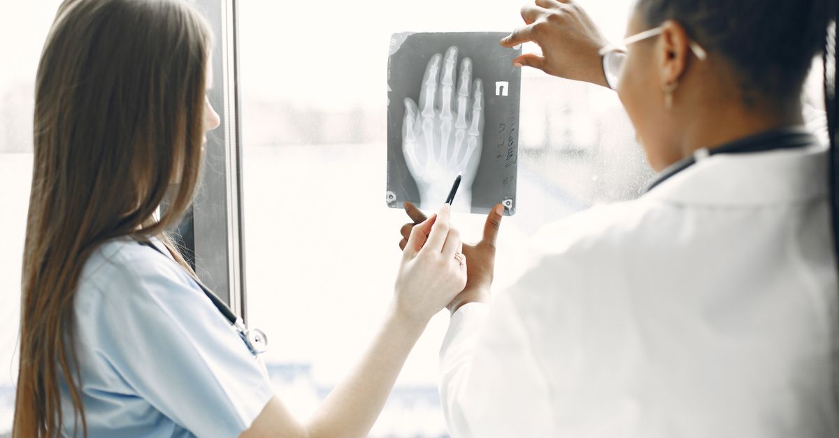
A precise diagnosis relies heavily on diagnostic imaging. Standard anterior-posterior and lateral X-rays of the humerus, including the shoulder and elbow joints, are the primary tools for identifying and characterizing bone fractures.
When X-rays appear normal but a severe soft tissue injury is suspected, advanced imaging is employed. Magnetic Resonance Imaging provides exceptional detail of the musculature and tendons. MRI is the gold standard for diagnosing partial or complete tendon ruptures and identifying hidden fluid collections within the muscle bellies.
In complex intra-articular fractures near the elbow or shoulder, a Computed Tomography scan may be ordered. The CT scan provides three-dimensional, cross-sectional views that allow the orthopedic surgeon to meticulously plan the placement of surgical hardware before entering the operating room.
12. Conservative Management and Immobilization
For many upper arm injuries, conservative non-surgical management yields excellent clinical results. Non-displaced proximal humerus fractures and the majority of humeral shaft fractures are successfully treated with external immobilization.
Physicians utilize specialized bracing systems, such as a Sarmiento brace for shaft fractures. This brace relies on the principle of soft tissue compression, gently squeezing the surrounding muscles to hold the bone ends in anatomical alignment while allowing the elbow and shoulder to move freely, preventing joint stiffness.
Pharmacological management involves a scheduled regimen of pain relievers and non-steroidal anti-inflammatory drugs to control local tissue swelling. The arm is often supported in a simple sling for comfort during the initial weeks of healing.
13. Surgical Interventions and Fixation
Surgical intervention is indicated when conservative measures cannot achieve adequate bone alignment or when a critical tendon is completely avulsed. For displaced humeral shaft fractures, an orthopedic surgeon may perform an open reduction and internal fixation, using a long titanium plate and screws to rigidly hold the bone together.
Alternatively, the surgeon may use an intramedullary nail. This procedure involves inserting a long titanium rod directly down the hollow center of the humerus bone. This provides robust internal stability while requiring smaller surgical incisions.
For distal biceps tendon ruptures at the elbow, the surgeon makes an incision over the forearm, locates the retracted tendon, and anchors it securely back into its precise anatomical footprint on the radius bone using specialized surgical buttons or suture anchors.
14. Physical Therapy and Rehabilitation
Rehabilitation is an essential phase of recovery designed to restore full functional capacity to the upper extremity. Following a period of immobilization, the shoulder and elbow joints inevitably become stiff, and the surrounding muscles experience significant disuse atrophy.
Physical therapy begins with gentle, passive range of motion exercises to break down restrictive scar tissue and restore joint flexibility. As bone and soft tissue healing progresses, the therapist introduces active-assisted exercises, allowing the patient to slowly engage their own muscles.
The final stage of rehabilitation focuses on progressive resistance training. Rebuilding the dynamic strength of the biceps, triceps, and rotator cuff is critical for ensuring the patient can safely return to heavy lifting, occupational duties, or athletic endeavors without a high risk of re-injury.
15. Frequently Asked Questions (FAQ)
1. How long does a broken humerus take to heal?
The bone typically takes six to eight weeks to fuse sufficiently for basic activities. However, regaining full strength and complete range of motion often requires three to six months of dedicated physical therapy.
2. What is a radial nerve palsy?
It is a temporary loss of function in the radial nerve, often caused by a broken upper arm bone pinching the nerve. It results in a condition called wrist drop, where you cannot lift your hand backward. It usually resolves naturally over several months.
3. Does a torn biceps muscle require surgery?
A tear of the biceps tendon at the shoulder often heals well without surgery, though a small cosmetic bulge may remain. However, a tear of the biceps tendon at the elbow usually requires surgical reattachment to restore normal arm strength.
4. Can I sleep on my arm if it is injured?
You should avoid sleeping directly on an injured upper arm, as the sustained pressure can compress nerves, disrupt bone alignment, and exacerbate tissue swelling. Most patients sleep most comfortably in a slightly reclined position.
5. Why is my entire arm bruised down to my wrist?
Gravity pulls the blood from a deep muscle tear or a bone fracture in the upper arm downward through the tissue layers. It is very common for bruising to appear around the elbow and track down the forearm days after the initial injury.
16. Bibliography
Disclaimer: The content is for informational purposes only and does not replace medical advice. Always consult your doctor for personalized treatment.