1. Introduction
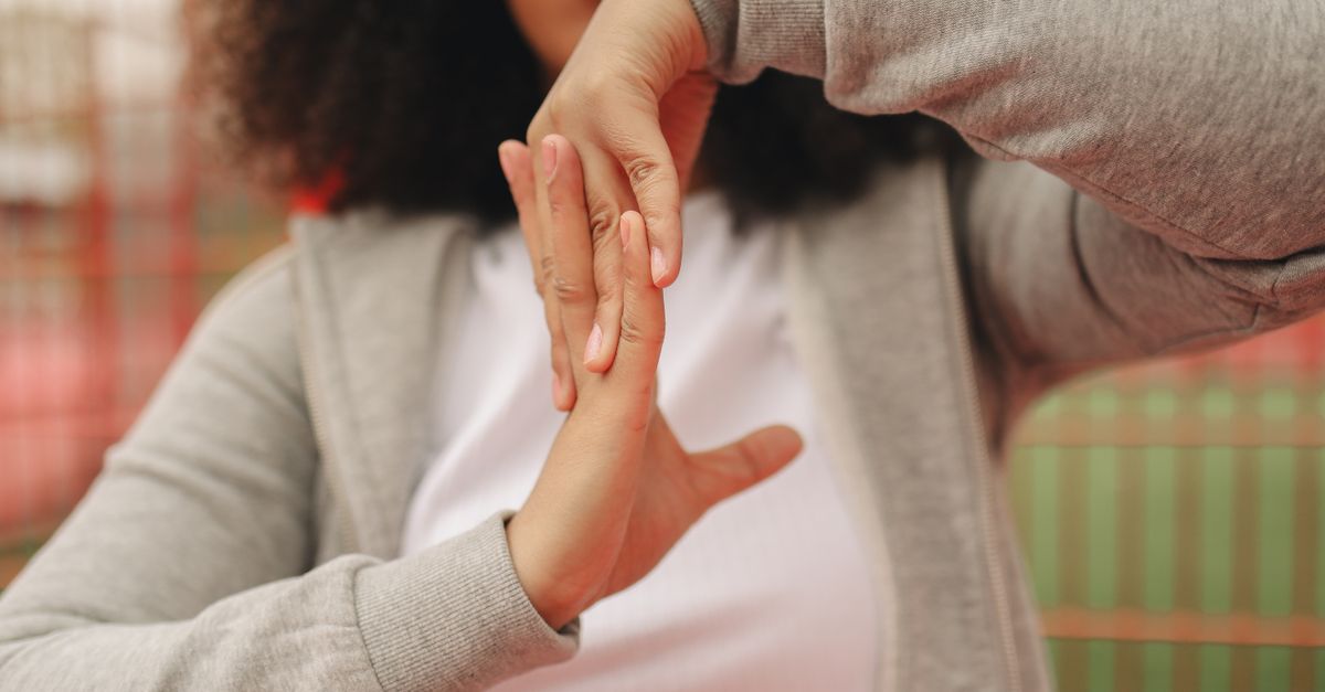
Fingers locking into a bent position upon waking is the classic clinical presentation of stenosing tenosynovitis, commonly known as trigger finger. This condition occurs when the flexor tendon and its protective synovial sheath become inflamed and swollen, forming a localized nodule. During the night, natural fluid shifts cause this nodule to expand further. When you attempt to straighten your fingers in the morning, the swollen tendon cannot glide smoothly through the rigid ligamentous tunnel at the base of the finger, resulting in a sudden, painful mechanical block.
The human hand achieves exceptional dexterity through a precise pulley system that anchors tendons closely to the bones. This system operates with negligible tolerances. Even microscopic swelling disrupts the biomechanics of movement. Understanding the architecture of the hand and the physiological changes that occur during sleep provides clarity on why this locking phenomenon is most severe immediately after waking.
2. Anatomy of the Flexor Tendons
The movement of closing the hand into a fist is powered by the flexor muscles located in the forearm. These muscles connect to the fingertips via long, tough, fibrous cords known as flexor tendons.
As these tendons travel from the wrist into the hand and down the fingers, they are enveloped in a protective, double-layered synovial sheath. This sheath secretes synovial fluid, a biological lubricant that allows the tendons to glide effortlessly back and forth with minimal friction during repetitive movements.
The tendons must be held tightly against the bones of the fingers to function efficiently. If they were not anchored, bending a finger would cause the tendon to bowstring away from the joint, resulting in a profound loss of mechanical leverage and grip strength.
3. The Pulley System of the Hand
To prevent bowstringing, the flexor tendons pass through a series of fibrous tunnels along the length of each finger. These tunnels are formed by annular ligaments, functioning precisely like the eyelets on a fishing rod.
There are five annular pulleys in each finger, designated A1 through A5. The most critical of these is the A1 pulley, located exactly at the base of the finger where it joins the palm over the metacarpophalangeal joint.
The A1 pulley is subjected to the highest degree of mechanical stress and friction during gripping tasks. The space inside this tunnel is exactingly calibrated to accommodate the healthy diameter of the flexor tendon and its synovial sheath, leaving no room for tissue expansion.
4. Stenosing Tenosynovitis Explained
Stenosing tenosynovitis develops when repetitive strain, micro-trauma, or systemic inflammation irritates the flexor tendon and its sheath at the site of the A1 pulley. The irritation prompts an inflammatory response, leading to localized tissue swelling.
As the tendon repeatedly struggles to pull through the tight A1 pulley, the friction causes a localized thickening of the tendon tissue. This thickening eventually forms a distinct, palpable nodule just proximal to the pulley.
The term “stenosing” refers to the narrowing of the functional space. The swollen nodule is physically too large to pass smoothly through the opening of the A1 pulley. The mechanical conflict between the expanded tendon and the rigid ligamentous tunnel is the fundamental cause of the locking sensation.
5. The Mechanics of Finger Locking
When you bend your finger to make a fist, the flexor muscle contracts and pulls the tendon toward the wrist. The swollen nodule is dragged forcibly through the A1 pulley. Because the muscle exerts significant power, it successfully pulls the nodule through the tight space.
However, the extensor muscles on the back of the hand, responsible for straightening the finger, are structurally much weaker than the flexors. When you attempt to open your hand, these weaker muscles lack the necessary force to pull the nodule back through the restrictive tunnel.
The nodule becomes physically wedged against the edge of the A1 pulley. The finger remains locked in a bent position. The lock is only resolved when enough force is generated, often requiring the use of the opposite hand, causing the nodule to pop forcefully through the tunnel with a painful, audible snap.
6. Nocturnal Fluid Accumulation
The locking is almost universally most severe upon first waking. This is due to physiological fluid shifts that occur during sleep. Throughout the day, the continuous movement of the hands acts as a mechanical pump, driving interstitial fluid and inflammatory exudate out of the extremities.
During sleep, the hands remain largely immobile for hours. Without the pumping action of the muscles, and influenced by a supine posture, fluid naturally pools in the dependent tissues of the hands.
This nocturnal fluid accumulation exacerbates the pre-existing swelling in the flexor tendon sheath. The nodule becomes engorged and reaches its maximum size by morning. Consequently, the mechanical discrepancy between the nodule and the A1 pulley is at its absolute worst precisely when you first attempt to move your fingers upon waking.
7. Underlying Risk Factors
Trigger finger is highly prevalent and can affect anyone, but certain risk factors significantly increase susceptibility. It occurs more frequently in women than in men and typically presents between the ages of forty and sixty.
Occupations or hobbies that demand repetitive, forceful gripping or sustained isometric finger flexion are primary mechanical triggers. Prolonged use of hand tools, heavy gardening, or playing specific musical instruments subjects the A1 pulley to continuous micro-friction, accelerating the inflammatory process.
Systemic metabolic conditions also play a profound role. Individuals with diabetes mellitus are significantly more likely to develop stenosing tenosynovitis. The exact mechanism involves the glycosylation of collagen, which stiffens the connective tissues and alters the gliding properties of the tendon sheaths, predisposing them to nodule formation.
8. Osteoarthritis and Rheumatoid Arthritis Links
Inflammatory joint diseases contribute heavily to tendon sheath pathology. Rheumatoid arthritis is a systemic autoimmune disease that directly attacks the synovial tissues of the joints and tendons.
In a patient with rheumatoid arthritis, the widespread tenosynovitis drastically narrows the space within all the annular pulleys, often leading to multiple fingers triggering simultaneously.
While osteoarthritis is primarily a degenerative disease of the cartilage, the resulting bone spurs and joint deformities can alter the biomechanical alignment of the flexor tendons. This misalignment increases the friction as the tendon approaches the A1 pulley, promoting secondary stenosing tenosynovitis.
9. Differentiating Trigger Finger from Dupuytren’s Contracture
It is imperative to distinguish trigger finger from Dupuytren’s contracture, as both conditions cause the fingers to rest in a bent position.
Trigger finger is a dynamic, mechanical tendon issue. The finger can be forcibly straightened, albeit with a painful pop. The locking is often intermittent and accompanied by acute tenderness at the base of the finger in the palm.
Dupuytren’s contracture is a progressive thickening and shrinking of the palmar fascia, the layer of tissue just beneath the skin. It forms rigid, painless cords that permanently anchor the fingers into a bent position. The finger cannot be mechanically straightened, and there is no sudden popping sensation. A physician can easily differentiate these through physical palpation.
10. Clinical Diagnosis and Evaluation
Diagnosis of stenosing tenosynovitis is primarily clinical, requiring no advanced imaging. A physician will observe the mechanical catching or locking as the patient opens and closes their hand.
The clinician will palpate the palm precisely over the A1 pulley, typically locating a tender, firm nodule that moves in tandem with the flexor tendon during finger motion. The severity is graded based on whether the finger catches but corrects actively, requires passive assistance to straighten, or remains fixed in a contracted position.
If the patient reports numbness or a burning sensation alongside the locking, the physician must also evaluate for concurrent carpal tunnel syndrome, as swelling in the flexor tendons can simultaneously compress the median nerve within the wrist.
11. Initial Conservative Management
The primary goal of conservative management is to reduce the localized inflammation and shrink the tendon nodule, restoring smooth biomechanical gliding.
| Management Strategy | Therapeutic Rationale |
|---|---|
| Nocturnal Splinting | Wearing a rigid splint at night keeps the finger straight, preventing the tendon from resting in a flexed, swollen state and avoiding the morning lock. |
| Activity Modification | Ceasing repetitive gripping tasks halts the continuous micro-friction against the A1 pulley, providing the tissue an opportunity to heal. |
| Non-Steroidal Anti-Inflammatories | Oral NSAIDs help mitigate systemic and localized prostaglandin production, reducing acute tissue edema and relieving pain. |
| Gentle Stretching | Performing slow, controlled finger extensions promotes healthy fluid exchange within the synovial sheath and maintains joint mobility. |
12. Corticosteroid Injections
If rest and splinting fail to resolve the locking, the next clinical step is a targeted corticosteroid injection. This is the most effective non-surgical intervention for trigger finger.
A physician injects a potent anti-inflammatory steroid directly into the flexor tendon sheath, taking care not to inject the tendon itself. The corticosteroid acts rapidly to suppress the local immune response, significantly shrinking the swollen nodule and the inflamed synovial lining.
Many patients experience complete resolution of symptoms within a few days of the injection. However, the efficacy may be temporary in individuals with severe diabetes or long-standing mechanical damage, requiring a second injection or evaluation for surgical options.
13. Surgical Interventions: A1 Pulley Release
When conservative measures and injections fail, or if the finger becomes permanently locked, a minor surgical procedure known as a trigger finger release is indicated.
The surgery is typically performed under local anesthesia. The surgeon makes a small incision in the palm and carefully locates the A1 pulley. The pulley is then intentionally severed. Cutting the pulley instantly opens the restrictive tunnel, completely removing the mechanical barrier.
Because the finger has four other intact annular pulleys, severing the A1 pulley does not result in significant bowstringing or loss of hand function. The flexor tendon is immediately free to glide smoothly, providing a permanent cure for the locking phenomenon.
14. Minimally Invasive Percutaneous Release
An alternative to open surgery is the percutaneous release. Using local anesthesia, the physician inserts a sturdy needle through the skin directly over the A1 pulley.
Using ultrasound guidance or precise anatomical landmarks, the bevel of the needle is used to blindly score and cut the fibers of the pulley from the inside. This technique leaves no surgical scar and allows for an exceptionally rapid recovery.
While highly effective in skilled hands, the percutaneous method carries a slightly higher risk of incomplete release or inadvertent injury to the nearby digital nerves compared to the direct visualization afforded by open surgery.
15. Frequently Asked Questions (FAQ)
1. Can cracking my knuckles cause trigger finger?
No. Cracking your knuckles involves the release of gas bubbles within the synovial joint capsule and does not affect the flexor tendons or the A1 pulley system. Trigger finger is strictly a result of repetitive tendon strain or inflammation.
2. Will massaging the painful lump in my palm make it go away?
Aggressive deep tissue massage directly over the A1 pulley is not recommended. The lump is an inflamed nodule, and applying intense mechanical pressure will only further irritate the tissue and worsen the swelling.
3. Does eating a specific diet help cure tenosynovitis?
While maintaining a balanced diet helps control systemic conditions like diabetes, there is no specific food that cures localized mechanical tendon inflammation. Blood sugar control is paramount for preventing recurrence in diabetic patients.
4. How long do I have to wear the splint at night?
Patients are typically advised to wear the nocturnal extension splint for four to six weeks. This duration allows sufficient time for the inflammation to subside and prevents the recurrent trauma of the morning snap.
5. Why is my thumb locking instead of my fingers?
Trigger thumb is identical in pathology to trigger finger. The thumb has its own flexor tendon (the flexor pollicis longus) and a corresponding pulley system. Because the thumb opposes the fingers in almost every gripping action, it is highly susceptible to stenosing tenosynovitis.
16. Bibliography
Disclaimer: The content is for informational purposes only and does not replace medical advice. Always consult your doctor for personalized treatment.