
1. Introduction to Corneal Irregular Astigmatism
Corneal irregular astigmatism is a complex refractive disorder characterized by an uneven, asymmetrical curvature of the cornea—the clear, dome-shaped front surface of the eye. In a healthy eye, the cornea is perfectly smooth and uniformly curved, allowing it to focus incoming light rays precisely onto a single point on the retina. When the corneal surface becomes structurally irregular, light rays are scattered in multiple unpredictable directions, resulting in profound visual distortion that cannot be corrected with standard eyeglasses.
This condition differs fundamentally from regular astigmatism, a very common condition where the cornea is shaped smoothly like a football rather than a basketball. In regular astigmatism, the curvature is symmetrical and easily corrected with standard cylindrical lenses. Irregular astigmatism, however, involves localized areas of steepening, flattening, or scarring that create an optically chaotic surface, severely degrading both the quantity and quality of the patient’s vision.
Medical and optometric management of corneal irregular astigmatism requires highly specialized diagnostic imaging and tailored interventions. Because the distorted optical surface cannot be corrected by simple spectacles, treatment often relies on advanced, custom-designed rigid contact lenses that artificially replace the uneven cornea with a perfect, smooth optical surface. In severe or progressive cases, surgical interventions are necessary to stabilize the corneal tissue, remove obscuring scars, or replace the damaged cornea entirely.
2. Anatomy and Optics of the Cornea
To comprehend the profound impact of irregular astigmatism, one must understand the vital optical role of the cornea. The cornea is a highly organized, transparent tissue devoid of blood vessels. It is composed of five distinct layers, the most prominent being the thick, central stroma, which consists of precisely aligned collagen fibers that provide the tissue with its remarkable clarity and structural strength.
The cornea is the eye’s primary refractive element. It provides approximately two-thirds of the total focusing power required to see clearly, making it far more powerful than the internal crystalline lens. Because the cornea is the first surface that light encounters when entering the eye, its physical shape is paramount.
Even microscopic alterations in the uniform curvature of the anterior corneal surface drastically alter the trajectory of incoming light photons. When the surface is irregular, light rays passing through different zones of the cornea are bent at entirely different angles. Consequently, instead of forming a sharp, single focal point on the retina, the light smears into a distorted, multiple-image blur, a phenomenon classified in optical physics as higher-order aberrations.
3. Pathophysiology of Keratoconus
The most prominent and clinically significant cause of naturally occurring corneal irregular astigmatism is keratoconus. This progressive, non-inflammatory ectatic disorder typically emerges during adolescence or early adulthood.
The pathophysiology of keratoconus involves a fundamental weakening of the corneal stroma. The highly organized collagen fibers lose their normal structural cross-linking, rendering the cornea mechanically unstable. As the tissue weakens, the normal intraocular pressure inside the eye pushes against the thinned cornea, causing it to bulge outward into an irregular, cone-like shape.
This conical protrusion frequently occurs asymmetrically, typically displacing inferiorly and decentering from the visual axis. The bulging stretches the tissue unevenly, generating severe irregular astigmatism. If the bulging progresses rapidly, the innermost layer of the cornea (Descemet’s membrane) can physically tear, allowing aqueous fluid to flood into the stroma. This results in acute corneal hydrops, causing sudden, profound swelling, severe pain, and the formation of dense, vision-obscuring central scars.
4. Trauma, Infection, and Corneal Scarring
Any significant physical or infectious insult to the cornea that damages the stroma will heal with the formation of a scar. Because the cornea cannot regenerate perfectly aligned collagen once the deep stroma is breached, the resulting scar tissue is inherently opaque and structurally uneven.
Penetrating ocular trauma, chemical burns, or deep lacerations heal with significant fibrosis. This thick, contracted scar tissue physically warps the surrounding healthy cornea, tugging the surface into a highly irregular contour.
Severe infectious keratitis, specifically corneal ulcers caused by aggressive bacteria (such as Pseudomonas aeruginosa), herpes simplex virus, or fungal pathogens, literally dissolve the corneal stroma. Even when the infection is successfully cured with potent antimicrobial drops, the resulting crater heals with a dense, flattened, and highly irregular scar (a macula or leukoma), leaving the patient with profound irregular astigmatism long after the active infection has resolved.
5. Post-Surgical Irregular Astigmatism
Paradoxically, surgical procedures designed to correct vision or treat ocular diseases can occasionally induce severe corneal irregular astigmatism. This is a recognized complication of earlier generations of refractive surgery, particularly Radial Keratotomy (RK), a procedure popular in the 1980s and 1990s where deep, spoke-like incisions were made into the cornea to flatten it. Over decades, these deep incisions frequently become structurally unstable, causing the cornea to warp irregularly.
Complications following modern laser refractive surgeries, such as LASIK or PRK, can also cause irregularities. If the excimer laser beam is decentered during the treatment, or if the patient heals asymmetrically, the resulting optical zone becomes irregular. A more severe, though rare, complication is post-LASIK ectasia, where the laser removes too much tissue, weakening the cornea and causing it to bulge forward, mimicking the pathology of keratoconus.
Furthermore, corneal transplant surgery (penetrating keratoplasty) often results in significant irregular astigmatism. The donor cornea must be sutured into the host eye. The uneven tension from the multiple surgical sutures, combined with asymmetric healing of the circular wound, almost invariably warps the new graft, requiring intensive postoperative astigmatic management.
6. Clinical Presentation and Visual Symptoms
The visual symptoms generated by corneal irregular astigmatism are highly distressing and difficult for patients to articulate, as the distortion is fundamentally different from a simple lack of focus. Because standard glasses only correct symmetrical refractive errors, patients frequently report that their glasses offer no improvement or even make the distortion worse.
A hallmark symptom is monocular diplopia or polyopia. Even with one eye completely covered, the patient sees double or multiple overlapping “ghost” images of a single object with the affected eye. This occurs because the uneven cornea splits the incoming light into multiple focal points on the retina.
Patients also experience profound difficulties in low-light environments. As the pupil dilates in the dark, it exposes more of the irregular, peripheral corneal surface to incoming light. This exacerbates the higher-order aberrations, resulting in severe glare, distinct starburst patterns around streetlights and headlights, and massive visual “halos,” making nighttime driving exceedingly hazardous or impossible.
7. Diagnostic Corneal Topography
The definitive diagnosis and precise quantification of irregular astigmatism rely entirely on advanced computerized imaging, specifically corneal topography. A standard manifest refraction (reading an eye chart through different lenses) is completely inadequate for identifying or measuring these complex surface irregularities.
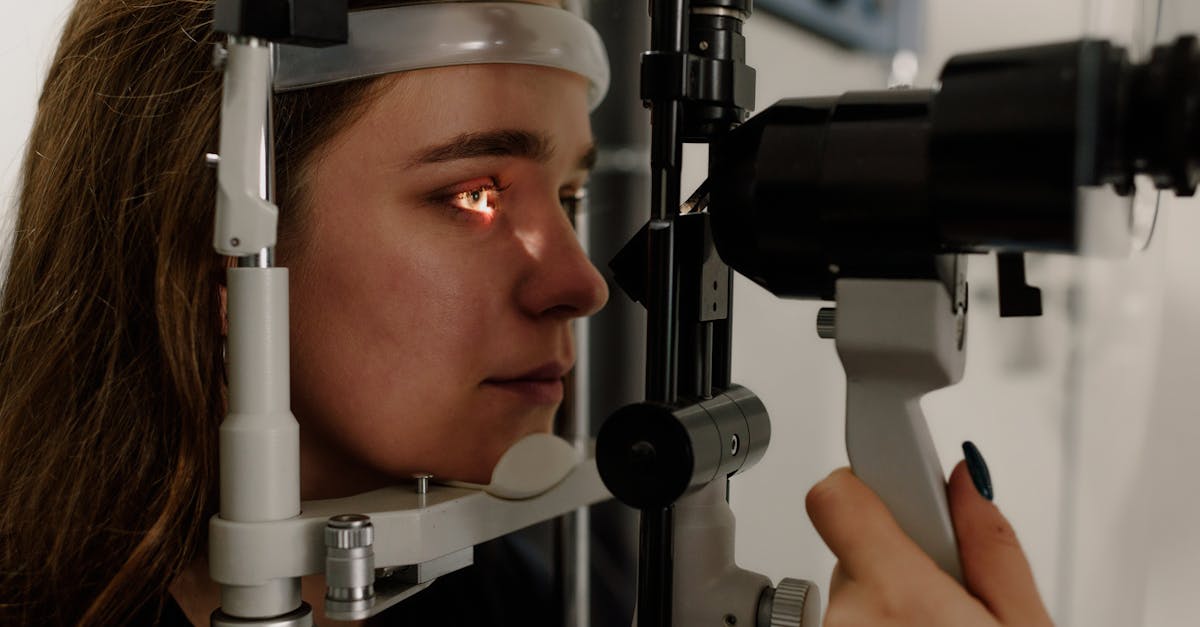
Corneal topography functions by projecting a series of illuminated concentric rings (a Placido disc) onto the tear film of the cornea. A digital camera captures the reflection of these rings. If the cornea is perfectly spherical, the reflected rings appear as perfect, evenly spaced circles. If the cornea is irregular, the reflected rings appear distorted, pinched, or severely compressed.
Sophisticated software analyzes this reflection to generate a highly detailed, color-coded topographical map of the entire corneal surface. Red and orange colors indicate areas of steep curvature (such as the cone in keratoconus), while blue colors indicate flattened areas. This topographical map provides the clinician with a precise, microscopic elevation profile, essential for diagnosing ectatic diseases early and for designing custom contact lenses.
8. Corneal Tomography and Pachymetry
While topography analyzes the front surface reflection, corneal tomography provides a comprehensive, three-dimensional reconstruction of the entire anterior segment of the eye. Utilizing Scheimpflug imaging or optical coherence tomography, tomography devices capture cross-sectional images of the cornea.
Tomography is critical because it accurately maps the posterior (back) surface of the cornea and provides a highly precise measurement of global corneal thickness, known as pachymetry. In early keratoconus, the initial bulging often occurs on the posterior surface of the cornea before the anterior surface is significantly affected.
By analyzing the posterior elevation and identifying areas of dangerous focal thinning, tomography allows ophthalmologists to diagnose keratoconus at its earliest, most subtle stages, long before the patient notices severe visual symptoms, allowing for early, vision-saving surgical interventions.
9. Categorizing Corneal Abnormalities
Understanding the specific cause of the irregularity dictates the treatment path. The table below outlines common sources of corneal irregular astigmatism.
| Etiology | Pathological Mechanism | Primary Diagnostic Finding |
|---|---|---|
| Keratoconus | Biomechanical weakening and thinning. | Inferior steepening and focal thinning on tomography. |
| Corneal Scarring | Post-infection/trauma fibrosis. | Visible opacity on slit-lamp; chaotic topography over scar. |
| Post-Refractive Ectasia | Structural failure after LASIK/PRK. | Bulging within the thinned laser treatment zone. |
| Pterygium | Fleshy growth pulling on the cornea. | Flattening of the cornea in the axis of the growth. |
10. Non-Surgical Management: Rigid Gas Permeable Lenses
Because eyeglasses cannot bend light to compensate for an asymmetrical, bumpy surface, the absolute gold standard for restoring vision in patients with moderate to severe corneal irregular astigmatism is the use of specialized contact lenses. Soft contact lenses are generally ineffective, as they simply drape over the irregular surface, taking on its distorted shape.
Rigid Gas Permeable (RGP) lenses are constructed from firm, highly oxygen-permeable plastics. When an RGP lens is placed on the eye, it does not conform to the irregular cornea. Instead, it maintains its perfect, smooth, spherical shape.
The space between the back of the rigid lens and the front of the bumpy cornea fills with a layer of natural tears. This “tear lens” optically neutralizes the uneven surface of the cornea completely. The incoming light rays refract smoothly through the front of the RGP lens and pass straight through the tear layer, entirely bypassing the corneal irregularities and delivering a sharp, focused image to the retina.
11. Advanced Treatment: Scleral Contact Lenses
For patients with severe keratoconus, massive surgical scarring, or extremely sensitive corneas, standard RGP lenses may be physically impossible to wear. Standard rigid lenses rest directly on the sensitive cornea; if the cornea is highly cone-shaped, the lens will rock, dislodge easily, and cause severe, painful friction on the peak of the cone.
Scleral contact lenses represent a massive technological leap in managing irregular corneas. These are large-diameter rigid lenses designed to vault entirely over the entire corneal surface. They rest exclusively on the sclera, the white part of the eye, which is much less sensitive than the cornea.
The massive space between the scleral lens and the cornea is filled with a sterile, non-preserved saline solution prior to insertion. This creates a deep, perfectly smooth, liquid optical vault. Scleral lenses provide unparalleled optical correction, effectively eliminating massive irregular astigmatism, while simultaneously offering exceptional comfort, as the rigid plastic never physically touches the diseased or scarred corneal tissue.
12. Surgical Intervention: Corneal Cross-Linking
If the irregular astigmatism is caused by a progressive ectatic disease, specifically keratoconus or post-LASIK ectasia, restoring vision with lenses is insufficient; the underlying biological weakening of the cornea must be halted.
Corneal cross-linking is a minimally invasive surgical procedure specifically designed to strengthen the corneal stroma. The ophthalmologist saturates the cornea with riboflavin (Vitamin B2) drops and subsequently exposes the tissue to targeted ultraviolet-A (UV-A) light for a specific duration.
The photochemical reaction between the UV light and the riboflavin creates robust, new chemical covalent bonds (cross-links) between the weak collagen fibers in the stroma. While this procedure does not cure the irregular astigmatism or significantly reverse the bulging, it dramatically stiffens the cornea. This effectively halts the progression of the disease, preventing the irregularity from worsening and avoiding the need for a future corneal transplant.
13. Surgical Interventions: PTK and Intracorneal Rings
For patients whose irregular astigmatism is caused by superficial corneal scarring (such as from a healed ulcer or previous trauma), an excimer laser procedure known as Phototherapeutic Keratectomy (PTK) may be employed. The surgeon uses the highly precise laser to physically vaporize and shave away the superficial opaque scar tissue, leaving a smoother, clearer underlying stromal bed, which significantly reduces light scattering and irregularity.
For specific cases of keratoconus, surgeons may implant Intracorneal Ring Segments (ICRS). These are tiny, semi-circular inserts made of a biocompatible plastic (PMMA). The surgeon uses a femtosecond laser to create micro-tunnels deep within the periphery of the corneal stroma and slides the rings into place.
The physical bulk of the rings acts as a structural scaffold, pulling and stretching the central cornea. This mechanical tension flattens the bulging cone and significantly improves the symmetry of the cornea, reducing the irregular astigmatism and making it much easier for the patient to tolerate standard contact lenses or glasses.
14. Penetrating and Deep Anterior Lamellar Keratoplasty
When corneal irregular astigmatism becomes so profound that scleral lenses can no longer provide adequate vision, or if dense, full-thickness scarring completely blocks the visual axis, replacing the damaged tissue through corneal transplantation is the definitive surgical option.
A Penetrating Keratoplasty (PK) is a full-thickness transplant. The surgeon uses a circular cutting tool (trephine) to remove a central “button” of the patient’s diseased cornea, encompassing all five layers, and sutures a perfectly sized, clear donor cornea in its place. While effective, a full-thickness graft carries a lifelong risk of immune rejection.
A more advanced technique is Deep Anterior Lamellar Keratoplasty (DALK). This procedure is ideal for keratoconus or superficial scars where the innermost endothelial layer of the patient’s cornea remains healthy. The surgeon meticulously dissects and removes only the diseased outer stroma, leaving the patient’s own endothelium intact, before suturing on the donor tissue. Because the endothelium is the primary target of immune rejection, DALK significantly lowers the long-term risk of graft failure compared to a full-thickness transplant.
15. Postoperative Care and Visual Rehabilitation
Following any surgical intervention for the cornea, the recovery and visual rehabilitation process is prolonged and requires fastidious patient compliance. Healing after a corneal transplant takes many months. The presence of surgical sutures temporarily induces significant astigmatism, meaning the patient’s vision will fluctuate significantly during the first year of healing.
The ophthalmologist manages the healing process by prescribing potent topical corticosteroid drops to suppress the immune system and prevent graft rejection, slowly tapering the medication over several months. To control the post-surgical astigmatism, the surgeon will selectively remove specific sutures over time to adjust the tension on the new graft, a process known as suture management.
Even after a successful, clear corneal transplant, patients frequently require specialized rigid or scleral contact lenses to achieve their best possible visual acuity, as the healed graft-host junction almost always retains a mild degree of irregular astigmatism that glasses cannot fully correct.
16. When to Consult a Cornea Specialist
Because standard eyeglasses cannot correct irregular astigmatism, patients experiencing unexplained visual distortions must seek evaluation by an eye care professional, particularly if their current glasses fail to sharpen their vision.
Immediate consultation with an ophthalmologist, specifically a fellowship-trained cornea specialist, is absolutely mandated if an individual experiences a rapid, progressive decline in vision, or the sudden onset of profound double vision in one eye, severe glare, or starbursts around lights at night.
Furthermore, any patient diagnosed with keratoconus, regardless of how mild the symptoms currently are, must be evaluated by a specialist to discuss corneal cross-linking. Halting the progression of the disease early is the single most critical intervention to prevent severe, irreversible irregular astigmatism and the eventual need for invasive corneal transplant surgery.
17. Frequently Asked Questions (FAQ)
1. Can LASIK fix my irregular astigmatism?
Generally, no. In fact, if you have irregular astigmatism caused by a disease like keratoconus, performing LASIK is highly dangerous. The laser removes tissue, which will further weaken the already fragile cornea, causing it to bulge massively and rapidly worsening your vision.
2. Why can’t I just get stronger glasses to fix the blurriness?
Glasses have a uniform, symmetrical curve. They cannot physically bend light to match the hundreds of different, chaotic bumps and curves on an irregular cornea. Only a rigid contact lens or scleral lens that creates a new, perfectly smooth surface over the eye can correct the distortion.
3. Are scleral lenses uncomfortable because they are so large?
No, surprisingly, they are incredibly comfortable. The cornea is highly sensitive, but scleral lenses do not touch the cornea at all. They vault over it and rest on the white part of the eye (sclera), which has very few nerve endings, making them much more comfortable than standard rigid lenses.
4. Will cross-linking improve my vision?
The primary goal of cross-linking is not to improve your vision, but to lock the cornea in its current shape and stop the disease from getting worse. While some patients experience a slight improvement as the cone flattens slightly, you will still need specialized contact lenses to see clearly after the procedure.
18. Bibliography
Disclaimer: The content is for informational purposes only and does not replace medical advice. Always consult your doctor for personalized treatment.

