1. Introduction
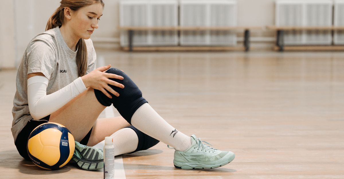
Osgood-Schlatter disease is a highly prevalent musculoskeletal condition that causes pain and swelling just below the knee joint. Despite possessing a name that sounds like a serious illness, it is a localized, benign inflammation of the growth plate located at the upper portion of the shinbone. This condition is a classic overuse injury, frequently striking active adolescents who participate in sports requiring repetitive running, jumping, and rapid changes of direction.
The condition arises due to a biomechanical mismatch between the strength of the developing muscles and the relative vulnerability of the actively growing bone. While the pain can be profoundly disruptive to an adolescent athlete’s training schedule, the prognosis is excellent. Understanding the mechanical triggers and implementing a structured program of rest, targeted stretching, and activity modification are the fundamental pillars of achieving full recovery and returning safely to athletic competition.
2. Anatomy of the Knee Extensor Mechanism
To understand the nature of this injury, it is necessary to examine the anatomy of the knee extensor mechanism. The quadriceps are a massive, powerful group of four muscles located on the front of the thigh. These muscles merge into the quadriceps tendon, which encases the patella, or kneecap.
From the bottom of the patella, the tissue continues as the strong patellar tendon. This thick, fibrous band stretches downward and anchors itself firmly into a small bony bump on the front of the upper tibia, known as the tibial tuberosity. When the quadriceps muscles contract, they pull on this entire mechanism, straightening the knee joint and propelling the body upward or forward.
3. The Tibial Tuberosity Growth Plate
In a mature adult, the tibial tuberosity is solid, fused bone. However, in children and adolescents, this bony bump features its own specialized area of developing cartilage, known as an apophysis. This growth center serves as the transition zone where new bone is generated, allowing the tibia to lengthen and mature.
Because this apophysis is composed of cartilage rather than solid bone, it is structurally the weakest link in the entire knee extensor mechanism. It is significantly softer and more vulnerable to mechanical stress than the powerful quadriceps muscles, the robust patellar tendon, and the main shaft of the tibia itself.
4. Pathophysiology of Apophysitis
Osgood-Schlatter disease is medically classified as a traction apophysitis. Every time an adolescent runs, jumps, or kicks, the massive quadriceps muscle contracts forcefully. This contraction transmits an immense pulling force through the patellar tendon directly onto the fragile cartilage of the tibial tuberosity apophysis.
When these forceful traction events occur repeatedly without adequate rest, they cause microscopic tearing and inflammation at the insertion site of the tendon. The body responds to this chronic microtrauma by rushing inflammatory fluid to the area and laying down new bone tissue in a disorganized attempt to reinforce the weakened attachment site.
5. Biomechanical Triggers in Sports
The mechanical forces generated during specific athletic movements are the primary triggers for this condition. Sports that demand explosive knee extension, such as basketball, volleyball, soccer, and gymnastics, place astronomical loads on the patellar tendon.
During a jump, the quadriceps must fire with maximum velocity to launch the athlete into the air. More critically, when the athlete lands, the quadriceps must perform a powerful eccentric contraction—lengthening while under tension—to absorb the shock and decelerate the body. This eccentric loading places the highest possible mechanical traction stress directly on the vulnerable tibial tuberosity.
6. The Role of Growth Spurts
The timing of this condition is intricately linked to the adolescent growth spurt, typically occurring between the ages of ten and fourteen for girls, and twelve and fifteen for boys. During a rapid growth phase, the long bones of the legs lengthen at an extraordinary rate.
Unfortunately, the muscles and tendons do not lengthen at the same speed. Consequently, the quadriceps and hamstring muscles become relatively tight, stretched taut across the newly lengthened femur and tibia. This baseline muscular tightness dramatically increases the resting tension on the patellar tendon, compounding the mechanical stress applied to the tibial tuberosity during physical activity.
7. Clinical Presentation and Symptoms
The hallmark symptom is a localized, aching pain situated precisely on the bony bump just below the kneecap. The pain is distinctly exacerbated by physical activities involving running, squatting, or climbing stairs. The adolescent typically points to the exact site of discomfort, noting that the pain significantly worsens during sports practice and subsides with rest.
Visually, the tibial tuberosity often appears noticeably swollen and enlarged compared to the unaffected knee. The bump is exquisitely tender to direct touch; simple acts like kneeling on a hard floor or lightly bumping the knee against a desk can provoke a sharp, intense pain response. If your child experiences persistent knee pain directly below the kneecap, an orthopedic assessment is recommended.
8. Diagnostic Physical Evaluation
The clinical diagnosis is usually straightforward and relies primarily on a targeted physical examination. The physician will observe the enlarged tibial tuberosity and apply gentle digital pressure directly to the site. Eliciting sharp, localized pain at this precise anatomical landmark strongly supports the diagnosis.
The examiner will also assess the flexibility of the lower extremity. Profound tightness in the quadriceps muscles on the front of the thigh, as well as the hamstrings on the back, is a nearly universal clinical finding in these patients. The physician will also test the stability of the knee ligaments to ensure the pain is not originating from a deeper joint injury.
9. Radiographic Assessment
While the diagnosis is primarily clinical, standard x-rays of the knee viewed from the side are frequently ordered. The primary purpose of the x-ray is to entirely rule out more severe bone pathologies, such as an occult fracture, a bone cyst, or a dangerous bone tumor, which can rarely present with similar localized pain.
In a patient with classic Osgood-Schlatter disease, the x-ray may appear completely normal. However, it often reveals fragmentation or irregular ossification of the tibial tuberosity. Small, distinct pieces of bone may be visible pulling slightly away from the main tibial shaft within the patellar tendon, confirming the chronic traction injury.
10. Differentiating from Other Knee Pathologies
Accurate diagnosis requires distinguishing this specific apophysitis from other common adolescent knee ailments. Patellofemoral pain syndrome, often called runner’s knee, presents with vague pain located underneath or around the edges of the kneecap itself, rather than strictly on the bump below it.
Sinding-Larsen-Johansson syndrome is an almost identical traction apophysitis, but it occurs at the inferior pole of the patella—the bottom tip of the kneecap—where the tendon originates, rather than where it inserts. Differentiating these conditions ensures that physical therapy exercises are targeted correctly.
11. Initial Conservative Management
The foundation of treatment is activity modification and the rigorous management of acute inflammation. Absolute immobilization is rarely required, but the adolescent must significantly decrease the intensity and frequency of the activities provoking the pain. Complete rest from high-impact sports may be necessary for several weeks in severe cases.
Applying cold therapy directly to the tibial tuberosity for fifteen minutes after any physical activity powerfully constricts blood vessels and reduces painful swelling. Short courses of over-the-counter nonsteroidal anti-inflammatory medications can assist in managing the acute discomfort during severe flare-ups.
12. Physical Therapy Protocols
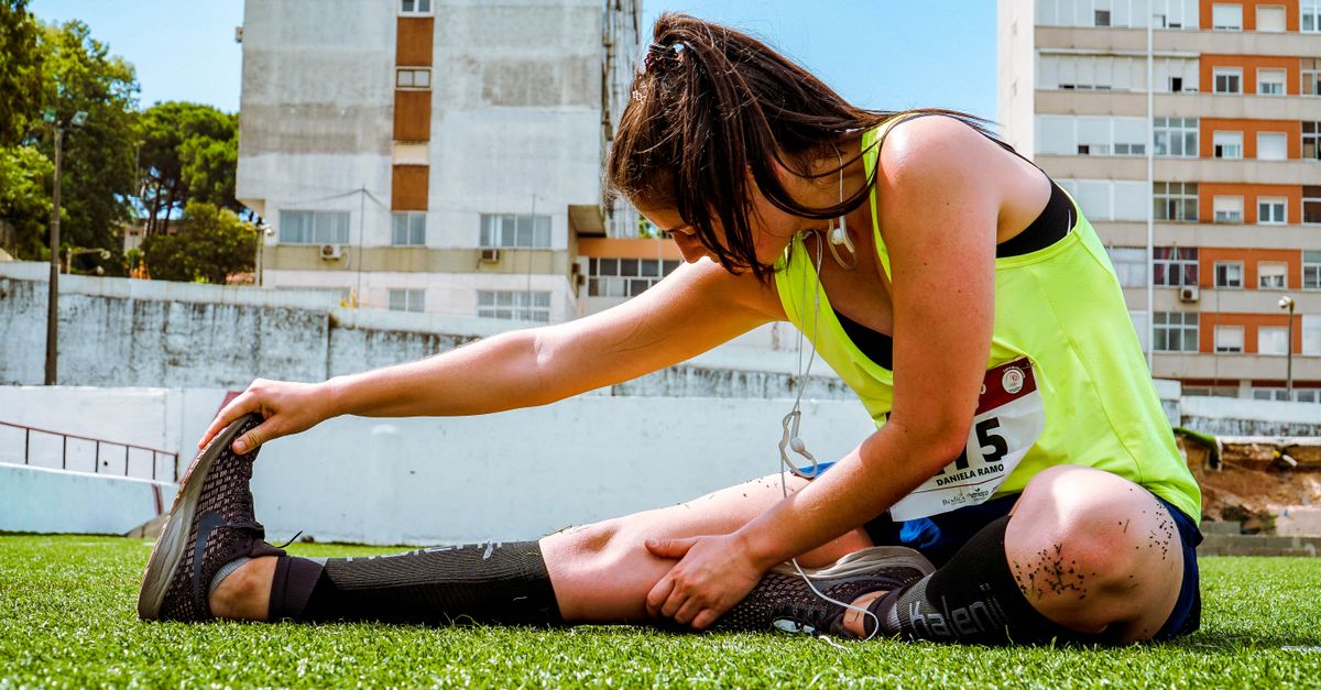
A structured physical therapy program is the most critical component for long-term resolution. The primary objective is to eliminate the biomechanical tightness that is placing excessive traction on the growth plate. The physical therapist will implement a rigorous, daily stretching routine focusing heavily on the quadriceps and hamstring muscles.
Once flexibility is restored, the program shifts toward strengthening the core and hip musculature. Weak hips often force the knee to absorb improper mechanical loads during running and jumping. Correcting overall leg alignment and movement mechanics significantly reduces the isolated strain on the patellar tendon.
13. Activity Modification Strategies
Returning to sports requires careful, negotiated activity modification. The athlete should avoid specializing in a single high-impact sport year-round, as continuous repetition prevents the growth plate from healing. Incorporating low-impact cross-training, such as swimming or cycling, maintains cardiovascular fitness without pounding the knee.
Using a specialized patellar tendon strap can provide mechanical relief. This narrow band is worn tightly around the leg just below the kneecap. It subtly alters the angle of the tendon insertion and dissipates some of the pulling force away from the inflamed tibial tuberosity during physical exertion.
14. Long-Term Outcomes and Bone Ossicles
The natural history of Osgood-Schlatter disease is highly favorable. The condition is self-limiting, meaning it resolves completely on its own once the adolescent finishes growing and the tibial tuberosity apophysis fuses into solid bone. This fusion typically occurs around age fourteen for girls and sixteen for boys.
However, the enlarged bony bump below the knee often remains a permanent anatomical feature into adulthood. In a small percentage of patients, a tiny piece of fragmented bone, known as an ossicle, fails to fuse and remains trapped within the tendon. This can cause chronic kneeling pain in adulthood and may rarely require a minor surgical procedure for removal.
15. Summary of Diagnostic Factors
Recognizing the classic clinical markers ensures prompt and accurate care for the adolescent athlete. The table below outlines these definitive features.
| Clinical Feature | Diagnostic Indicator |
|---|---|
| Patient Demographic | Active adolescents, typically aged 10 to 15 years |
| Pain Location | Strictly on the bony bump (tibial tuberosity) below the kneecap |
| Aggravating Factors | Running, jumping, kneeling, squatting |
| Physical Exam Finding | Severe tenderness and prominent swelling at the insertion site |
16. Prevention and Safe Training
While the rapid bone growth driving this condition cannot be prevented, the severity of the symptoms can be significantly mitigated through proactive athletic training. Coaches and parents must emphasize the importance of comprehensive warm-up routines and daily lower-body stretching.
Monitoring training volume is crucial. Adolescents should not abruptly increase their sports participation or run mileage. A gradual, incremental approach allows the muscles and tendons adequate time to adapt to new mechanical stresses, reducing the likelihood of overwhelming the vulnerable growth plates.
17. Frequently Asked Questions
1. Will this condition stunt my child’s growth?
No. The inflammation is strictly localized to the front surface of the bone where the tendon attaches. It does not affect the main horizontal growth plates responsible for the lengthening of the leg bones. Your child will reach their normal, genetically predetermined height.
2. Does my child need to wear a knee brace or a cast?
Rigid casts are universally avoided as they cause severe muscle atrophy. A large knee brace is also unnecessary. A small, targeted patellar tendon strap worn during sports is often all the mechanical support required.
3. Can cortisone shots cure the inflammation quickly?
Corticosteroid injections are strictly contraindicated for this condition. Injecting strong steroids into a load-bearing tendon in a growing child significantly weakens the collagen fibers and drastically increases the risk of a catastrophic tendon rupture.
4. Should my child push through the pain to toughen up?
Absolutely not. Pushing through severe pain will only increase the microscopic tearing, enlarge the bony lump, and prolong the total recovery time. Pain is a biological signal that the tissue is sustaining damage and requires rest.
5. Is surgery ever required to fix the bump?
Surgery is never performed on a growing child for this condition. In very rare cases, if an adult continues to have severe pain when kneeling due to a loose bone fragment trapped in the tendon, a minor outpatient surgery can be performed to remove the fragment.
18. Bibliography
Disclaimer: The content is for informational purposes only and does not replace medical advice. Always consult your doctor for personalized treatment.