
1. Introduction
A forehead laceration is a traumatic disruption of the facial skin and underlying subcutaneous tissues, frequently resulting from blunt force trauma, falls, or sharp instrument injuries. Because the forehead acts as the leading edge of the cranium, it is exceptionally vulnerable to impact during physical accidents. The highly vascular nature of the facial tissue means that even minor, superficial lacerations can produce a dramatic and alarming amount of hemorrhage.
Managing a forehead laceration extends far beyond simply stopping the bleeding. The primary clinical objectives include ruling out concomitant traumatic brain injury, assessing for underlying skull fractures, and identifying any damage to the intricate network of cranial nerves and facial muscles. Because the face is central to human identity and communication, achieving a meticulous, aesthetically pleasing cosmetic closure is a paramount concern for both the patient and the physician.
Through rigorous clinical evaluation, meticulous wound decontamination, and precise surgical techniques utilizing fine suturing materials, medical professionals can effectively repair these injuries. Modern protocols ensure that the risk of infection is minimized and that the healing tissue leaves the least perceptible scar possible, restoring the patient normal appearance and functional integrity.
2. Anatomy of the Forehead and Scalp
To appreciate the complexity of a forehead laceration, one must understand the stratified anatomical architecture of this region. The tissue layers of the forehead and scalp are classically remembered by the mnemonic SCALP: Skin, Connective tissue, Aponeurosis, Loose areolar tissue, and Pericranium.
The skin of the forehead is thick and richly supplied with blood vessels and sweat glands. Immediately beneath it lies a dense layer of subcutaneous connective tissue that anchors the skin tightly to the underlying musculoaponeurotic layer. This layer contains the frontalis muscle, which is responsible for elevating the eyebrows and creating horizontal forehead wrinkles.
Deep to the frontalis muscle is the loose areolar tissue, a spongy layer that allows the scalp to glide smoothly over the skull. The deepest layer is the pericranium, a tough, fibrous membrane firmly attached to the frontal bone. A laceration that penetrates through the aponeurosis into the loose areolar space tends to gape widely and requires a highly specific, multi-layered surgical closure.
3. Common Mechanisms of Facial Trauma
The mechanism of injury provides critical diagnostic clues regarding the depth, contamination, and potential complications of the laceration. Blunt force trauma is the most frequent cause. When the head strikes a solid object, such as a steering wheel or a concrete floor, the skin is violently crushed and split against the rigid underlying frontal bone. These bursting lacerations often possess ragged, irregular edges and are surrounded by significant swelling and contusion.
Sharp trauma, resulting from broken glass, metal edges, or knife wounds, produces clean, linear incisions. While these wounds are mechanically easier to approximate during suturing, sharp objects carry a higher risk of penetrating deeply through all tissue layers, potentially severing major nerves or breaching the cranium.
Avulsion injuries involve a tangential shearing force that peels a flap of skin and muscle away from the skull. These injuries are notoriously difficult to repair because the blood supply to the avulsed flap is often severely compromised, increasing the risk of subsequent tissue necrosis and massive scarring.
4. Neurological Evaluation and Concussion Risk
Because the forehead directly overlies the frontal lobes of the brain, any impact substantial enough to lacerate the skin carries a significant risk of inducing a traumatic brain injury. Before focusing on the cosmetic repair of the wound, the clinician must perform a rapid and thorough neurological assessment.
The physician evaluates the patient level of consciousness, orientation, and memory of the event. A Glasgow Coma Scale score is frequently documented to establish a neurological baseline. Symptoms such as a loss of consciousness, persistent vomiting, severe lethargy, or profound amnesia following the impact strongly suggest a concussion or deeper intracranial hemorrhage.
If you have experienced significant cranial trauma resulting in localized cranial discomfort, reading about headache pathologies can provide context for post-concussive symptoms. Any patient exhibiting altered mental status or focal neurological deficits requires an immediate non-contrast computed tomography scan of the head prior to wound management.
5. Vascular Supply and Profuse Hemorrhage
The face and scalp possess one of the most robust vascular networks in the human body. The forehead is primarily supplied by the supraorbital and supratrochlear arteries, which branch directly from the internal carotid system. This exceptional blood supply is physiologically beneficial, as it promotes rapid wound healing and confers a remarkable resistance to clinical infection.
However, this extensive vascularity also means that lacerations in this region bleed profusely. The blood vessels in the dense connective tissue layer of the scalp are tethered open and cannot easily spasm and retract to stem the flow of blood.
A patient with a seemingly small forehead laceration may present covered in blood, which can be highly distressing. Achieving rapid hemostasis is the first step in wound management, typically accomplished through direct, firm pressure applied continuously over the wound bed for ten to fifteen minutes.
6. Assessing for Underlying Skull Fractures
Once the bleeding is controlled, the clinician must meticulously explore the depths of the laceration under bright lighting to evaluate the structural integrity of the frontal bone. The physician gently palpates the wound bed with a sterile, gloved finger or a blunt surgical instrument.
The presence of a bony step-off, a palpable crack, or a noticeable depression in the skull indicates a potential underlying skull fracture. Furthermore, the physician examines the deep wound for the presence of cerebrospinal fluid leakage, which presents as a clear, watery fluid mixed with the blood, signaling a direct breach of the central nervous system.
If a skull fracture is suspected, digital exploration must be halted immediately to prevent inadvertently driving bone fragments into the brain tissue, and advanced radiological imaging is immediately procured.
7. The Importance of Wound Decontamination
Preparing the wound bed is the most critical step in preventing subsequent infection and minimizing the inflammatory response that leads to excessive scarring. The laceration must be meticulously cleaned to remove all coagulated blood, environmental debris, and microscopic pathogens.
High-pressure irrigation is the clinical standard of care. The physician uses a syringe to deliver a forceful stream of sterile normal saline directly into the wound depths. This mechanical flushing dislodges foreign bodies far more effectively than passive soaking.
In cases where the patient fell on gravel or asphalt, aggressive surgical scrubbing or sharp debridement may be necessary. Leaving even microscopic particles of dirt embedded in the dermis will result in a permanent, dark “traumatic tattoo” that is cosmetically disfiguring and exceptionally difficult to remove later.
8. Clinical Assessment in the Emergency Setting
A comprehensive clinical assessment involves evaluating specific cranial nerves that traverse the forehead region. The temporal branch of the facial nerve innervates the frontalis muscle. The physician tests this by asking the patient to raise their eyebrows against resistance. An inability to wrinkle one side of the forehead indicates a potential nerve transection.
The supraorbital and supratrochlear nerves provide sensory innervation to the forehead and anterior scalp. The clinician assesses sensation using a two-point discrimination test or light touch comparison above the laceration. Sensory loss is common if these superficial nerves are severed.
The physician also checks the anatomical alignment of the eyebrows and the natural skin tension lines, known as Langer lines. Lacerations that run parallel to these horizontal lines tend to heal with superior cosmetic results, whereas vertical lacerations constantly pull against the natural muscle tension, resulting in wider, more prominent scars.
9. Data Summary: Layers of the Forehead
Understanding the tissue layers helps clinicians determine the necessary depth and complexity of the surgical closure.
| Anatomical Layer | Characteristics | Clinical Implication for Laceration |
|---|---|---|
| Epidermis / Dermis | Highly vascular, rich in sweat glands | Profuse bleeding, requires precise cosmetic approximation |
| Subcutaneous Connective Tissue | Dense, tethers blood vessels open | Vessels cannot retract, contributing to heavy hemorrhage |
| Musculoaponeurotic Layer (Galea) | Contains frontalis muscle | If severed, wound gapes widely; requires deep suturing |
| Loose Areolar Tissue | Spongy, avascular plane | High risk area for rapid spread of severe infection |
10. Local Anesthesia and Nerve Blocks
To achieve the meticulous cleansing and precise suturing required for facial wounds, profound anesthesia is necessary. The standard approach involves injecting a local anesthetic, such as lidocaine mixed with epinephrine, directly into the wound margins. The epinephrine constricts local blood vessels, reducing bleeding and significantly extending the duration of pain relief.
Because injecting directly into a wound can distort the tissue edges and complicate precise alignment, clinicians frequently utilize regional nerve blocks for extensive forehead lacerations.
A supraorbital nerve block involves injecting anesthetic just above the eyebrow, where the nerve exits the skull. This technique provides profound, uninterrupted anesthesia to the entire ipsilateral forehead and anterior scalp without causing any tissue distortion at the actual laceration site, allowing for optimal surgical approximation.
11. Techniques for Primary Closure
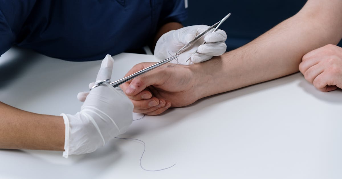
The primary objective of suturing a forehead laceration is to achieve perfect anatomical alignment with minimal tension on the skin edges. If the laceration breaches the galea aponeurotica (the muscle layer), a multi-layered closure is mandatory.
The physician first places absorbable sutures deep within the muscle and connective tissue. These deep sutures serve two critical functions: they eliminate “dead space” where blood could pool and form an infection-prone hematoma, and they absorb the mechanical tension of the surrounding muscles.
By taking the tension off the superficial skin layer, the deep sutures prevent the final scar from stretching and widening over time. Once the deep structural integrity is restored, the physician focuses on the precise approximation of the epidermal layer.
12. Suture Selection for Optimal Cosmesis
The choice of suturing material and technique significantly impacts the final cosmetic outcome. For the delicate skin of the face and forehead, clinicians utilize very fine, non-absorbable synthetic sutures. These thin threads minimize the inflammatory tissue reaction that causes scarring.
The sutures are placed very close to the wound edges and spaced closely together. The clinician meticulously everts the skin edges, ensuring they are slightly pushed outward. This eversion compensates for the natural contraction of the scar tissue during the remodeling phase, preventing the formation of a depressed, shadow-casting scar.
Special attention is paid to lacerations that cross the eyebrow. The physician must perfectly align the hair margins. Crucially, the eyebrow is never shaved prior to repair, as the hair provides vital anatomical landmarks for alignment, and eyebrow hair regrowth is notoriously unpredictable and slow.
13. Alternatives to Traditional Sutures
For small, clean, superficial lacerations that do not gap widely, alternative closure methods may be employed to reduce the trauma of needle punctures and eliminate the need for a follow-up removal visit.
Medical-grade tissue adhesives (cyanoacrylate formulations) are frequently used in pediatric patients. The adhesive acts as a strong, waterproof bridge that holds the skin edges together perfectly. It naturally sloughs off over five to ten days as the epidermis heals beneath it.
Reinforced adhesive strips (Steri-Strips) are also highly effective for linear, low-tension wounds on the flat surface of the forehead. These alternative methods provide excellent cosmetic results when used appropriately by a skilled clinician.
14. Postoperative Care and Scar Management
Proper wound care at home is essential for preventing infection and optimizing the final scar. The patient is instructed to keep the sutured wound clean and covered with a thin layer of antibiotic ointment or petroleum jelly. Maintaining a moist wound environment promotes faster epithelialization and significantly reduces scarring compared to letting the wound dry out and scab over.
Non-absorbable sutures on the face are typically removed very early, usually between four to six days post-injury. Removing them early prevents the formation of “railroad track” scarring, where the skin heals around the suture entry points.
Once the sutures are removed, the patient must strictly protect the immature scar from ultraviolet radiation. Exposure to the sun will cause the new, delicate collagen to permanently hyperpigment, creating a dark, highly noticeable scar. Daily application of high-SPF sunscreen or wearing a hat is mandatory for at least six to twelve months.
15. Tetanus Prophylaxis and Antibiotic Therapy
Confirming the patient tetanus immunization status is a standard component of all laceration management. Clostridium tetani is an environmental bacterium that causes severe neurological spasms if it proliferates in a wound. If the patient has not received a tetanus booster within the last five to ten years, depending on wound contamination, a vaccine is administered.
Due to the excellent vascular supply of the face, prophylactic oral antibiotics are generally not required for simple, clean lacerations that have been adequately irrigated.
However, antibiotics are strictly indicated if the laceration was caused by an animal or human bite, if the wound was heavily contaminated with soil or organic matter, or if the patient is severely immunocompromised, such as those with poorly controlled diabetes or those undergoing chemotherapy.
16. When to Seek Emergency Medical Care
While minor cuts can occasionally be managed at home, any significant trauma to the head requires immediate professional evaluation. Emergency medical care is absolutely necessary if the patient experienced a loss of consciousness, is persistently vomiting, or exhibits increasing confusion or lethargy following the impact.
A forehead laceration requires urgent closure if the wound gapes open, exposes yellow fat or white bone, or bleeds profusely and pulsatilely without slowing after ten minutes of direct pressure.
Furthermore, if a previously repaired laceration begins to demonstrate spreading redness, increasing warmth, severe throbbing pain, or starts draining foul-smelling purulent fluid, these are definitive signs of a developing infection requiring immediate medical and pharmacological intervention.
17. Frequently Asked Questions (FAQ)
1. Should I shave my eyebrow if I get a cut through it?
No, absolutely never shave an eyebrow. The hair serves as a critical visual guide for the physician to perfectly align the tissue edges during suturing. Furthermore, eyebrow hair grows very slowly and sometimes fails to grow back entirely if shaved.
2. How long do I have to wait before going to the emergency room for stitches?
To minimize the risk of infection and achieve the best cosmetic result, facial lacerations should ideally be evaluated and closed within eight to twelve hours. However, due to the excellent blood supply on the face, physicians can sometimes safely close facial wounds up to twenty-four hours after the injury.
3. Will I have a permanent scar on my forehead?
Yes, any laceration that cuts through the full thickness of the skin will leave a scar. However, prompt professional suturing, early suture removal, and strict sun protection will ensure the scar is as thin and unnoticeable as physiologically possible.
4. Can I use commercial superglue to close the cut at home?
No. Commercial superglue is highly toxic to human tissue, generates damaging heat as it cures, and can cause severe inflammatory reactions. It also risks sealing dangerous bacteria deep inside the wound. Always seek professional medical adhesives applied by a clinician.
5. Why did the doctor remove my facial stitches so early?
Stitches on the face are removed early, typically within 4 to 6 days, to prevent the skin from healing around the thread, which causes permanent, visible puncture marks known as “railroad track” scars. The underlying deep sutures continue to hold the wound together.
18. Bibliography
Disclaimer: The content is for informational purposes only and does not replace medical advice. Always consult your doctor for personalized treatment.
