1. Introduction and Clinical Overview
Late tooth eruption, clinically termed delayed dentition, occurs when a child’s primary (baby) or permanent teeth fail to emerge into the oral cavity within the established, normal chronological timeframes. Tooth eruption is a highly complex, genetically regulated biological process involving the precise coordination of bone resorption, root formation, and mucosal penetration. While parents frequently express significant anxiety when a child’s teeth do not appear exactly on schedule, a substantial degree of chronological variation is entirely normal and biologically expected.
However, when the delay extends beyond standard physiological variations—typically defined as an eruption occurring more than twelve months past the average age range for a specific tooth—a thorough clinical investigation is warranted. Delayed eruption can be a localized issue affecting a single tooth, often due to a physical obstruction in the jaw, or a generalized condition affecting the entire dentition, which may serve as an early indicator of underlying systemic diseases, endocrine disorders, or genetic syndromes.
Clinical management requires a meticulous evaluation by a pediatric dentist or orthodontist. The goal is to differentiate a harmless, late-blooming developmental pattern from a pathological delay that requires interceptive intervention. Early identification of structural blockages or systemic causes allows clinicians to utilize targeted orthodontic or surgical strategies to guide the teeth into their proper positions, preventing long-term malocclusion, impaction, and aesthetic complications.
2. Understanding Normal Eruption Chronology
To identify a delay accurately, one must first understand the baseline biological schedule. The eruption of human dentition occurs in two distinct phases: the primary (deciduous) dentition and the permanent dentition.
The primary dentition usually begins to emerge around six months of age, starting with the lower central incisors, and is typically complete with the eruption of the second molars by the age of two and a half to three years. The permanent dentition phase begins around age six, with the eruption of the first permanent molars behind the primary teeth, and the lower central incisors replacing their primary counterparts.
This process continues sequentially until approximately age twelve to thirteen, culminating with the second permanent molars. The third molars, or wisdom teeth, exhibit massive biological variation, typically erupting between the ages of seventeen and twenty-one, though they frequently remain impacted. It is essential for clinicians to evaluate the child’s overall dental age rather than strictly their chronological age, utilizing established dental development charts to assess the symmetry and sequence of eruption.
3. Primary Versus Permanent Tooth Delay
The clinical implications and underlying causes of delayed eruption differ significantly depending on whether the primary or permanent teeth are involved.
Delayed eruption of the primary dentition is relatively rare. When it occurs, particularly if no teeth have emerged by twelve to fifteen months of age, it is frequently linked to systemic factors, prematurity, low birth weight, or specific nutritional deficiencies. Because the primary teeth act as essential space maintainers and guides for the developing permanent teeth beneath them, prolonged delays can mildly impact early speech development and masticatory function.
Delayed eruption of permanent teeth is substantially more common and is frequently localized. The most commonly delayed or impacted permanent teeth are the maxillary (upper) canines, mandibular (lower) second premolars, and third molars. Localized delays in the permanent dentition are heavily driven by mechanical factors, such as a lack of physical space in the dental arch or early loss of a primary tooth leading to unfavorable shifting of adjacent teeth.
4. Genetic and Familial Factors
Genetics play the most prominent role in determining the timeline of dental development. Eruption schedules are highly heritable. If a child’s parents or older siblings experienced a late eruption pattern, it is highly probable that the child will follow a similar, delayed trajectory.
This familial pattern is generally considered a variation of normal physiological development rather than a pathology. In these cases, the entire dentition is typically delayed symmetrically. Radiographic evaluation will confirm that the teeth are developing normally within the jawbone, possessing healthy root structures and forming at an appropriate, albeit slower, pace compared to population averages.
Genetic variations also dictate the physical size of the teeth and the corresponding size of the jawbones. A genetic mismatch—inheriting large teeth and a small jaw—inevitably leads to severe crowding, physically blocking teeth from erupting on schedule and necessitating early orthodontic space management.
5. Nutritional Deficiencies and Environmental Factors
Proper tooth formation and eruption require significant metabolic energy and highly specific nutritional building blocks. Severe, chronic nutritional deficiencies during the critical windows of early childhood development can profoundly slow the biological engine driving tooth eruption.
Vitamin D deficiency, leading to clinical rickets, is a historically well-documented cause of generalized delayed dentition. Vitamin D is essential for calcium and phosphorus metabolism, which are the primary minerals required for the calcification of the dental hard tissues and the surrounding alveolar bone. Without adequate mineralization, the eruptive forces are compromised.
Similarly, severe protein-energy malnutrition or chronic gastrointestinal diseases causing severe malabsorption, such as untreated celiac disease, can stunt overall physical growth, mirroring a corresponding delay in dental age. Correcting the underlying nutritional deficits usually restores a normal, though shifted, developmental timeline.
6. Endocrine and Metabolic Causes
The endocrine system acts as the master regulator of physical growth, and systemic hormonal imbalances frequently manifest in the oral cavity as generalized delayed tooth eruption.
Hypothyroidism is the most common endocrine disorder associated with late dentition. Thyroid hormones are critical for bone remodeling and cellular maturation. Children with untreated or inadequately managed congenital hypothyroidism consistently demonstrate a significant delay in both the shedding of primary teeth and the emergence of permanent teeth. The teeth often remain fully formed but trapped within the jawbone due to a failure of the surrounding bone to resorb properly.
Hypopituitarism, or growth hormone deficiency, also causes proportional delays in dental age, corresponding with the child’s delayed skeletal maturation. Less commonly, hypoparathyroidism, which severely disrupts calcium homeostasis, can cause profound delays and distinct malformations in the enamel structure of the teeth that do eventually erupt.
7. Structural and Localized Obstructions
When the delay affects only one or two specific teeth while the rest of the mouth develops normally, the cause is almost invariably local and structural. A physical barricade within the jawbone prevents the tooth from migrating upward.
Supernumerary teeth, or extra teeth that form abnormally in the jaw, are a frequent culprit. The most common is the mesiodens, a small, peg-like tooth that forms directly between the upper central incisors, physically blocking the permanent incisors from erupting.
Odontogenic cysts and benign tumors, such as odontomas, can form around the crown of a developing tooth, creating a dense, physical barrier that halts the eruption pathway. Additionally, gingival fibromatosis, a condition characterized by exceptionally thick, fibrous gum tissue, can act as a literal ceiling, preventing a fully formed tooth from successfully piercing the mucosa.
8. Early Primary Tooth Loss and Space Loss
The premature loss of primary teeth due to severe dental caries (cavities) or trauma is a leading, preventable cause of delayed permanent tooth eruption. Primary teeth serve a critical biological function: they act as rigid space maintainers, holding the exact amount of space open for the underlying permanent tooth.
If a primary molar is extracted years before the permanent premolar is ready to emerge, the adjacent teeth will naturally drift and tilt into the empty space. When the permanent tooth eventually attempts to erupt, it finds the pathway completely blocked by the roots and crowns of the shifted teeth.
This mechanical blockage frequently results in an impacted tooth. The tooth may remain buried deep in the bone, or it may erupt at a severe angle, emerging out of the palate or high up on the gum line, requiring complex orthodontic traction to pull it back into the correct anatomical position.
9. Tooth Ankylosis
Ankylosis is a specific pathological condition where the roots of a tooth fuse directly to the surrounding alveolar bone, entirely bypassing the flexible periodontal ligament that normally suspends the tooth.
When a primary tooth becomes ankylosed, it ceases to erupt vertically as the child’s jaw continues to grow around it. It appears to “sink” below the level of the adjacent healthy teeth. Because it is fused to the bone, its roots will not naturally resorb.
This creates a rigid, immovable barricade directly above the developing permanent tooth. The permanent tooth is physically trapped and cannot erupt. The ankylosed primary tooth must be surgically extracted by a pediatric dentist to clear the pathway and allow the permanent tooth a chance to emerge normally.
10. Syndromic Associations
In pediatric medicine, generalized delayed tooth eruption is recognized as a classical clinical feature of several prominent genetic and chromosomal syndromes. When a severe delay is accompanied by other physical or developmental anomalies, a syndromic etiology must be evaluated.
Cleidocranial dysplasia is a rare genetic disorder characterized by the underdevelopment of the collarbones and a profound disruption in dental development. Patients typically retain their primary teeth well into adulthood, possess numerous supernumerary teeth, and experience a nearly complete failure of the permanent teeth to erupt without extensive surgical intervention.
Down syndrome (Trisomy 21) is consistently associated with significant delays in the eruption of both the primary and permanent dentitions. The teeth often emerge in an irregular, asymmetrical sequence. Other conditions, including Amelogenesis Imperfecta and Gardner Syndrome, also feature prominent eruptive delays alongside structural dental defects or jaw osteomas.
11. Clinical Dental Examination
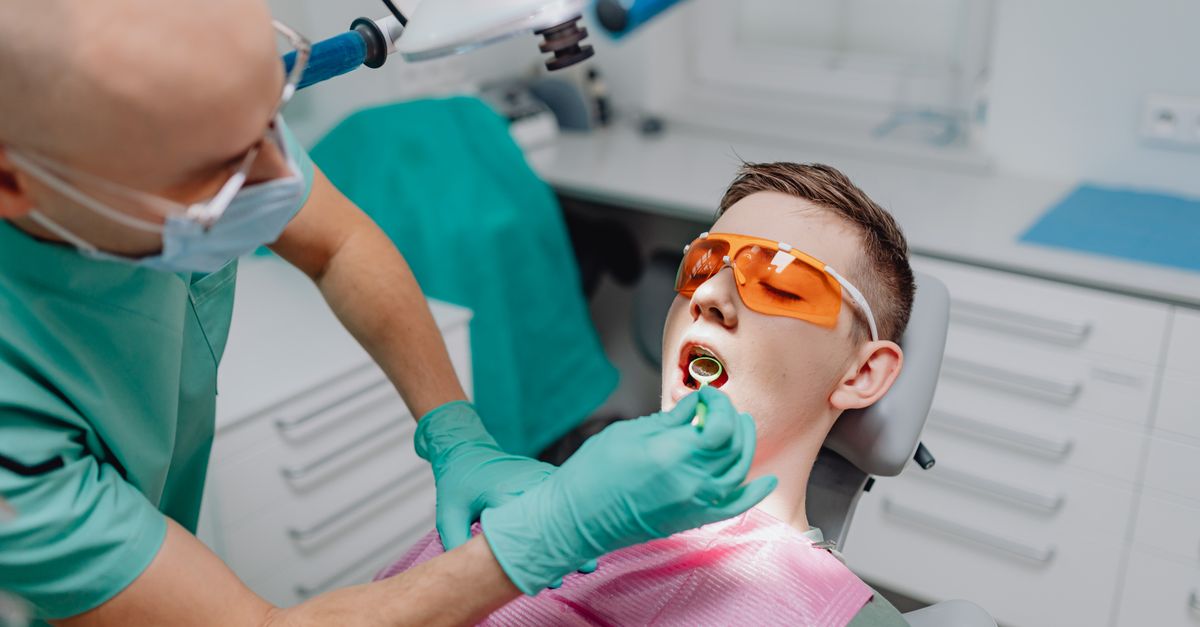
The diagnostic process begins with a meticulous clinical examination by a dental professional. The dentist assesses the exact sequence and symmetry of the teeth present in the oral cavity, comparing this data against standard chronological and dental age charts.
Palpation is a crucial diagnostic technique. The dentist will feel the gum ridges to determine if the crown of the missing tooth can be felt just below the mucosal surface, suggesting it is on its proper trajectory but slightly delayed.
The dentist also meticulously examines the surrounding soft tissues, looking for signs of exceptionally dense, fibrotic gingiva, localized swelling indicating an eruption cyst, or the tilting of adjacent teeth that signifies significant space loss and mechanical impaction. The child’s overall medical history is reviewed to identify potential systemic illnesses, nutritional deficits, or a family history of late-blooming dentition.
12. Radiographic Imaging and Assessment
Clinical observation alone cannot determine the status of teeth hidden within the jawbone. Comprehensive radiographic imaging is the definitive diagnostic tool for evaluating late tooth eruption.
A panoramic radiograph is typically ordered for children exhibiting generalized or multiple localized delays. This broad image displays the entire upper and lower jaws, allowing the clinician to count the developing permanent teeth, ensuring none are congenitally missing (agenesis).
The radiograph reveals the exact developmental stage of the roots. A tooth generally requires at least two-thirds of its root to be fully formed before it generates enough biological force to pierce the gums. The X-ray also immediately highlights structural blockages, clearly visualizing supernumerary teeth, dense bone sclerosis, abnormal angulation, or the presence of cysts enveloping the delayed crowns.
13. Differentiating Normal Variation from Pathology
The critical clinical decision is differentiating a harmless physiological delay from a pathological condition requiring intervention. The standard rule of thumb is the “rule of six months” for primary teeth and twelve months for permanent teeth. If a tooth is delayed beyond this window compared to population averages, intervention is considered.
Symmetry is a highly reliable indicator. If the lower right permanent molar erupts, the corresponding lower left permanent molar should erupt within a few months. A delay of more than six months between the eruption of corresponding teeth on opposite sides of the mouth strongly suggests a localized pathological obstruction on the delayed side, rather than a generalized, systemic delay.
If radiographic analysis shows that the root of the delayed tooth is fully formed and the apex (tip) is closed, yet the tooth remains deep in the bone, it has lost its eruptive potential. This is a definitive sign of mechanical impaction or ankylosis, requiring surgical and orthodontic management.
14. Interceptive Orthodontic Interventions
When a lack of space is identified as the primary cause of delayed or impacted permanent teeth, early interceptive orthodontics (Phase I treatment) is deployed to modify the jaw growth and create the necessary room.
Palatal expanders are frequently utilized in growing children to physically widen a narrow upper jaw. This creates additional arch perimeter, providing the necessary space for crowded, delayed incisors or canines to drop into place naturally without the need for extraction.
If primary teeth are lost prematurely, the immediate placement of a space maintainer is the standard of care. This small, custom-made wire appliance is cemented to the adjacent teeth, physically preventing them from drifting into the empty space, thereby holding the eruption pathway open for the permanent tooth hiding below.
15. Surgical Management Strategies
When radiographic imaging identifies a hard, physical obstruction, or when a tooth is severely impacted and has lost its eruptive force, oral surgical intervention becomes necessary.
If the delay is caused by exceptionally thick, fibrotic gum tissue, a simple surgical procedure called an operculectomy or a gingivectomy is performed. The surgeon uses a laser or scalpel to carefully remove the thick “ceiling” of gum tissue covering the crown, removing the physical barrier and allowing the tooth to emerge rapidly.
For severely impacted teeth, such as an upper canine blocked by dense bone or a supernumerary tooth, a combined surgical-orthodontic approach is required. An oral surgeon removes the obstruction and exposes the crown of the impacted tooth. An orthodontic bracket with a small gold chain is immediately bonded to the exposed crown. Over several months, the orthodontist applies gentle, continuous traction to the chain, physically pulling the stubborn tooth through the bone and into its correct position in the dental arch.
16. Frequently Asked Questions (FAQ)
1. Should I worry if my baby has no teeth at ten months old?
Usually, no. While the first tooth often appears around six months, it is entirely normal for some babies not to get their first tooth until twelve to fifteen months of age. If no teeth have emerged by fifteen months, a brief check-up with a pediatric dentist is recommended.
2. Why did my older child’s adult tooth come in perfectly, but the younger one is delayed?
Genetics dictate tooth eruption, and siblings can inherit different timelines. However, if a younger child lost a baby tooth early due to a cavity or trauma, the surrounding teeth may have shifted, blocking the adult tooth from coming in.
3. Does a late eruption mean the adult tooth is missing entirely?
It is a possibility. Congenitally missing teeth (agenesis) is a common genetic trait, most often affecting the lower second premolars or upper lateral incisors. A simple panoramic X-ray can immediately confirm if the adult tooth is actually developing under the gums.
4. Will late teeth cause my child to need braces?
If the late eruption is simply a natural slow biological timeline, braces may not be needed. However, if the delay is caused by severe crowding, an obstruction, or a tooth coming in at a severe angle, early orthodontic treatment will be required to guide the teeth into place.
5. What is a “shark tooth,” and is it related to delayed eruption?
A “shark tooth” occurs when a permanent tooth erupts directly behind a primary tooth that has failed to fall out on time. The baby tooth acts as a roadblock. The baby tooth typically needs to be extracted by a dentist to allow the adult tooth to move forward into its proper spot.
17. Bibliography
Disclaimer: The content is for informational purposes only and does not replace medical advice. Always consult your doctor for personalized treatment.