
1. Defining Visual Field Loss
A loss of a part of the visual field represents a structural or neurological deficit in the pathway that transmits visual information from the eye to the brain. The visual field encompasses the entire area that can be seen when the eyes are focused on a central point, including both central and peripheral vision. When a section of this field is obliterated or dimmed, it indicates a disruption at a specific anatomical location within the visual system.
Visual field defects vary significantly in their presentation. A scotoma is an isolated island of blindness surrounded by normal vision. More extensive losses, such as hemianopsia, involve the loss of an entire half of the visual field in one or both eyes. The specific pattern of the visual field loss serves as a highly precise neurological map, allowing clinicians to trace the symptom back to the exact site of the lesion.
Immediate clinical evaluation is critical. A sudden loss of a visual field frequently signifies a severe acute event, such as a cerebrovascular accident or a retinal detachment, requiring emergency intervention to prevent permanent blindness. Gradual loss often points toward progressive ocular diseases or slow-growing intracranial tumors.
2. Anatomy of the Visual Pathway
Understanding visual field defects requires a thorough knowledge of the visual pathway. Light enters the eye and strikes the retina, where photoreceptors convert the light into electrical signals. These signals travel via the optic nerve toward the brain.
Just behind the eyes, the optic nerves meet at a crucial intersection called the optic chiasm. Here, a specific sorting of nerve fibers occurs. The fibers carrying information from the nasal (inner) half of each retina cross over to the opposite side of the brain. The fibers from the temporal (outer) half of each retina stay on the same side.
After the chiasm, the re-sorted fibers travel as the optic tract, pass through the lateral geniculate nucleus in the thalamus, and fan out into the optic radiations before finally reaching the primary visual cortex in the occipital lobe at the back of the brain. A lesion at any point along this complex pathway produces a distinct, predictable pattern of visual loss.
3. Retinal Detachment and Pre-Chiasmal Lesions
Lesions occurring before the optic chiasm (pre-chiasmal) affect only the eye on the side of the lesion. A retinal detachment is a critical ocular emergency where the neurosensory retina peels away from its underlying supportive tissue. This separation deprives the photoreceptor cells of oxygen and nourishment.
Patients with a retinal detachment typically describe a shadow or a dark curtain descending over a portion of their peripheral vision. This visual field loss is often preceded by a sudden shower of floaters or bright flashes of light. If the detachment progresses to involve the macula, central vision is permanently destroyed.
Other pre-chiasmal causes include severe optic neuritis, an inflammation of the optic nerve frequently associated with multiple sclerosis, and ischemic optic neuropathy, where blood flow to the optic nerve is abruptly compromised. These conditions produce profound visual field defects, usually in a single eye, and require rapid medical intervention to preserve nerve function.
4. Glaucoma and Peripheral Field Constriction
Glaucoma is a progressive optic neuropathy characterized by the slow, irreversible destruction of the retinal ganglion cells and their axons, which make up the optic nerve. This damage is most frequently associated with elevated intraocular pressure, although it can occur at normal pressures.
The hallmark of glaucomatous damage is the insidious, gradual loss of the peripheral visual field. Because the loss is slow and initially affects only the outermost edges of vision, the brain compensates, and the patient remains entirely unaware of the deficit until the disease is highly advanced.
As glaucoma progresses, the peripheral loss advances inward, eventually resulting in tunnel vision, where only a small central island of sight remains. If left untreated, even this central vision is extinguished. Regular, comprehensive eye examinations featuring automated perimetry are the only way to detect and arrest this silent blinding disease.
5. Macular Degeneration and Central Scotomas
While glaucoma attacks the peripheral vision, age-related macular degeneration specifically targets the macula, the small central area of the retina responsible for sharp, detailed, and color vision. As the macular cells deteriorate due to age, oxidative stress, and poor blood flow, a central visual field defect develops.
This defect presents as a central scotoma—a dark, blurry, or completely missing area in the direct center of the patient’s vision. Peripheral vision remains intact, allowing the patient to navigate their environment, but they lose the ability to read, recognize faces, or drive.
In the “wet” form of macular degeneration, abnormal blood vessels grow beneath the macula and leak fluid or blood, causing rapid and severe central visual field loss. This requires immediate clinical intervention with intraocular injections of anti-vascular endothelial growth factor medications to halt the abnormal vessel growth and preserve central sight.
6. Bitemporal Hemianopsia and the Optic Chiasm
A highly specific pattern of visual field loss is bitemporal hemianopsia. In this condition, the patient loses the outer (temporal) half of the visual field in both eyes. This exact pattern points directly to a lesion compressing the center of the optic chiasm, where the crossing nerve fibers are located.
The most common cause of bitemporal hemianopsia is a pituitary adenoma, a benign tumor of the pituitary gland, which sits anatomically directly beneath the optic chiasm. As the tumor grows upward, it stretches and compresses the crossing nasal fibers.
Patients often do not notice the missing outer fields immediately but may report repeatedly bumping into doorframes or vehicles on their sides. Diagnosis requires a prompt magnetic resonance imaging scan of the brain to visualize the tumor, which typically requires surgical resection or targeted pharmacological therapy to relieve the pressure on the optic chiasm and restore vision.
7. Homonymous Hemianopsia and Post-Chiasmal Lesions
Lesions occurring after the optic chiasm (post-chiasmal) within the optic tract, optic radiations, or occipital lobe produce a pattern known as homonymous hemianopsia. In this condition, the patient loses the same half of the visual field in both eyes. For example, a right-sided homonymous hemianopsia means the patient cannot see the right half of the visual world with either eye.
This pattern occurs because the visual pathways have already sorted the information; the left side of the brain processes the entire right half of the visual world, and vice versa. Therefore, a lesion on the left side of the brain produces a right-sided visual field defect.
The most frequent cause of sudden homonymous hemianopsia is a cerebrovascular accident, specifically an ischemic stroke affecting the posterior cerebral artery which supplies the occipital lobe. Other causes include traumatic brain injuries and intracranial neoplasms.
8. Quadrantanopsia and the Optic Radiations
As the visual fibers travel from the thalamus to the occipital lobe, they fan out into a broad pathway known as the optic radiations. These radiations pass through different lobes of the brain. The superior fibers pass through the parietal lobe, while the inferior fibers sweep through the temporal lobe (Meyer’s loop).
A localized lesion in these specific lobes produces a visual field defect known as quadrantanopsia, where exactly one-quarter of the visual field is lost in both eyes.
| Lesion Location | Visual Field Defect | Common Etiology |
|---|---|---|
| Parietal Lobe | Inferior Quadrantanopsia (loss of lower quarter) | Ischemic stroke, tumor |
| Temporal Lobe | Superior Quadrantanopsia (loss of upper quarter) | Temporal lobe epilepsy surgery, stroke |
Identifying whether the lost quadrant is superior or inferior allows the neurologist to precisely localize the brain lesion to either the temporal or parietal lobe before confirming with advanced neuroimaging.
9. Macular Sparing in Occipital Lobe Strokes
A fascinating clinical phenomenon is observed in some patients with homonymous hemianopsia caused by a stroke in the occipital lobe. The patient loses half of their visual field, but a small, precise circle of central vision remains intact on the affected side. This is termed macular sparing.
The macula has a massive representation at the very tip (pole) of the occipital lobe. This specific area often receives dual blood supply from both the posterior cerebral artery and the middle cerebral artery. Therefore, if the posterior cerebral artery is blocked by a stroke, the middle cerebral artery can sometimes provide enough collateral blood flow to keep the macular representation alive.
The presence of macular sparing strongly indicates that the lesion causing the visual field loss is located at the very back of the visual pathway in the primary visual cortex, rather than further forward in the optic tract or radiations.
10. Transient Ischemic Attacks and Amaurosis Fugax
A loss of part or all of the visual field that lasts for only a few minutes before completely resolving is a critical medical emergency. This transient visual loss in one eye is termed amaurosis fugax. It is typically described as a shade being pulled down over the eye and then raised back up.
Amaurosis fugax is the ocular equivalent of a transient ischemic attack. It is most commonly caused by a small cholesterol plaque or blood clot breaking off from a diseased carotid artery in the neck, traveling up to the eye, and temporarily blocking the ophthalmic or central retinal artery.
Although the vision returns, the underlying vascular disease remains. The patient is at an extraordinarily high risk for a massive, permanent stroke in the immediate future. Immediate vascular imaging of the carotid arteries and the initiation of potent antiplatelet therapy are mandatory.
11. Diagnostic Visual Field Testing (Perimetry)
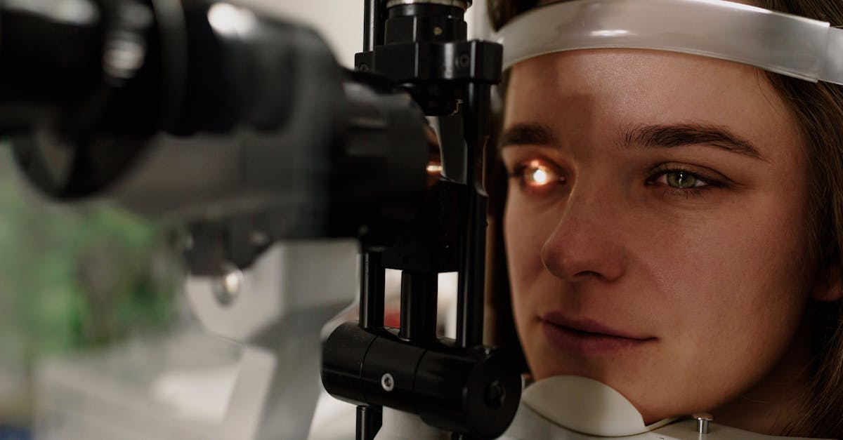
The clinical evaluation of visual field loss requires precise, objective mapping using automated perimetry. During this test, the patient stares at a central fixation target inside a specialized illuminated bowl. Small flashes of light of varying intensities are presented in different areas of the peripheral and central fields.
The patient presses a button whenever they perceive a light. The computer records the exact location and intensity of every light the patient could not see, generating a highly detailed topographical map of the visual field. This map precisely delineates the borders of scotomas and the exact patterns of hemianopsias.
Automated perimetry is not only essential for initial diagnosis but is heavily utilized to monitor the progression of diseases like glaucoma, ensuring that therapeutic interventions are successfully halting further visual decay.
12. Neurological Examination and Neuroimaging
When a visual field defect points to a neurological rather than a purely ocular cause, a comprehensive neurological examination is required. The clinician will assess cranial nerve function, motor strength, and sensory perception to identify any accompanying deficits that help localize the brain lesion.
Magnetic resonance imaging of the brain and orbits with gadolinium contrast is the gold standard diagnostic tool. It provides exquisite soft-tissue detail, allowing the neuroradiologist to visualize optic nerve inflammation in multiple sclerosis, compressions of the optic chiasm by pituitary tumors, or areas of infarction in the occipital lobes.
In cases of suspected vascular pathology, such as an impending stroke causing amaurosis fugax, a computed tomography angiogram or magnetic resonance angiogram is necessary to evaluate the patency of the carotid and cerebral arteries.
13. Rehabilitation and Clinical Management
Treatment for visual field loss depends entirely on reversing or halting the underlying cause. While vision lost to glaucoma or a completed stroke is typically permanent, visual loss caused by compressive tumors or acute retinal detachments can often be restored if surgical intervention is prompt.
For patients with permanent hemianopsia, clinical management shifts toward intensive vision rehabilitation. Patients lose the ability to see half of their environment, leading to frequent collisions and significant safety risks. Occupational therapists teach scanning techniques, training the patient to actively and continuously turn their eyes and head into the blind field to compensate for the neurological deficit.
Specialized optical devices, such as prisms mounted on eyeglasses, can also be utilized to artificially shift the image from the blind visual field into the seeing field, enhancing the patient’s spatial awareness and improving their ability to safely navigate their surroundings.
14. Frequently Asked Questions (FAQ)
1. What does a blind spot in the middle of my vision mean?
A blind spot directly in the center of your vision is called a central scotoma. It usually points to a problem with the macula, the center of the retina. The most common cause in older adults is macular degeneration, which requires immediate evaluation by an ophthalmologist.
2. Why did I lose vision on the right side of both eyes?
This pattern is called homonymous hemianopsia. It happens because the visual pathways in the brain cross over. A problem on the left side of your brain—most commonly an ischemic stroke—will knock out the right half of the visual world in both of your eyes.
3. Can glaucoma cause me to lose part of my vision?
Yes. Glaucoma slowly destroys the optic nerve due to high eye pressure. It typically attacks the outermost peripheral vision first, causing tunnel vision. Because it happens slowly, you may not notice the loss until it is severe and permanent.
4. Is a sudden dark shadow over my vision dangerous?
A sudden dark shadow or curtain descending over your vision is a classic sign of a retinal detachment. This is a critical ocular emergency. If not repaired surgically within hours or days, it will lead to permanent blindness in that eye.
5. Why is temporary vision loss a medical emergency?
If you lose vision in one eye and it comes back a few minutes later, you likely had a transient ischemic attack, or a “mini-stroke” in the eye. This means a blood clot temporarily blocked the artery and indicates you are at an extremely high risk for a major brain stroke very soon.
15. Bibliography
Disclaimer: The content is for informational purposes only and does not replace medical advice. Always consult your doctor for personalized treatment.
