1. Introduction
Determining whether lower back pain originates from a musculoskeletal issue or a kidney-related pathology requires analyzing the exact location, quality of pain, and the presence of associated systemic symptoms. Musculoskeletal pain typically affects the lower lumbar region, fluctuates with movement or posture, and often feels like a dull ache or sharp spasm. Conversely, kidney pain is generally located higher in the back, specifically in the flank area beneath the rib cage, presents as a constant, deep ache or severe, wave-like pain, and is frequently accompanied by urinary symptoms or fever. Accurate differentiation is critical for directing appropriate clinical intervention.
The lumbar region is a complex anatomical structure comprising vertebrae, intervertebral discs, muscles, ligaments, and nerves, any of which can generate nociceptive signals. Situated just anterior to the paraspinal muscles in the retroperitoneal space are the kidneys. Because of this anatomical proximity, pathological processes in either system can refer pain to overlapping areas, creating a diagnostic challenge for both patients and clinicians.
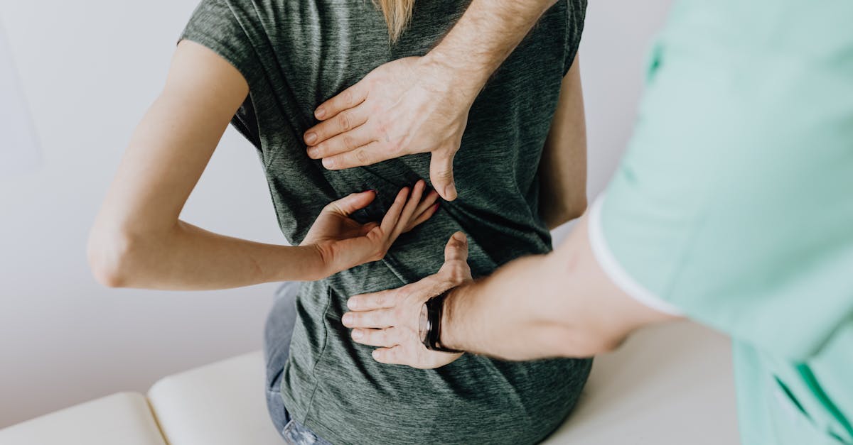
A structured clinical evaluation focuses on precise history taking and physical examination maneuvers to isolate the source of the discomfort. Understanding the distinct neurological pathways and physiological responses of the renal and musculoskeletal systems provides the foundation for an accurate diagnosis and the prevention of chronic complications.
2. The Anatomy of the Lumbar Region
The lower back, or lumbar spine, is engineered for strength, flexibility, and load-bearing. It consists of five massive lumbar vertebrae separated by shock-absorbing intervertebral discs. A complex network of ligaments and robust paraspinal muscles supports this bony column, controlling movement and maintaining posture.
The kidneys are bilateral, bean-shaped organs located in the retroperitoneal space, outside the peritoneal cavity. They reside against the posterior abdominal wall, roughly between the levels of the twelfth thoracic and third lumbar vertebrae. The upper portions of the kidneys are partially protected by the eleventh and twelfth ribs.
Understanding this topographical anatomy is essential. Because the kidneys are positioned higher and deeper than the structures typically involved in mechanical lower back strain, the epicenter of renal pain generally differs from the epicenter of musculoskeletal lumbar pain.
3. Understanding Renal Pain
Renal pain, often referred to as renal colic or capsular pain, arises primarily from the distension of the renal capsule or the stretching of the renal pelvis and ureter. The renal parenchyma itself lacks nociceptors. It is the sudden expansion of the highly innervated capsule, usually due to inflammation or obstruction, that generates the pain signal.
Visceral pain fibers transmit these nociceptive signals from the kidneys to the spinal cord at the T10 to L1 levels. Due to the convergence of these visceral fibers with somatic nerve fibers in the spinal cord, the brain often perceives the pain as originating from the corresponding skin dermatomes, a phenomenon known as referred pain.
This mechanism explains why kidney pain is typically felt in the flank, the fleshy area between the ribs and the hip, and can radiate anteriorly toward the abdomen, groin, or even the inner thigh, following the course of the ureter.
4. Mechanisms of Musculoskeletal Back Pain
Musculoskeletal back pain is predominantly somatic in origin. It results from mechanical injury, inflammation, or degenerative changes in the structures of the lumbar spine. Muscle strains and ligament sprains are the most common culprits, often triggered by improper lifting, sudden twisting movements, or prolonged poor posture.
Another significant mechanism is radiculopathy, which involves the compression or irritation of a spinal nerve root. Conditions like a herniated intervertebral disc or spinal stenosis can encroach on the neural foramina. This causes sharp, shooting pain that radiates down the trajectory of the affected nerve, classically known as sciatica.
Unlike the visceral pain of the kidneys, somatic musculoskeletal pain is usually highly localized and mechanically sensitive. The pain receptors in the muscles, fascia, and periosteum respond directly to mechanical stress, making the symptoms exquisitely responsive to movement and positioning.
5. Location and Radiation of the Pain
The location of the pain is a primary differentiating factor. Kidney pain is characteristically located in the costovertebral angle, the region formed by the lower border of the twelfth rib and the spine. It is typically unilateral, affecting only one side, although bilateral involvement is possible in certain systemic conditions.
Musculoskeletal back pain is most frequently located lower down, in the lumbosacral region, near the beltline or the sacroiliac joints. It can be central, bilateral, or unilateral. When a nerve root is involved, the pain often radiates below the knee, following a specific dermatomal distribution.
Renal pain may radiate, but it typically travels anteriorly and inferiorly toward the lower abdomen, the groin, or the external genitalia. It does not radiate down the posterior leg, which is a hallmark of lumbar radiculopathy often associated with sciatica pain.
6. Nature and Quality of the Discomfort
The qualitative description of the pain provides significant diagnostic insight. Kidney pain associated with an acute obstruction, such as a calculus, is often described as excruciating, sharp, and colicky. It comes in intense waves that fluctuate in severity but rarely resolve completely without intervention.
In contrast, kidney pain from an infection, such as pyelonephritis, presents as a constant, deep, and heavy ache in the flank. It lacks the severe, colicky nature of an obstruction but is profoundly debilitating and often associated with systemic malaise.
Musculoskeletal pain is frequently described as a dull ache, a stiffness, or a feeling of being locked in place. In cases of muscle spasm, the pain can be acute and stabbing, but it is intrinsically tied to movement. The intensity of musculoskeletal pain is generally constant while maintaining a specific posture but changes immediately upon shifting position.
7. Urinary Symptoms Associated with Renal Issues
The presence of urinary symptoms is a powerful indicator of renal pathology. Since the kidneys are the primary organs of the urinary system, any localized inflammation or infection frequently alters urinary function. Dysuria, a painful or burning sensation during urination, strongly suggests a urinary tract infection that may have ascended to the kidneys.
Changes in the macroscopic appearance of the urine are also critical indicators. Hematuria, the presence of blood in the urine, can make the urine appear pink, red, or cola-colored. This is a common consequence of a kidney stone damaging the urothelium or severe inflammation from a renal infection.
Increased urinary frequency and urgency, or the sensation of incomplete bladder emptying, further corroborate a urinary tract etiology. These symptoms are entirely absent in isolated musculoskeletal lower back pain.
8. Systemic Signs and Fever
Systemic manifestations help distinguish infectious renal processes from mechanical back pain. Acute pyelonephritis, a bacterial infection of the kidney parenchyma, elicits a robust systemic inflammatory response. This clinically manifests as a high-grade fever, often accompanied by severe chills and rigors.
Nausea and vomiting are also frequently associated with acute renal colic or renal infections. The intense visceral pain and the shared autonomic innervation of the kidneys and the gastrointestinal tract often trigger significant gastrointestinal upset.
Musculoskeletal back pain, unless secondary to a rare spinal infection such as diskitis or osteomyelitis, is an isolated mechanical issue. It does not produce a fever, systemic chills, or significant gastrointestinal disturbances.
9. Mechanical Triggers and Movement
The response to mechanical loading and movement is arguably the most reliable clinical differentiator. Musculoskeletal back pain is highly mechanical. It is exacerbated by specific movements, such as bending forward, twisting, or lifting. Prolonged sitting or standing may also worsen the discomfort.
Conversely, changing positions, resting in a supine position with the knees elevated, or applying localized heat often provides temporary relief for mechanical lower back pain. The pain is directly linked to the biomechanical stress placed on the lumbar structures.
Kidney pain, being visceral in origin, is completely independent of body movement. The pain remains constant and severe regardless of whether the patient is standing, sitting, or lying down. Patients with renal colic are often observed pacing continuously, unable to find a comfortable position.
10. Common Causes of Kidney Pain
Nephrolithiasis, or kidney stones, are a leading cause of acute renal pain. These crystalline structures form in the renal pelvis and cause severe obstruction and ureteral spasm as they attempt to pass through the narrow urinary tract. The resulting pressure buildup behind the stone causes agonizing pain.
Pyelonephritis is a serious bacterial infection that typically ascends from the lower urinary tract into the renal parenchyma. It causes severe inflammation, swelling of the kidney tissue, and distension of the renal capsule, leading to a constant, throbbing flank ache.
Other less common causes include renal cysts, which can cause chronic discomfort if they enlarge significantly, and renal infarction, a rare condition where blood flow to a portion of the kidney is obstructed, causing acute, severe tissue ischemia and pain.
11. Common Causes of Lumbar Strain
Lumbar muscle strains are the most ubiquitous cause of lower back pain. These occur when the muscle fibers are abnormally stretched or torn due to sudden force, improper lifting techniques, or repetitive microtrauma. The resulting inflammation leads to localized pain and protective muscle spasms.
Degenerative disc disease is a chronic condition where the intervertebral discs lose hydration and structural integrity over time. This reduces their shock-absorbing capacity, leading to increased mechanical stress on the facet joints and surrounding soft tissues, resulting in chronic, aching back pain.
Lumbar herniated discs occur when the soft inner core of the disc extrudes through the tough outer layer. If this extruded material impinges on a neighboring spinal nerve root, it triggers intense local inflammation and radicular pain that extends into the lower extremity.
12. Clinical Differentiation
A comparative breakdown aids in rapidly assessing the likely origin of the patient’s symptoms during a clinical encounter.
| Symptom Category | Kidney-Related Pain | Musculoskeletal Back Pain |
|---|---|---|
| Typical Location | High in the back (flank), costovertebral angle, usually one-sided. | Lower lumbar region, beltline, can be central or bilateral. |
| Pain Quality | Sharp, severe waves (colic) or a constant, deep ache. | Dull ache, stiffness, or sharp catching pain with movement. |
| Effect of Movement | No relief with position changes; patient often paces. | Worsens with bending or lifting; improves with rest. |
| Associated Symptoms | Fever, chills, nausea, painful urination, hematuria. | Muscle spasms, restricted range of motion, leg numbness. |
| Radiation Path | Anteriorly to the lower abdomen or groin. | Posteriorly down the buttocks and back of the leg. |
13. Diagnostic Imaging and Urinalysis
When clinical presentation alone is insufficient, specific diagnostic investigations are warranted. Urinalysis is a fundamental, non-invasive test that can quickly identify signs of urinary tract pathology. The presence of leukocytes and nitrites suggests infection, while red blood cells indicate potential trauma or stones.
Renal ultrasound is a safe and effective imaging modality to evaluate the kidneys. It can detect structural abnormalities, identify the presence of hydronephrosis indicative of obstruction, and locate larger calculi within the renal pelvis.
For musculoskeletal pain, plain radiographs can identify bone fractures or significant degenerative changes. However, Magnetic Resonance Imaging provides superior visualization of soft tissues, including intervertebral discs and nerve roots, making it the modality of choice for diagnosing radiculopathy.
14. Treatment Modalities
The management of kidney pain depends on the underlying etiology. Uncomplicated nephrolithiasis is often managed conservatively with aggressive hydration, antiemetics, and potent analgesics until the stone passes. Severe infections require targeted antibiotic therapy and, occasionally, hospitalization for intravenous administration.
Treating musculoskeletal back pain focuses on reducing mechanical stress and inflammation. Initial management involves short periods of rest, nonsteroidal anti-inflammatory drugs, and the application of heat or ice. Physical therapy is a cornerstone of long-term management, focusing on strengthening the core musculature to support the lumbar spine.
In cases where mechanical back pain is refractory to conservative measures, epidural steroid injections or surgical interventions, such as a microdiscectomy, may be considered to relieve nerve root compression and restore function.
15. Red Flag Symptoms Requiring Immediate Care
Certain clinical presentations signify a medical emergency requiring immediate evaluation. The presence of lower back or flank pain accompanied by a high fever, severe nausea, and an inability to tolerate oral fluids suggests a severe systemic infection that demands immediate medical intervention.
A sudden loss of bowel or bladder control, accompanied by numbness in the saddle region (the groin and inner thighs) and severe lower back pain, is a hallmark of cauda equina syndrome. This is a critical neurological emergency requiring urgent surgical decompression to prevent permanent paralysis.
Additionally, excruciating flank pain that is sudden in onset and unremitting, particularly if accompanied by macroscopic hematuria, requires emergent evaluation to rule out a severe obstructing stone or vascular compromise of the kidney.
16. Frequently Asked Questions (FAQ)
1. Can drinking enough water prevent kidney pain?
Adequate hydration is crucial for preventing the formation of kidney stones, which are a primary cause of severe kidney pain. By diluting the urine, water prevents the crystallization of minerals. However, hydration will not resolve musculoskeletal back pain.
2. Does applying heat help with kidney pain?
While a heating pad can soothe tense back muscles, it is generally ineffective for the deep, visceral pain caused by kidney stones or infections. Systemic analgesics are typically required to manage true renal pain.
3. Can a kidney infection cause muscle spasms in the back?
While the primary pain is visceral, the intense nerve signals from a severe kidney infection can sometimes cause reflex muscle guarding or spasms in the surrounding paraspinal muscles, complicating the clinical picture.
4. How long does a pulled back muscle take to heal?
Most acute mechanical lower back strains resolve significantly within a few weeks with appropriate conservative care, including gentle movement and anti-inflammatory medications. Chronic pain lasting longer than three months requires further clinical evaluation.
5. Is it possible to have both kidney issues and back pain simultaneously?
Yes. A patient can have chronic lumbar degenerative disc disease and simultaneously develop an acute kidney stone or infection. Careful clinical assessment is required to identify and treat both distinct pathologies.
17. Bibliography
Disclaimer: The content is for informational purposes only and does not replace medical advice. Always consult your doctor for personalized treatment.