1. Introduction
To tell if a cut is infected and requires antibiotics, closely monitor the wound for expanding redness, escalating pain, localized warmth, and the presence of thick, purulent discharge or pus. While mild, localized redness and a small amount of clear fluid are normal parts of early healing, a spreading red streak, a foul odor, or systemic signs such as a fever indicate a pathogenic bacterial invasion. These progressive signs necessitate prompt medical evaluation and targeted antibiotic treatment to prevent deeper tissue damage or a severe systemic infection.
The human skin is a formidable physical and immunological barrier. When this barrier is breached by a laceration, puncture, or abrasion, the underlying sterile tissues are instantly exposed to the myriad of bacteria residing on the skin surface and in the external environment.
The body immediately initiates a highly orchestrated healing cascade to close the defect and repel invaders. However, when the bacterial load overwhelms the local immune defenses, a clinical infection develops. Differentiating the normal inflammatory phase of wound healing from an active, tissue-destroying infection is a critical clinical skill necessary for preventing complications and ensuring optimal tissue repair.
2. The Normal Phases of Wound Healing
Understanding what constitutes an infection requires a baseline knowledge of normal wound healing. The process occurs in four distinct, overlapping phases: hemostasis, inflammation, proliferation, and remodeling.
Hemostasis begins instantly upon injury. Blood vessels constrict, and platelets aggregate to form a clot, stopping the bleeding and providing a temporary scaffold for incoming immune cells.
The inflammatory phase begins within hours. Blood vessels dilate, increasing vascular permeability to allow white blood cells, primarily neutrophils and macrophages, to flood the wound site. These cells aggressively scavenge debris and destroy invading bacteria. Clinically, this normal physiological inflammation produces mild redness, slight swelling, warmth, and tenderness strictly localized to the immediate edges of the cut.
In the proliferative phase, new blood vessels form, and fibroblasts lay down collagen to rebuild the tissue. Finally, during remodeling, the collagen is reorganized to increase tensile strength, forming a mature scar.
3. Bacterial Colonization versus Infection
Every open wound quickly becomes colonized by bacteria. Colonization means that bacteria are present and multiplying on the surface of the wound, but they are not actively invading the living tissue or triggering an escalating immune response. A colonized wound will heal normally without antibiotics.
A clinical infection occurs when the bacteria penetrate deeper into the viable tissue, multiply rapidly, and begin to release toxins and enzymes that damage the host cells. The host’s immune system responds with an overwhelming, escalating inflammatory attack.
The transition from colonization to infection is characterized by a shift from static, expected healing signs to progressive, worsening symptoms. An infected cut does not improve day by day; it actively deteriorates, becoming more painful, more swollen, and more visually concerning.
4. Expanding Erythema and Heat
Erythema, or redness, is the hallmark of inflammation. In a normally healing cut, the redness is faint and confined to a very narrow margin—usually less than a few millimeters—directly adjacent to the wound edge. As days pass, this redness gradually fades.
In an infected cut, the redness becomes deep and angry. More importantly, it actively expands. The erythema spreads outward from the wound, increasing in diameter over hours or days. This indicates that the bacterial invasion and the resulting inflammatory response are moving deeper into the surrounding healthy tissue, a condition known as cellulitis.
Palpation reveals localized heat. The infected area feels noticeably warmer to the touch than the surrounding, uninjured skin. This heat is generated by the massive influx of warm arterial blood and the high metabolic activity of millions of fighting immune cells.
5. Escalating Pain and Tenderness
Pain is a highly sensitive indicator of tissue status. A fresh cut is inherently painful, but in normal healing, the pain typically peaks within the first twenty-four to forty-eight hours and then steadily diminishes. It transitions from a sharp pain to a mild, tolerable ache.
If a cut is infected, the pain trajectory reverses. The cut becomes increasingly painful days after the initial injury. The pain is often described as throbbing or severe, and it may radiate outward from the wound site.
The area also becomes exquisitely tender to the touch. Even light pressure on the skin surrounding the wound elicits significant pain. This escalating discomfort is a direct result of the rising pressure in the tissues caused by localized edema and the chemical irritation of sensory nerves by bacterial toxins.
6. Purulent Discharge and Odor
The nature of the fluid weeping from a wound provides excellent diagnostic clues. A normally healing wound often produces a small amount of exudate. This fluid, called serous drainage, is typically thin, clear, or slightly pale yellow. It is a normal byproduct of the inflammatory phase.
An infected wound produces purulent discharge, commonly known as pus. Pus is a thick, opaque fluid that can be white, yellow, green, or brown. It consists of millions of dead white blood cells, dead tissue cells, living and dead bacteria, and protein-rich fluid. The presence of pus is a definitive clinical sign of bacterial infection.
Furthermore, infected wounds often emit a distinct, foul odor. Different bacteria produce specific metabolic byproducts that generate unpleasant smells. A sweet, grape-like odor is sometimes associated with Pseudomonas, while a putrid, rotten smell strongly suggests an infection involving anaerobic bacteria.
7. Swelling and Induration
Mild swelling, or edema, is expected immediately following an injury. However, as the initial inflammatory phase resolves, this swelling should visibly decrease.
In an active infection, the swelling is pronounced and persistent. The tissue surrounding the cut becomes engorged with fluid and immune cells. This causes the skin to look tight and shiny.
Palpation of the area often reveals induration. Induration means the normally soft, pliable subcutaneous tissue has become abnormally firm or hard. This firmness indicates profound, deep tissue inflammation and suggests that the infection has established a strong foothold beneath the epidermis.
8. Identifying Lymphangitis
A critical clinical warning sign is the appearance of red streaks spreading away from the infected cut. These distinct, linear red lines typically run proximally, meaning they travel up a limb toward the center of the body.
This finding indicates lymphangitis, an infection and inflammation of the lymphatic vessels. The invading bacteria have breached the local tissue defenses and have entered the lymphatic system, which is actively attempting to drain the infection toward the regional lymph nodes.
Lymphangitis is a clinical red flag. It demonstrates that the infection is no longer localized but is rapidly spreading through the body’s transport networks. This presentation requires immediate medical evaluation and systemic antibiotic therapy to prevent the bacteria from entering the bloodstream.
9. Systemic Signs of Infection
A localized skin infection can quickly escalate into a systemic illness. The presence of systemic symptoms indicates that the local immune response is failing to contain the bacteria, and bacterial toxins or the bacteria themselves are affecting the entire body.
A fever, particularly one above 38 degrees Celsius, is a primary systemic sign. The patient may also experience severe chills or rigors.
Profound malaise, body aches, and significant fatigue are common. If an individual with a cut develops a fever, feels generally ill, or experiences an unexplained rapid heart rate, the infection has progressed to a dangerous systemic level, raising the clinical concern for impending sepsis.
10. High-Risk Wounds
Certain types of wounds carry a substantially higher risk of infection and require a lower threshold for initiating prophylactic or therapeutic antibiotics.
Puncture wounds, such as stepping on a rusty nail or a deep animal bite, are notoriously dangerous. These wounds drive bacteria deep into the tissues but have a tiny surface opening. This creates a closed, anaerobic environment highly conducive to bacterial proliferation, making them difficult to clean effectively.
Bite wounds, whether from a human or an animal, are universally heavily contaminated with complex oral flora and almost always require immediate antibiotic therapy. Wounds sustained in highly contaminated environments, such as a farm or muddy water, are also at an elevated risk for severe, unusual bacterial infections.
11. Patient Risk Factors
The patient’s underlying physiological status dramatically influences their ability to fight off a wound infection. Individuals with compromised immune systems are highly vulnerable to rapid, severe infections.
Patients with poorly controlled diabetes mellitus are particularly at risk. High blood glucose impairs the function of white blood cells, hindering their ability to reach the wound and destroy bacteria. Furthermore, diabetic neuropathy may prevent the patient from feeling the escalating pain of an infection, leading to dangerous delays in seeking care.
Patients taking immunosuppressive medications, those undergoing chemotherapy, or individuals with chronic vascular disease resulting in poor peripheral circulation all require exceedingly vigilant wound monitoring, as their intrinsic defenses are compromised.
12. Clinical Differentiation Matrix
The following table contrasts the signs of a normal, healing inflammatory response with the signs of an active bacterial infection.
| Clinical Sign | Normal Healing Phase | Active Clinical Infection |
|---|---|---|
| Redness | Faint, narrow margin around the cut. | Deep red, actively expanding outward. |
| Pain | Sharp initially, then steadily decreases. | Escalating, throbbing, excruciating tenderness. |
| Discharge | Small amount of clear or pale yellow fluid. | Thick, opaque pus (yellow, green, white); foul odor. |
| Swelling | Mild, improves after a few days. | Pronounced, skin feels tight, firm, and hard (indurated). |
| Systemic Signs | None. Patient feels well. | Red streaks, fever, chills, profound fatigue. |
13. Proper Wound Care and Cleaning
The most effective way to prevent a cut from becoming infected is meticulous initial cleaning. Immediately following the injury, the wound should be aggressively irrigated. Copious amounts of clean, running tap water or sterile saline should be used to physically flush out dirt, debris, and bacteria.
Harsh chemicals, such as hydrogen peroxide, rubbing alcohol, or iodine, should generally be avoided inside the wound. While they kill bacteria, they are highly cytotoxic and destroy the delicate host fibroblasts and macrophages essential for wound healing, ultimately delaying the repair process.
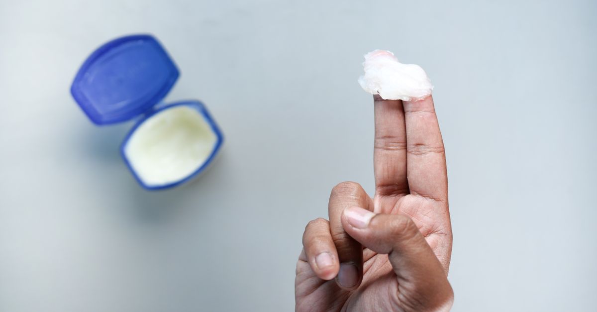
After cleaning, a thin layer of petrolatum ointment can keep the wound bed moist, which promotes rapid epithelial cell migration, covered by a sterile, breathable bandage to protect it from further environmental contamination.
14. When Antibiotics are Indicated
Topical over-the-counter antibiotic ointments are often unnecessary for minor, clean cuts and can occasionally cause allergic contact dermatitis. Their primary role is maintaining a moist environment rather than effectively treating an established infection.
Systemic oral antibiotics are strictly indicated when clinical signs of an active, spreading infection are present. If a cut exhibits expanding cellulitis, significant purulence, or if the patient develops a fever, a physician will prescribe targeted antibiotics, typically covering common skin pathogens like Staphylococcus aureus and Streptococcus pyogenes.
Antibiotics must be taken exactly as prescribed, completing the entire course even if the redness disappears rapidly. Stopping early promotes the survival of resistant bacterial strains.
15. Emergency Medical Interventions
Certain clinical presentations associated with an infected cut constitute a medical emergency. If the redness is spreading extremely rapidly—visibly expanding by the hour—or if the skin begins to turn purple, black, or develops large, fluid-filled hemorrhagic blisters, immediate emergency evaluation is required.
These are potential signs of necrotizing fasciitis, a rare but devastating, rapidly progressive “flesh-eating” bacterial infection that destroys the deep fascia and muscle tissue.
Additionally, if a patient with an infected wound becomes profoundly lethargic, confused, or experiences a sudden drop in blood pressure resulting in dizziness or fainting, they are likely developing sepsis. This requires immediate transport to an emergency department for intravenous antibiotics and aggressive fluid resuscitation.
16. Frequently Asked Questions (FAQ)
1. Is it normal for a cut to itch while healing?
Yes. Mild itching is a very common and normal part of the later stages of wound healing. It is caused by the mechanical pulling of the shrinking tissue and the release of histamine by immune cells during the remodeling phase. It is not a sign of infection.
2. Should I let my cut “breathe” to heal faster?
No. Extensive dermatological research proves that wounds heal much faster and with less scarring in a moist environment. Leaving a cut exposed to the air dries out the tissue, causing a hard scab to form, which acts as a physical barrier that delays new skin cells from closing the wound.
3. If I see a red streak, do I have blood poisoning?
A red streak indicates lymphangitis, which means the infection has entered your lymphatic vessels. While serious and requiring immediate antibiotics, it is not yet “blood poisoning” (sepsis). However, if left untreated, the bacteria can travel through the lymphatics into the bloodstream, leading to sepsis.
4. Why did my doctor prescribe antibiotics for an animal bite that doesn’t look infected yet?
Animal mouths harbor highly aggressive, complex bacteria (like Pasteurella in cat bites). Because puncture wounds from teeth inject these bacteria deep into the tissue where they are impossible to wash out, the risk of severe infection is almost guaranteed. Antibiotics are given prophylactically to prevent the infection from taking hold.
5. Can I use leftover antibiotics for an infected cut?
Never. Leftover antibiotics may be the wrong type of drug for skin bacteria, they may be expired, and taking a partial course will not kill the infection. It only contributes to creating dangerous, antibiotic-resistant “superbugs.” Always get a fresh medical evaluation.
17. Bibliography
Disclaimer: The content is for informational purposes only and does not replace medical advice. Always consult your doctor for personalized treatment.