
1. Introduction to Subcutaneous Foreign Bodies
The retention of a foreign body within the subcutaneous tissue represents a complex and frequently encountered clinical challenge in soft tissue trauma management. When a penetrating injury possesses sufficient velocity or force, an external object—ranging from glass shards and metallic fragments to biological debris like wood or thorns—can bypass the protective epidermal and dermal layers, embedding itself deep within the adipose (fat) tissue and superficial fascia.
Unlike superficial epidermal splinters that are easily visualized and extracted, subcutaneous foreign bodies reside in a highly compliant, loose tissue environment. This anatomical space allows objects to migrate, hide from visual inspection, and incite insidious, long-term inflammatory responses. A retained object acts as a permanent biological irritant, triggering a cascade of immune defenses that culminate in chronic granulomatous inflammation, localized abscess formation, and persistent, deep-seated pain.
Clinical management of subcutaneous foreign bodies demands a highly structured approach. Blind surgical exploration is universally condemned, as it invariably leads to extensive collateral tissue damage and frequently fails to locate the object. Successful resolution requires precise preoperative anatomical mapping utilizing advanced diagnostic imaging, followed by meticulous, targeted surgical excision under appropriate anesthesia, ensuring complete eradication of the foreign material and the surrounding pathological tissue.
2. Anatomy of the Subcutaneous Tissue Layer
To comprehend the behavior and clinical trajectory of a deep foreign body, an understanding of the subcutaneous anatomy—also known as the hypodermis—is essential. Situated immediately beneath the dense, fibrous dermis, the subcutaneous layer is composed primarily of loose areolar connective tissue and lobules of adipose (fat) tissue.
This layer serves crucial physiological functions: it acts as a thermal insulator, a nutritional reservoir, and a mechanical shock absorber protecting the underlying deep fascia, skeletal muscles, and bone. The subcutaneous tissue is richly vascularized with larger branching arteries and veins, and it contains significant nerve trunks that transmit sensory information from the overlying skin to the central nervous system.
Because the structural composition of fat is incredibly soft and yielding compared to the rigid dermis above, a foreign body that enters this space encounters minimal physical resistance. This loose anatomical architecture facilitates the silent embedding of objects and permits significant positional shifting or migration of the object over time, complicating subsequent localization and extraction efforts.
3. Mechanisms of Deep Penetration
The depth to which a foreign body penetrates is directly proportional to the kinetic energy of the impact and the structural density of the object. High-velocity mechanisms are frequently responsible for deep subcutaneous impactions. For example, metallic shavings expelled from grinding machinery, or shattered glass propelled during a motor vehicle collision, possess immense kinetic energy, allowing them to effortlessly slice through the tough dermal barrier and bury deeply into the fat layer.
Conversely, low-velocity mechanisms typically involve substantial mechanical force applied over a small surface area. Stepping heavily on a sewing needle or falling forcefully onto a sharp wooden branch drives the object deep into the tissue. In these scenarios, the object often breaks off beneath the surface of the skin, leaving a minute, rapidly healing entry wound that completely conceals the massive fragment hidden in the subcutaneous space below.
The entry trajectory is rarely a perfectly straight line. As an object penetrates, it deflects off denser anatomical structures, such as thick fascial bands or tendons, altering its path. Consequently, the final resting place of a subcutaneous foreign body is frequently not directly beneath the visible entry wound, underscoring the absolute necessity for precise diagnostic imaging prior to surgical intervention.
4. Chronic Foreign Body Reaction
When a foreign object establishes residence in the subcutaneous tissue, the innate immune system recognizes the profound breach and launches a relentless, albeit frustrated, attack. Because macrophages and neutrophils cannot enzymatically dissolve inorganic materials like metal or glass, or dense organic matter like wood, the acute inflammatory phase fails to eliminate the threat.
The immune system subsequently transitions to a chronic containment protocol. Over several weeks to months, a specialized cellular response constructs a biological prison around the object. Macrophages fuse into massive, multinucleated giant cells that tightly adhere to the foreign material. Surrounding fibroblasts lay down thick, disorganized concentric layers of collagen.
This pathological structure is known as a foreign body granuloma. While the thick fibrous capsule successfully isolates the object, preventing the spread of localized infection, the granuloma itself becomes a persistent, firm, and highly tender subcutaneous mass. This chronic inflammatory environment disrupts local blood flow and causes debilitating pain whenever mechanical pressure is applied to the overlying skin.
5. Material Degradation and Toxicity
The specific material composition of the retained object drastically influences its long-term stability and toxicity within the subcutaneous environment. Inorganic materials like surgical-grade steel, clean glass, and certain plastics are exceptionally inert. They do not degrade or release toxins, often remaining encapsulated silently within a mature granuloma for decades without causing systemic illness.
Organic materials, however, are highly reactive and unstable. Wood, plant thorns, and unsterilized biological matter absorb tissue fluid, swelling substantially and undergoing slow, putrefactive degradation. This decay releases highly irritating chemical compounds into the surrounding fat tissue, continuously fueling a massive, aggressive inflammatory response that prevents the granuloma from ever fully stabilizing.
Certain heavy metals pose a distinct, systemic toxicological threat. A retained lead fragment, such as a bullet or buckshot lodged in a highly vascularized subcutaneous space, can slowly dissolve. Over years, the lead enters the systemic circulation, leading to chronic lead poisoning, characterized by profound neurological deficits, severe abdominal pain, and intractable anemia, mandating urgent surgical removal of the metallic source.
6. Clinical Symptoms and Palpation
The clinical presentation of a subcutaneous foreign body varies widely based on the duration of retention and the depth of the object. In the acute setting, the patient typically recalls the traumatic event and presents with a visible puncture wound, localized erythema, and sharp, pinpoint pain.
However, in delayed presentations, the entry wound has completely healed. The patient presents with a chronic, firm, subcutaneous mass. The hallmark symptom is highly localized, exquisite tenderness that is strictly aggravated by applying direct, deep pressure over the mass. If the granuloma is situated over a joint or a muscle belly, the patient will experience sharp pain restricting their range of motion.
During physical examination, the clinician systematically palpates the soft tissue. Unlike superficial dermal splinters, a deep subcutaneous object may not be visibly apparent. The clinician feels for a distinct, hard, rubbery nodule within the fat layer. If the object is a long, linear structure like a needle, the clinician may occasionally be able to palpate the firm, rigid outline of the object beneath the skin.
7. Migration of Subcutaneous Objects
A highly unique and clinically challenging aspect of subcutaneous foreign bodies is their capacity for migration. Because the adipose tissue is loose and highly compliant, an object is not always fixed in its original anatomical position.
Smooth, sharp objects, particularly sewing needles or glass shards, can actively travel through the subcutaneous planes over time. The continuous, dynamic contractions of the underlying skeletal muscles during normal daily activities act as a mechanical pump, slowly but inexorably pushing the sharp object along the path of least resistance through the fat layer.
This migration transforms the clinical scenario. An object that initially penetrated the forearm may slowly migrate toward the elbow or wrist, potentially intersecting with and lacerating major nerves or blood vessels long after the initial trauma. This migratory behavior reinforces the medical rule that an extraction procedure must never be attempted based solely on the location of the original entry wound; real-time imaging is strictly required.
8. Differential Diagnosis of Soft Tissue Masses
When a patient presents with a firm, tender subcutaneous mass without a clearly recalled history of a puncture wound, the clinician must accurately differentiate a foreign body granuloma from an array of benign and malignant soft tissue tumors.
A lipoma is the most common benign tumor of the subcutaneous tissue. Composed of mature fat cells, a lipoma feels soft, doughy, and highly pliable, lacking the firm, distinct fibrous capsule and the sharp tenderness characteristic of a foreign body granuloma.
An epidermal inclusion cyst forms when surface skin cells are trapped in the dermis or subcutaneous space, filling with cheesy keratin. While cysts can become inflamed and tender, they frequently feature a central dark punctum (pore) connecting them to the surface. Furthermore, the clinician must maintain a high index of suspicion to rule out malignant soft tissue sarcomas, which present as firm, rapidly expanding deep masses requiring specialized oncological biopsy.
| Subcutaneous Condition | Primary Origin | Distinguishing Clinical Features |
|---|---|---|
| Foreign Body Granuloma | Retained external object. | Firm, highly tender nodule, history of trauma. |
| Lipoma | Benign adipose (fat) tumor. | Soft, doughy, painless, slowly growing over years. |
| Epidermal Inclusion Cyst | Trapped skin cells. | Dome-shaped, contains foul-smelling keratin, central pore. |
| Soft Tissue Sarcoma | Malignant cellular growth. | Firm, deep, rapidly expanding mass, painless initially. |
9. Advanced Diagnostic Imaging
Accurate anatomical mapping is the most critical step prior to attempting the surgical excision of a subcutaneous foreign body. The choice of imaging modality is dictated entirely by the suspected material, as different substances interact uniquely with X-rays and high-frequency sound waves.
Plain radiography (X-rays) in multiple views is the standard first-line test. It is highly effective for identifying radiopaque objects. Nearly all metallic objects, stones, and glass fragments larger than two millimeters are easily visualized as bright white shapes on a standard X-ray, instantly revealing their size and orientation.
However, biological materials (wood, thorns) and plastics are entirely radiolucent. They possess the same physical density as human soft tissue and are completely invisible on standard X-rays. For these dangerous materials, high-resolution ultrasound is the definitive gold standard. Ultrasound visualizes the precise physical outline of a wooden splinter, assesses the size of the surrounding fluid-filled abscess or granuloma capsule, and accurately measures the object’s depth from the skin surface.
10. Surgical Planning and Considerations
Surgical excision of a deep subcutaneous foreign body is a meticulous procedure that requires careful preoperative planning. The surgeon must evaluate the imaging to understand the object’s exact proximity to critical anatomical structures, such as major nerves, arteries, or underlying tendons.
Blind exploration—creating an incision and blindly probing the fat tissue with forceps—is universally condemned in modern surgical practice. The subcutaneous fat is a visually homogenous, yellow, bloody environment. A tissue-colored wooden splinter is virtually indistinguishable from surrounding tissue during a blind search. Furthermore, aggressive probing severely crushes the fat lobules, causing extensive collateral tissue damage, severe postoperative scarring, and almost guarantees failure to locate the object.
Therefore, the extraction is meticulously planned. In cases involving small, difficult-to-locate radiolucent objects, the surgeon frequently utilizes real-time, intraoperative ultrasound guidance. This allows the surgeon to visualize the object continuously on a monitor while simultaneously navigating the surgical instruments directly to the target.
11. Excision Procedures and Bloodless Fields
To ensure a successful and safe extraction, the procedure is typically performed under profound local or regional anesthesia. For objects located on the extremities, achieving a completely bloodless surgical field is absolutely paramount. Even minor capillary bleeding instantly fills the surgical cavity, entirely obliterating the surgeon’s view of microscopic fragments.
A bloodless field is achieved by elevating the limb to exsanguinate the venous blood, followed by the inflation of a sterile, pneumatic tourniquet proximal to the surgical site. This temporarily halts all arterial blood flow, providing the surgeon with a pristine, perfectly clear view of the deep subcutaneous structures.
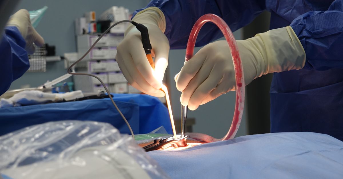
The surgeon makes a precise incision over the radiologically confirmed location of the object. Meticulous, sharp dissection is used to navigate through the fat lobules. If a chronic granuloma has formed, the surgeon does not attempt to pull the object out of the scar tissue; instead, the entire fibrous capsule, containing the foreign body intact within it, is excised en bloc (as a single unit). This guarantees that no microscopic fragments or infected fluid are left behind.
12. Wound Closure and Drainage
Following the successful extraction of the object and its surrounding granuloma, the management of the surgical cavity depends entirely on the nature of the foreign body and the presence of active infection.
If the object was a clean, non-reactive material (like sterile glass) and no pus was present, the surgeon aggressively irrigates the cavity with sterile saline and closes the incision primarily using deep subcutaneous dissolving sutures and superficial skin sutures, ensuring optimal cosmetic healing.
However, if the object was organic, heavily contaminated, or surrounded by a purulent abscess, primary closure is strictly contraindicated. Suturing an infected cavity closed guarantees the rapid reformation of a massive, destructive abscess. Instead, the wound is left intentionally open and packed with sterile gauze. This allows the cavity to continuously drain infectious fluid and heal naturally from the bottom up, a process known as secondary intention.
13. Infection Control and Tetanus Prophylaxis
Any penetrating injury reaching the subcutaneous tissue introduces a substantial risk of Clostridium tetani, the bacterium responsible for tetanus. This aggressive pathogen thrives in deep, oxygen-deprived puncture wounds. The clinician must review the patient’s immunization history; if a tetanus booster has not been received within the last five to ten years, a prophylactic vaccination is administered immediately prior to discharge.
Systemic oral antibiotics are strictly prescribed if the object was organic, heavily contaminated with soil, associated with an animal bite, or if the patient presents with established signs of localized cellulitis. A standard regimen typically involves a first-generation cephalosporin or amoxicillin-clavulanate to target common skin and environmental flora.
Routine, uncomplicated removals of clean, inorganic objects generally do not require prophylactic antibiotics, provided the surgical cavity was aggressively irrigated and meticulously debrided during the operative procedure.
14. Long-Term Prognosis
The long-term prognosis following the complete, successful excision of a subcutaneous foreign body and its associated granuloma is excellent. Once the persistent biological irritant is entirely eradicated, the chronic inflammatory cascade ceases immediately.
The residual surgical cavity slowly fills with healthy granulation tissue and reorganizes into normal, flexible dermal and subcutaneous scar tissue. Patients typically experience complete resolution of the localized, sharp, and throbbing pain that characterized the granuloma, rapidly regaining full, pain-free functional mobility in the affected limb or region.
However, if the extraction was incomplete, or if a highly friable wooden object shattered and left microscopic fragments behind, the inflammatory process will inevitably recur. The patient will present weeks or months later with a new, painful nodule at the exact same surgical site, necessitating a secondary, more expansive surgical exploration.
15. Frequently Asked Questions (FAQ)
1. Can a piece of glass stay in my arm forever?
Yes. If the glass does not introduce bacteria, your body will eventually build a hard scar tissue capsule around it. It can stay deep in the fat tissue permanently without causing systemic harm, though the lump may be painful when you press on it.
2. Will soaking it in hot water or Epsom salts draw a deep splinter out?
No. Soaking in warm water only affects the very top layer of the skin. It will not magically “draw out” a splinter that is deeply embedded in the fat layer beneath the skin; deep objects require surgical removal.
3. Why did the doctor order an ultrasound instead of an X-ray?
Standard X-rays are excellent for seeing metal and glass, but wood, thorns, and plastics are almost invisible on an X-ray. High-resolution ultrasound uses sound waves to display the exact location of wooden objects deep in the tissue.
4. Can a needle travel through my body if I don’t remove it?
Yes. Smooth, sharp objects like sewing needles can slowly migrate through the loose fat tissue over time, pushed by the movement of your muscles. This makes them very dangerous, as they can eventually hit a nerve or blood vessel.
5. Does the surgery to remove the object hurt?
The skin and deep tissue are completely numbed with local anesthesia before the procedure begins. While you may feel some firm pressure or pulling during the surgery, you will not feel any sharp pain.
6. What happens if part of the splinter is left inside?
If even a microscopic piece of organic material like wood or a thorn is left behind, your immune system will continue to attack it. The wound will not heal properly, and it will form a chronic, painful lump or an abscess until the remaining piece is surgically removed.
16. Bibliography
Disclaimer: The content is for informational purposes only and does not replace medical advice. Always consult your doctor for personalized treatment.
