
1. Introduction
Mechanical low back pain represents the most common cause of disability and work-related absence globally. From an orthopedic and clinical perspective, the term “mechanical” signifies that the pain originates from the structural components of the lumbar spine—specifically the muscles, ligaments, intervertebral discs, or facet joints—due to abnormal stress, strain, or age-related degeneration. Crucially, mechanical low back pain is distinguished by the absence of severe nerve root compression or serious systemic underlying diseases.
The human lumbar spine is an engineering marvel designed to bear the majority of the body’s weight while remaining highly flexible. However, this flexibility comes at a cost. The intricate network of soft tissues and articulating joints is inherently vulnerable to injury from improper lifting mechanics, poor postural habits, and the repetitive microtrauma associated with modern sedentary lifestyles.
When a patient presents with sudden, sharp lower back pain following a strenuous activity, the clinical objective is not simply to provide pain relief. The goal is to accurately triage the symptoms, rule out dangerous neurological compromise, identify the specific anatomical structure driving the pain, and implement a targeted rehabilitation program. This article details the complex anatomy of the lower back, the biomechanics of spinal injury, and the evidence-based medical and physical therapies used to restore lumbar function.
2. Anatomy of the Lumbar Spine
The lumbar spine consists of five massive vertebrae, labeled L1 through L5, stacked upon the sacrum. These vertebrae are robust, designed to absorb heavy compressive loads. Between each bony vertebra lies an intervertebral disc, a crucial shock-absorbing structure composed of a tough, fibrous outer ring (the annulus fibrosus) and a soft, gel-like center (the nucleus pulposus).
At the back of the vertebrae are the facet joints, small cartilage-lined joints that link each vertebra to the one above and below it. These joints guide and restrict the movement of the spine, preventing excessive twisting or sliding. Surrounding this bony column is a dense, multi-layered corset of muscles and tough connective ligaments.
The paraspinal muscles run parallel to the spine, maintaining upright posture, while the deep core muscles and the abdominal muscles provide dynamic stability. Mechanical low back pain occurs when any of these individual components are stretched beyond their physiological limits, inflamed, or structurally degraded.
3. Lumbar Muscle Strain and Ligament Sprain
The vast majority of acute mechanical low back pain episodes are caused by a muscle strain or a ligamentous sprain. A strain occurs when the muscle fibers of the lower back are forcefully overstretched or torn, typically during a sudden, forceful movement such as lifting a heavy box with poor biomechanics. A sprain involves similar damage to the tough ligaments connecting the vertebrae.
When these soft tissues tear, the body initiates an immediate inflammatory response. Blood vessels dilate, causing localized swelling, and specialized immune cells flood the area to clear damaged tissue. This inflammation heavily sensitizes the local pain receptors, causing severe, localized aching.
Furthermore, the surrounding uninjured muscles undergo reflex spasms. The muscles contract violently and rigidly in an involuntary attempt to splint the spine and prevent further movement that could exacerbate the injury. This muscle spasm is often the primary source of the severe, sharp pain patients feel when attempting to stand up or change positions.
4. Discogenic Pain and Annular Tears
Mechanical pain can also originate directly from the intervertebral discs, a condition known as discogenic pain. While a large disc herniation can press on a nerve root causing sciatica, a disc can cause profound localized back pain without ever compressing a nerve.
The outer ring of the disc, the annulus fibrosus, is heavily innervated with pain-sensing nerve fibers. Over time, or following a traumatic event, small microscopic tears can develop in this tough outer ring. These annular tears allow the highly inflammatory proteins from the center of the disc to leak into the outer layers.
This chemical irritation triggers severe, deep, aching pain in the central lower back. Discogenic pain is characteristically worsened by activities that increase the pressure inside the disc, such as prolonged sitting, bending forward, or coughing. Unlike a muscle strain, pain originating from a torn disc can take many months to heal due to the notoriously poor blood supply to the disc tissues.
5. Facet Joint Arthropathy
The facet joints at the posterior of the spine are in constant motion during daily activities. Like the knee or hip joints, these small spinal joints are lined with smooth cartilage to reduce friction. With advancing age, or due to abnormal mechanical stress from a misaligned spine, this cartilage gradually wears away, a condition known as facet joint osteoarthritis or arthropathy.
As the cartilage degrades, the underlying bone becomes exposed and irritated. Bone spurs (osteophytes) form around the joint margins, and the joint capsule becomes inflamed and thickened. This mechanical degradation causes significant pain and stiffness.
Facet joint pain has a distinct clinical presentation. It is typically described as a deep, dull ache on one or both sides of the lower back. Crucially, the pain is exacerbated by extending the spine backward or twisting to the side, movements which compress the inflamed facet joints together. The pain often improves when the patient bends forward, as this opens up the joint space.
6. Postural Factors and Core Instability
Chronic, recurrent mechanical low back pain is frequently driven by underlying postural deficits and core muscle instability. The spine relies on the coordinated firing of the deep abdominal muscles, the pelvic floor, and the paraspinal muscles to maintain a neutral, shock-absorbing alignment.
When a patient leads a highly sedentary lifestyle, the core muscles become weak and deconditioned. Additionally, prolonged sitting causes the hip flexor muscles to become chronically tight and shortened. This combination pulls the pelvis forward, creating an exaggerated curve in the lower back known as hyperlordosis.
Hyperlordosis forces the facet joints to bear a disproportionate amount of the body’s weight and places the paraspinal muscles under constant, exhausting tension. Treating this type of mechanical pain cannot be achieved with medication alone; it requires a dedicated physical therapy regimen to stretch tight anterior muscles and strengthen the posterior and core stabilizing structures.
7. Diagnostic Triage: Ruling Out Red Flags
When evaluating a patient with low back pain, the physician’s immediate priority is diagnostic triage. The clinician must ensure the pain is truly mechanical and not a symptom of a dangerous, underlying systemic disease or severe neurological compromise. Physicians use a specific checklist of clinical “red flags.”
Red flags include a history of significant physical trauma (suggesting a fracture), unexplained weight loss or a history of cancer (suggesting a spinal metastasis), fever or recent bacterial infection (suggesting a spinal epidural abscess), and prolonged use of corticosteroids (increasing fracture risk).
The most critical neurological red flags involve the sudden onset of bowel or bladder incontinence, numbness in the saddle or groin region, or severe, progressive weakness in both legs. These symptoms indicate cauda equina syndrome, a massive compression of the spinal nerve bundle that requires emergency surgical decompression to prevent permanent paralysis.
8. The Clinical Physical Examination
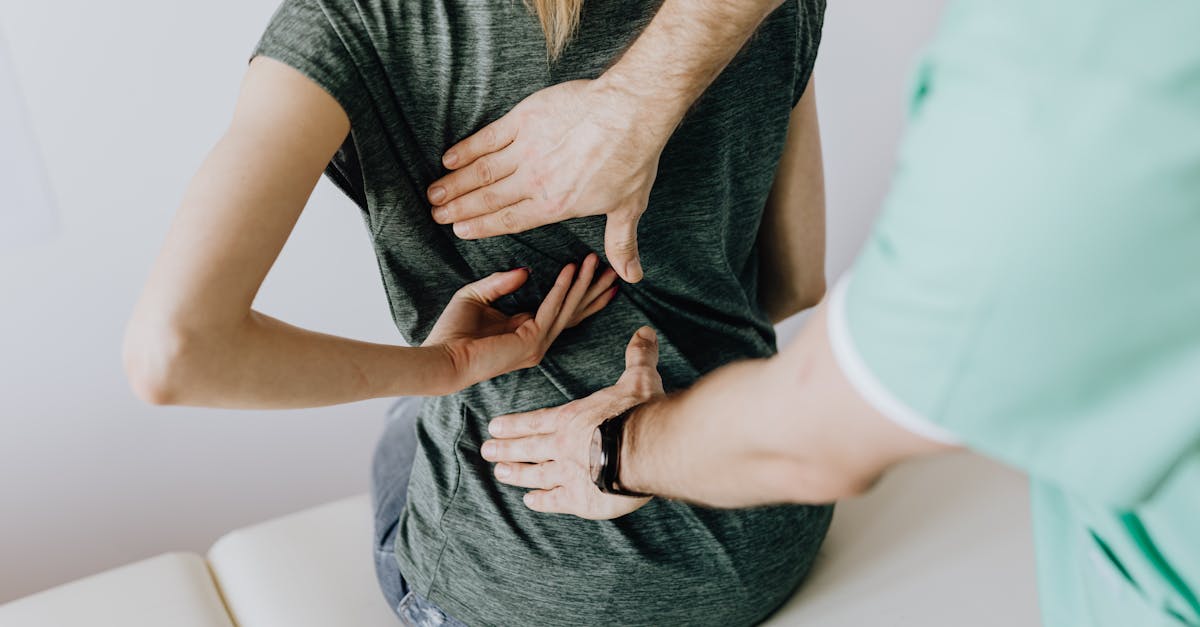
If no red flags are present, the physician proceeds with a focused physical examination of the musculoskeletal system. The clinician observes the patient’s gait, posture, and spinal alignment. They palpate the lower back to identify specific areas of muscle spasm, point tenderness over the vertebral spinous processes, or pain over the sacroiliac joints.
Range of motion testing is critical. The physician notes which specific movements—flexion, extension, or lateral bending—reproduce the patient’s primary pain. This biomechanical assessment helps differentiate between disc-related pain (worse with flexion) and facet joint pain (worse with extension).
A thorough neurological exam is also performed. The physician tests the strength of specific leg muscles, checks the deep tendon reflexes at the knee and ankle, and assesses sensation in the feet. Normal neurological findings confirm that the pain is purely mechanical and that the spinal nerve roots are healthy and uncompressed.
9. The Role and Limits of Radiological Imaging
A common misconception among patients is that a sudden episode of severe back pain requires an immediate MRI or X-ray. Clinical guidelines strongly recommend against routine imaging for acute mechanical low back pain within the first four to six weeks, provided no red flags are present.
Early imaging rarely alters the initial treatment plan, which consists of conservative management and physical therapy. Furthermore, imaging often reveals incidental, age-related degenerative changes—such as bulging discs or mild arthritis—that are completely asymptomatic in the general population. Presenting a patient with an MRI report detailing “degenerative disc disease” often induces unnecessary fear and anxiety, hindering their recovery.
Imaging is reserved for patients whose pain fails to improve after six weeks of dedicated physical therapy, or for those who develop new, radiating nerve pain or weakness in the legs. In these specific scenarios, an MRI is the gold standard for visualizing the soft tissues, discs, and nerves to guide advanced interventions.
10. Pharmacological Pain Management
The initial pharmacological management of mechanical low back pain aims to reduce acute inflammation and relieve debilitating muscle spasms, allowing the patient to remain mobile. Non-Steroidal Anti-Inflammatory Drugs, known as NSAIDs, such as ibuprofen or naproxen, are the first-line treatment.
If muscle spasms are severe and prevent the patient from sleeping or walking, a short course of skeletal muscle relaxants may be prescribed. These medications act on the central nervous system to reduce muscle rigidity. Acetaminophen may be used for general pain relief, though it lacks the anti-inflammatory properties of NSAIDs.
Opioid pain medications are strictly avoided for standard mechanical back pain. They offer no long-term benefit for musculoskeletal healing and carry severe risks of dependence, tolerance, and systemic side effects. The focus of modern pain management is facilitating functional movement rather than completely erasing the sensation of pain.
11. Physical Therapy and Active Rehabilitation
The absolute cornerstone of treating mechanical low back pain is active physical therapy. Resting in bed for more than a day or two is strongly contraindicated, as prolonged inactivity rapidly leads to muscle atrophy, joint stiffness, and delayed healing.
A physical therapist designs a targeted program based on the patient’s specific mechanical deficits. The initial phase focuses on pain modulation through manual therapy, gentle stretching, and teaching the patient specific movements that centralize or reduce their pain, such as the McKenzie method for discogenic pain.
The secondary phase focuses heavily on core stabilization. Patients are taught how to consciously engage their transverse abdominis and multifidus muscles—the deep stabilizers of the spine. Strengthening these specific muscles creates a natural, internal brace that protects the spine from abnormal rotational and compressive forces during daily activities.
12. Data Summary: Mechanical Pain Sources
The following table summarizes the common anatomical sources of mechanical low back pain and their typical clinical characteristics.
| Anatomical Source | Primary Pathology | Typical Aggravating Movements |
|---|---|---|
| Muscle / Ligament | Strain, microscopic tearing, spasm. | Sudden twisting, heavy lifting, transitions from sitting to standing. |
| Intervertebral Disc | Annular tear, internal inflammation. | Bending forward, prolonged sitting, coughing or sneezing. |
| Facet Joint | Osteoarthritis, cartilage degradation. | Bending backward, twisting, prolonged standing. |
| Postural Core | Muscle weakness, hyperlordosis. | Prolonged static postures, lack of supportive seating. |
This targeted biomechanical assessment allows the physical therapist to prescribe the exact exercises needed to unload the irritated structures.
13. Interventional Procedures: Injections
For patients whose mechanical pain is severe and fails to respond to oral medications and physical therapy, targeted interventional procedures may be considered to break the pain cycle. These are performed by specialized pain management physicians under fluoroscopic X-ray guidance.
If the pain is primarily driven by facet joint arthritis, the physician may perform a medial branch block, injecting a local anesthetic near the tiny nerves that supply the facet joints. If this temporarily erases the pain, a more permanent procedure called radiofrequency ablation can be used to burn the nerve endings, providing long-lasting relief.
Epidural steroid injections are occasionally used if there is profound inflammation within the spinal canal contributing to the mechanical pain. These injections deliver powerful anti-inflammatory medication directly to the irritated structures, often providing a window of pain relief that allows the patient to finally participate fully in their physical therapy exercises.
14. Ergonomics and Lifestyle Modifications
Preventing the recurrence of mechanical low back pain requires significant lifestyle and ergonomic adjustments. Patients must be educated on proper lifting mechanics, emphasizing bending at the hips and knees while maintaining a neutral, straight spine, rather than flexing the lumbar spine under load.
Workplace ergonomics are critical. Patients who sit at a desk must utilize chairs that support the natural lumbar curve. Computer monitors must be placed at eye level to prevent slouching, and frequent breaks to stand, stretch, and walk are essential to relieve the constant compressive pressure on the intervertebral discs.
Weight management is also a critical component of long-term spine health. Excess abdominal weight pulls the center of gravity forward, drastically increasing the mechanical strain on the lower back muscles and facet joints, exacerbating any underlying structural vulnerabilities.
15. The Psychological Impact of Chronic Pain
When mechanical low back pain persists for months, it transitions from an acute injury to a chronic pain syndrome. Chronic pain fundamentally alters the nervous system, making it hypersensitive, and deeply impacts the patient’s psychological well-being.
Patients often develop fear-avoidance behaviors. They become terrified that normal movement will cause further spinal damage, leading them to restrict their activities severely. This inactivity breeds further muscle weakness and stiffness, creating a vicious cycle of increasing pain and disability.
Addressing the psychological component is essential. Clinicians must reassure patients that hurt does not equal harm. Cognitive-behavioral therapy is frequently integrated into chronic back pain management to help patients manage pain-related anxiety, overcome fear-avoidance, and reclaim their functional independence, similar to the psychological support required when managing a permanent meaningfulness alteration or chronic neurological deficit.
16. Frequently Asked Questions (FAQ)
1. Should I stay in bed if I throw my back out?
No. While resting for the first few hours is fine, staying in bed for days is the worst thing you can do. Bed rest makes your muscles stiffen and weaken rapidly, which prolongs the healing process. Gentle, frequent movement like walking is highly recommended.
2. Does a slipped disc always need surgery?
No. First, a disc does not actually “slip”; it bulges or tears. The vast majority of disc injuries causing mechanical pain heal over time with physical therapy and proper mechanics. Surgery is rarely considered unless the disc is actively crushing a nerve and causing severe leg weakness.
3. Why won’t my doctor order an MRI for my severe back pain?
Medical guidelines dictate that for standard back pain without dangerous nerve symptoms, an MRI is not needed immediately. It will not change the fact that physical therapy is the best first step. Early MRIs often show normal aging changes that scare patients unnecessarily.
4. Can a weak stomach cause back pain?
Absolutely. Your deep abdominal and core muscles act like a natural corset that holds your spine upright. If they are weak, all the physical stress of moving and lifting falls directly onto the small joints and ligaments of your lower back, leading to chronic pain.
5. What is the best sleeping position for mechanical lower back pain?
Sleeping on your back with a pillow under your knees helps flatten your lower back and relieves pressure on the joints. If you sleep on your side, putting a firm pillow between your knees keeps your hips aligned and stops your spine from twisting during the night.
17. Bibliography
Disclaimer: The content is for informational purposes only and does not replace medical advice. Always consult your doctor for personalized treatment.


