1. Introduction
To relieve mid-back pain that hurts when taking a deep breath, one must employ gentle thoracic mobility exercises, apply targeted thermal therapy, and utilize nonsteroidal anti-inflammatory medications. This pain typically originates from a musculoskeletal strain of the intercostal muscles or an inflamed costovertebral joint, both of which are mechanically stressed during lung expansion. Resting the affected area while maintaining shallow, even breathing allows the acute inflammation to subside.
Pain localized in the thoracic spine that sharpens upon inspiration is a distressing symptom. Because breathing is an involuntary and continuous physiological process, any mechanical dysfunction in the respiratory apparatus results in a relentless cycle of discomfort. Patients often restrict their breathing to avoid the sharp twinges of pain, which unfortunately leads to secondary muscular stiffness and decreased oxygenation.
Addressing this specific type of back pain requires distinguishing between simple mechanical strains and more complex pulmonary conditions. By understanding the anatomical relationship between the rib cage, the thoracic vertebrae, and the lungs, clinicians can guide patients toward targeted physical and pharmacological interventions that restore painless, full-capacity respiration.
2. Anatomy of the Thoracic Spine
The thoracic spine forms the central segment of the vertebral column, consisting of twelve vertebrae designated T1 through T12. Unlike the highly mobile cervical and lumbar regions, the thoracic spine is structurally designed for stability. This rigidity protects the vital organs housed within the chest cavity.
Each thoracic vertebra articulates with a pair of ribs. These connections form the costovertebral and costotransverse joints. A dense network of ligaments encapsulates these small joints, providing structural integrity while allowing the slight gliding motions necessary for breathing.
The spaces between the ribs are filled by the intercostal muscles. These muscles are essential for expanding and contracting the rib cage. The intricate anatomical overlap of bone, joint capsule, and muscle means that any localized inflammation will instantly affect the mechanics of the entire thoracic segment.
3. The Mechanics of Respiration
Breathing relies on a complex coordination of muscular effort and joint mobility. During inspiration, the diaphragm contracts and moves downward, while the external intercostal muscles contract to lift the rib cage upward and outward. This expansion creates a negative pressure within the thoracic cavity, drawing air into the lungs.
For the rib cage to expand, every single costovertebral joint must glide smoothly. The thoracic spine must also extend slightly. This mechanical chain reaction occurs thousands of times a day.
When a patient takes a particularly deep breath, this mechanical system is pushed to its maximum range of motion. If there is a sprained ligament, a strained muscle, or an inflamed joint anywhere along the thoracic wall, this maximal expansion will stretch the compromised tissue, triggering an immediate and sharp pain signal.
4. Intercostal Muscle Strain
An intercostal muscle strain is the most frequent musculoskeletal cause of pain upon deep inspiration. These strains occur when the muscle fibers between the ribs are stretched beyond their anatomical limits or subjected to sudden, forceful contractions.
Common triggers include a violent bout of coughing, aggressive twisting motions, or lifting a heavy object awkwardly. The resulting micro-tears in the muscle belly initiate a localized inflammatory response. The tissue becomes swollen and exquisitely tender to the touch.
Because the intercostal muscles must contract to expand the chest, taking a deep breath demands active work from the injured tissue. The brain registers this mechanical strain as a sharp, localized pain, forcing the patient to adopt short, shallow breaths to minimize muscular engagement.
5. Costovertebral Joint Sprain
The costovertebral joints connect the heads of the ribs to the thoracic vertebrae. Like any joint in the human body, they are susceptible to sprains and capsular inflammation. A sudden, awkward twisting of the torso can overstretch the delicate ligaments supporting these joints.
When a costovertebral joint is sprained, it becomes acutely inflamed and stiff. The joint capsule swells, reducing the space available for movement. This condition is often referred to as a rib dysfunction or a locked rib.
During a deep breath, the rib must pivot at this exact joint to elevate. If the joint is inflamed, this necessary pivoting motion creates intense friction and stretching of the injured capsule. The pain is typically felt as a sharp, stabbing sensation directly next to the spine, radiating slightly outward along the path of the rib.
6. Rib Subluxation
A rib subluxation occurs when a rib slips slightly out of its normal alignment at the costovertebral or costotransverse joint. While not a complete dislocation, this subtle misalignment disrupts the smooth mechanics of the thoracic cage.
Subluxations can result from poor posture, chronic coughing, or blunt trauma to the back. The misaligned rib places continuous mechanical stress on the surrounding ligaments and local nerve endings.
When the patient inhales deeply, the misaligned rib fails to move synchronously with the rest of the rib cage. This asynchronous movement causes a sharp, catching pain in the mid-back. Manual therapy or specific mobilization techniques are often required to restore proper joint alignment and alleviate the mechanical block.
7. Pleurisy and Pulmonary Causes
While musculoskeletal issues are common, sharp pain upon inspiration is the hallmark symptom of pleurisy. The pleura is a double-layered membrane that surrounds the lungs and lines the inner chest wall. A thin layer of lubricating fluid sits between these layers, allowing them to glide friction-free during breathing.
If a viral or bacterial infection causes the pleura to become inflamed, the surfaces become rough and swollen. When the patient takes a deep breath, these inflamed layers rub directly against each other.
This friction generates a severe, sharp pain known as pleuritic chest pain, which often radiates directly to the mid-back. Differentiating pleurisy from a muscle strain requires clinical evaluation, as pleurisy is frequently accompanied by a fever, a persistent cough, and a generalized feeling of systemic illness.
8. Postural Triggers and Thoracic Kyphosis
Chronic poor posture contributes significantly to thoracic pain. Sitting slouched at a desk for hours induces a posture known as hyperkyphosis, an exaggerated forward rounding of the mid-back.
This posture physically compresses the anterior structures of the chest while chronically overstretching the posterior muscles of the mid-back. The costovertebral joints are forced into a sustained, unnatural angle, accelerating joint fatigue and localized inflammation.
Furthermore, slouched posture physically prevents the rib cage from expanding fully. When a patient with hyperkyphosis attempts to take a deep breath, the restricted joints and tight anterior muscles offer immense resistance, often resulting in a sharp, restrictive pain across the mid-back. Understanding related cervical alignment issues can also be explored in literature detailing cervical nerve irritation.
9. Diagnostic Assessment
Determining the exact source of mid-back pain requires a systematic clinical evaluation to rule out pulmonary emergencies and isolate the mechanical fault.
A clinician will perform palpation to identify specific areas of tenderness along the ribs and paraspinal muscles. The patient will be asked to perform various twisting and bending motions to see if movement replicates the pain independently of breathing.
Auscultation of the lungs using a stethoscope is a critical step. A pleural friction rub, a distinct grating sound heard during respiration, strongly indicates pleurisy rather than a muscular strain. If a pulmonary embolism or severe infection is suspected, advanced imaging such as a chest radiograph or a computed tomography scan is immediately ordered.
10. Conservative Management Strategies
Implementing a structured approach to conservative care yields the fastest recovery from acute thoracic musculoskeletal pain.
| Therapeutic Strategy | Method of Application | Clinical Objective |
|---|---|---|
| Thermal Therapy | Apply moist heat for 20 minutes | Increases localized blood flow and relaxes intercostal muscle spasms. |
| Modified Respiration | Practice slow, diaphragmatic breathing | Maintains oxygenation while minimizing the mechanical expansion of the upper rib cage. |
| Postural Correction | Utilize lumbar support while seated | Prevents thoracic kyphosis and reduces mechanical strain on the costovertebral joints. |
| Activity Modification | Avoid twisting or heavy lifting | Protects the healing ligaments and muscle fibers from secondary micro-tears. |
11. Pharmacological Management
When conservative measures are insufficient, pharmacological agents provide necessary relief to break the pain cycle and allow for comfortable respiration. Nonsteroidal anti-inflammatory drugs, such as ibuprofen or naproxen, are the primary medical intervention.
These medications actively reduce the inflammatory prostaglandins within the sprained joint capsule or the strained muscle belly. By lowering the localized swelling, the mechanical friction during breathing is significantly reduced.
In cases of severe muscular spasm where the patient is entirely unable to take a deep breath, a physician may prescribe a short course of muscle relaxants. These drugs suppress the central nervous system reflexes that keep the intercostal muscles locked in a rigid, protective state.
12. Thoracic Mobility Exercises
Once the acute, sharp pain has transitioned into a dull ache, restoring mobility to the thoracic spine is essential for complete recovery. Gentle stretching prevents the formation of restrictive scar tissue within the joint capsules.
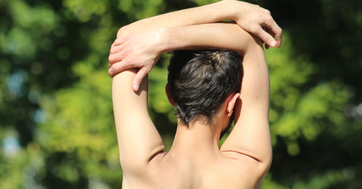
The thoracic extension stretch involves sitting in a chair with a supportive backrest, placing the hands behind the head, and gently leaning the upper back over the top edge of the chair. This opens the anterior chest wall and mobilizes the stiff thoracic vertebrae.
Cat-cow stretches, performed on the hands and knees, encourage smooth, sequential flexion and extension of the entire spine. These movements must be performed slowly and strictly within a pain-free range of motion, stopping immediately if the sharp, catching pain returns.
13. Breathing Techniques for Pain Relief
Patients suffering from pain upon inspiration instinctively adopt a shallow, rapid breathing pattern. While this minimizes immediate pain, it can lead to hyperventilation and anxiety. Retraining the respiratory pattern is a vital component of pain management.
Diaphragmatic breathing, or belly breathing, focuses on contracting the diaphragm to draw air deep into the lower lungs. The patient places one hand on their abdomen and concentrates on making the hand rise with each inhalation, keeping the chest and shoulders completely still.
This technique allows for adequate oxygen exchange without forcing the upper rib cage to expand maximally, thereby avoiding the mechanical stretching of the injured thoracic joints or intercostal muscles.
14. Ergonomic Modifications
Preventing recurrent episodes of mid-back pain requires optimizing the patient’s daily ergonomic environment. Office chairs must provide adequate mid-back support to maintain a neutral spinal alignment and prevent the slumping posture that compresses the rib cage.
Computer monitors should be positioned at eye level to prevent the user from leaning forward. When lifting objects, individuals must utilize their leg muscles and maintain a straight spine, avoiding any simultaneous lifting and twisting motions, which are exceptionally damaging to the costovertebral joints.
During sleep, patients should avoid sleeping on their stomach, as this forces the thoracic spine into an unnatural rotation. Sleeping on the back with a supportive pillow under the knees maintains the natural curvature of the spine and allows the rib cage to expand symmetrically.
15. When to See a Doctor
Medical evaluation is an absolute necessity if the pain upon breathing is accompanied by shortness of breath, a rapid heart rate, or coughing up blood. These are critical warning signs of a pulmonary embolism, a life-threatening blood clot in the lungs.
Patients must seek immediate emergency care if the mid-back pain radiates to the jaw, the left arm, or is accompanied by severe chest pressure and sweating, as this can indicate an acute myocardial infarction referring pain to the posterior thoracic region.
If the pain persists for more than two weeks despite anti-inflammatory medications and rest, or if it is accompanied by a persistent fever or unexplained weight loss, a comprehensive medical workup is required to rule out occult pulmonary infections or systemic inflammatory conditions.
16. Frequently Asked Questions FAQ
1. Can a cracked rib cause pain only when I take a deep breath?
Yes. A hairline fracture in a rib might not cause constant pain while you are resting. However, taking a deep breath forces the rib to flex and move, which pulls on the fractured bone segments and triggers a sudden, sharp spike in pain.
2. Is it safe to use a heating pad on my mid-back?
Applying a heating pad is generally very safe and effective for relieving muscle spasms and joint stiffness in the mid-back. Ensure the heat is not too intense to prevent skin burns, and limit application to twenty minutes per session.
3. Why does my back hurt when I sneeze?
Sneezing is one of the most forceful actions the respiratory muscles can perform. It causes a sudden, violent expansion and contraction of the rib cage. If you have a strained intercostal muscle or a sprained joint in your back, this explosive mechanical force will severely stretch the injured tissue.
4. Can acid reflux cause sharp pain in the mid-back when breathing?
While acid reflux can cause a burning sensation that radiates to the mid-back, it does not typically produce a sharp, catching pain that strictly worsens only upon taking a deep breath. That specific mechanical pattern strongly suggests a musculoskeletal or pleural issue.
17. Bibliography
Disclaimer: The content is for informational purposes only and does not replace medical advice. Always consult your doctor for personalized treatment.