
1. Introduction
Pink eye caused by a virus or bacteria typically presents with a thick or watery discharge, often starting in one eye before spreading to the other. Seasonal allergies cause intense itching, watery eyes, and simultaneous bilateral redness without thick pus. Distinguishing between an infectious process and an allergic reaction dictates whether a patient requires antimicrobial therapy or targeted histamine blockade.
Eye redness is a frequent and often alarming clinical presentation. The conjunctiva, the clear membrane covering the white part of the eye, reacts to a wide variety of irritants by dilating its dense network of surface blood vessels. This vasodilation creates the characteristic red or bloodshot appearance, which can look remarkably similar regardless of the underlying etiology.
An accurate diagnosis relies on evaluating the primary sensation experienced by the patient, the specific nature of the ocular discharge, and the precise timeline of symptom onset. Understanding the fundamentally different inflammatory pathways driving these conditions allows individuals to apply the correct therapeutic interventions and prevent unnecessary transmission of contagious pathogens.
2. Anatomy of the Conjunctiva
The conjunctiva is a thin, translucent mucous membrane that lines the inner surfaces of the eyelids and folds back to cover the anterior sclera, which is the white portion of the globe. Its primary physiological role is to provide lubrication by producing mucus and tears. This lubrication is essential to prevent dryness and mechanical friction during blinking.
This delicate membrane also serves as an immunological barrier. It contains numerous small blood vessels and resident immune cells designed to trap and destroy foreign debris, allergens, and microbial pathogens before they can penetrate deeper into the ocular structures. The superficial location of these vessels makes them uniquely reactive to external stimuli.
When an irritant or pathogen breaches this barrier, the immune system initiates an inflammatory cascade. The local blood vessels dilate to deliver additional white blood cells to the area. This sudden engorgement of the conjunctival microvasculature transforms the normally white sclera into a striking, vivid red.
3. Pathophysiology of Viral Conjunctivitis
Viral conjunctivitis is the most prevalent form of infectious pink eye. It is predominantly caused by adenoviruses, the same pathogens responsible for common upper respiratory tract infections. The virus is exceptionally contagious, spreading through direct contact with contaminated surfaces or respiratory droplets.
Once the virus infects the conjunctival epithelial cells, it hijacks the cellular machinery to replicate, causing localized cell death and shedding. The immune system responds by releasing inflammatory cytokines. This response generates a pronounced, diffuse redness across the eye surface.
Unlike bacterial infections, viral conjunctivitis primarily stimulates the production of a clear, watery fluid rather than dense pus. The inflammation often extends beyond the eye, causing the preauricular lymph nodes located just in front of the ears to swell and become tender, a classic sign of an active viral immune response.
4. Pathophysiology of Bacterial Conjunctivitis
Bacterial conjunctivitis occurs when opportunistic pathogens, such as Staphylococcus aureus or Streptococcus pneumoniae, colonize the ocular surface. These bacteria rapidly multiply, overwhelming the local mucosal defenses and triggering a severe, localized battle with host immune cells.
The defining characteristic of a bacterial infection is the production of mucopurulent discharge. As neutrophils rush to the site to engulf the bacteria, the accumulation of dead cells, bacterial debris, and proteinaceous fluid forms a thick, opaque pus. This discharge is typically yellow, green, or thick white.
This dense pus tends to accumulate at the lid margins and corners of the eye, especially during sleep. Patients frequently awaken to find their eyelids firmly glued together by dried crusts of this bacterial discharge, necessitating a warm compress to pry the eyes open safely.
5. The Allergic Inflammatory Cascade
Ocular allergies, or allergic conjunctivitis, represent a hypersensitivity reaction to harmless environmental proteins. Common triggers include pollen, pet dander, mold spores, and dust mites. When these allergens contact the conjunctiva of a sensitized individual, they bind to antibodies attached to resident mast cells.
This binding causes the mast cells to rapidly degranulate, releasing copious amounts of histamine and other potent inflammatory mediators directly into the ocular tissues. Histamine binds to specific receptors on the conjunctival blood vessels and local nerve endings.
The binding of histamine causes the blood vessels to leak fluid, resulting in mild to moderate swelling of the conjunctiva, a condition known clinically as chemosis. More importantly, the stimulation of the local sensory nerves produces the defining symptom of ocular allergies, which is an unrelenting, intense sensation of itching.
6. Assessing Pruritus and Itching
The presence and severity of itching serve as the most reliable diagnostic discriminator. In allergic conjunctivitis, pruritus is the dominant and most bothersome symptom. The urge to rub the eyes is profound and constant. Rubbing mechanically exacerbates the condition by physically breaking open more mast cells, releasing further histamine and intensifying the reaction.
Viral and bacterial conjunctivitis do not typically cause significant itching. Instead, patients with infectious pink eye generally describe a gritty, burning, or scratching sensation. They often feel as though a foreign body, like a grain of sand, is trapped under their eyelid.
If a patient states that their eyes are overwhelmingly itchy and they cannot stop rubbing them, the diagnosis heavily favors an allergic etiology. If the primary complaint is a burning discomfort or a gritty foreign body sensation, the clinical suspicion shifts toward an infectious process.
7. Evaluating Ocular Discharge
The nature of the fluid leaking from the eye provides a direct window into the underlying pathology. Allergic conjunctivitis produces tears. The eyes become excessively watery and may overflow, but the fluid remains completely clear and thin, without any opacity or stickiness.
Viral conjunctivitis also produces a watery discharge, but it is often more profuse and may contain thin, stringy mucus. The discharge is generally clear and does not form thick, crusted cement on the eyelashes overnight.
Bacterial conjunctivitis is distinguished by its thick, opaque, mucopurulent discharge. The pus is continuously produced throughout the day, requiring frequent wiping. The hallmark presentation involves waking up with the eyelids completely crusted shut. The color and consistency of this discharge are undeniable markers of bacterial colonization.
8. Unilateral Versus Bilateral Onset
The geometric progression of the symptoms aids in pinpointing the cause. Airborne allergens bathe the entire face simultaneously. Therefore, seasonal allergies almost universally affect both eyes at the exact same time. The redness, itching, and tearing are symmetrical from the moment of onset.
Infectious conjunctivitis, whether viral or bacterial, usually begins in just one eye. A patient touches a contaminated surface and subsequently rubs their right eye, inoculating the conjunctiva. The right eye becomes red and symptomatic while the left eye remains completely clear.
Because infectious pink eye is profoundly contagious, the patient inevitably transfers the pathogen to the unaffected eye within a few days via their hands or a shared pillowcase. A sequential onset, where one eye turns red followed a day or two later by the second eye, strongly indicates a transmissible infection.
9. Systemic and Associated Symptoms
The surrounding clinical context is vital. Viral conjunctivitis frequently occurs in tandem with a systemic upper respiratory infection. A patient presenting with a red, watery eye who also complains of a sore throat, profound nasal congestion, and a mild fever is likely suffering from an adenoviral infection affecting the entire mucosal tract.
Bacterial conjunctivitis is often an isolated event, though in pediatric populations it can occasionally accompany an ear infection. Systemic fever or profound fatigue is uncommon in isolated bacterial pink eye.
Allergic conjunctivitis is intrinsically linked to allergic rhinitis. Patients will typically exhibit concurrent symptoms such as sneezing, a runny nose with clear fluid, and a tickle in the back of the throat. For further details on evaluating related respiratory symptoms, read our article on the runny nose. These symptoms fluctuate predictably based on environmental exposure.
10. Environmental and Seasonal Triggers
The timeline of the symptoms provides critical clues. Allergic conjunctivitis often follows a distinct seasonal pattern, flaring up reliably in the spring with tree pollen or in the autumn with ragweed. The symptoms often worsen when the patient spends time outdoors and improve significantly in air-conditioned or filtered indoor environments.
Infectious pink eye does not adhere to seasonal pollen counts. While viral pink eye peaks slightly during cold and flu season due to increased indoor crowding, it can occur at any time of the year. Bacterial conjunctivitis is entirely sporadic and unrelated to environmental allergens.
If a patient reports that their red eyes occur every April and resolve when they take over-the-counter antihistamines, the diagnosis of seasonal allergies is clear. Sudden, unprovoked redness in the middle of winter strongly suggests an infectious pathogen.
11. Clinical Diagnostic Evaluation
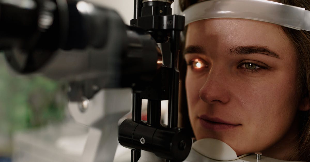
A formal clinical evaluation begins with a meticulous slit-lamp examination. The clinician uses targeted magnification and bright illumination to inspect the conjunctival surface, the cornea, and the anterior chamber of the eye. This rules out more severe causes of a red eye, such as acute glaucoma or uveitis.
During the slit-lamp exam, the clinician looks for follicles or papillae on the inside of the eyelids. Viral infections produce small, dome-shaped follicles, giving the tissue a cobblestone appearance. Allergic reactions typically produce flattened papillae.
Fluorescein dye is frequently utilized to assess the integrity of the cornea. If the infection or allergic rubbing has caused micro-abrasions on the delicate corneal surface, the dye will highlight these defects under a cobalt blue light, guiding the necessity for prophylactic treatments.
12. Data Structure: Differentiating Red Eye
The following table outlines the principal clinical features differentiating the common causes of a red eye.
| Clinical Feature | Viral Conjunctivitis | Bacterial Conjunctivitis | Allergic Conjunctivitis |
|---|---|---|---|
| Primary Sensation | Gritty, burning | Gritty, foreign body feeling | Intense itching |
| Eye Discharge | Watery, clear, thin mucus | Thick, yellow or green pus | Watery, stringy, clear |
| Onset Pattern | Starts in one eye, spreads to the other | Starts in one eye, spreads to the other | Both eyes simultaneously |
| Crusted Eyelids | Mild morning crusting | Severe, eyelids glued shut | None |
| Associated Symptoms | Sore throat, swollen lymph nodes | Usually isolated | Sneezing, clear runny nose |
13. Management of Infectious Conjunctivitis
Viral conjunctivitis is a self-limiting condition. Much like a common cold, there are no specific antiviral medications designed to cure typical adenoviral pink eye. Management relies on supportive care. Cold compresses applied to the closed eyelids soothe the burning sensation and reduce vascular engorgement. Artificial tears provide essential lubrication to wash away cellular debris. Strict hygiene is mandatory to prevent transmission.
Bacterial conjunctivitis requires targeted antimicrobial therapy. Clinicians prescribe topical antibiotic eye drops or ointments. These medications eradicate the colonizing bacteria, rapidly reducing the production of pus and shortening the duration of contagion. Patients must complete the entire prescribed course to prevent bacterial resistance.
For both infectious types, patients must discard contaminated cosmetics, thoroughly wash all pillowcases in hot water, and refrain from wearing contact lenses until the infection has completely resolved and the eye is perfectly white.
14. Management of Ocular Allergies
The foundational treatment for allergic conjunctivitis is absolute avoidance of the triggering allergen. When exposure is unavoidable, pharmacological intervention halts the inflammatory cascade. Topical antihistamine eye drops provide rapid, targeted relief directly to the ocular surface, neutralizing the intense itching within minutes.
Mast cell stabilizers are often combined with antihistamines in modern ophthalmic formulations. These medications prevent the mast cells from degranulating in the first place, providing long-term prophylactic control during peak allergy seasons.
Oral antihistamines can be utilized to manage concurrent nasal symptoms, but they possess a systemic drying effect. This can occasionally reduce natural tear production, exacerbating ocular friction. Preservative-free artificial tears help flush airborne allergens from the conjunctival surface before they can initiate an immune response.
15. When to Seek Urgent Ophthalmic Care
While most cases of red eye are benign and easily managed, specific clinical red flags necessitate immediate, emergency evaluation by an ophthalmologist. If the red eye is accompanied by severe, deep, aching pain rather than superficial grittiness, it indicates potential involvement of internal ocular structures.
Any sudden alteration in visual acuity, such as profound blurriness, double vision, or a noticeable decrease in peripheral vision, is an absolute medical emergency. Sensitivity to light, known as photophobia, where normal indoor lighting causes sharp pain, is a hallmark of corneal inflammation or uveitis, neither of which are simple conjunctivitis.
Furthermore, if the redness is concentrated in a tight ring directly around the colored part of the eye, or if the pupil appears irregular or fixed, prompt specialized intervention is required to prevent permanent, irreversible damage to the patient’s vision.
16. Frequently Asked Questions (FAQ)
1. Can you have pink eye and allergies at the same time?
Yes. Intense rubbing from seasonal allergies can damage the protective barrier of the eye, making it easier for opportunistic bacteria from the hands to enter and cause a secondary bacterial conjunctivitis.
2. Does pink eye always produce green pus?
No. Only bacterial pink eye produces thick yellow or green pus. Viral pink eye, which is the most common infectious type, typically only produces a clear, watery discharge that feels gritty.
3. Are seasonal allergies contagious?
No, seasonal allergies are an individual immune system overreaction to harmless pollen or dust. They cannot be spread from person to person, whereas viral and bacterial pink eye are both highly contagious.
4. Can I wear my contact lenses if my eyes are just itchy from allergies?
It is best to remove contact lenses during a severe allergy flare. Lenses can trap airborne pollen against the surface of the eye, worsening the reaction, and constant rubbing can displace the lens or scratch the cornea.
5. How long is viral pink eye contagious?
Viral pink eye is contagious as long as the eye remains red and watery, which typically lasts between one and two weeks. Strict hand hygiene is required during this entire period to protect others.
17. Bibliography
Disclaimer: The content is for informational purposes only and does not replace medical advice. Always consult your doctor for personalized treatment.
