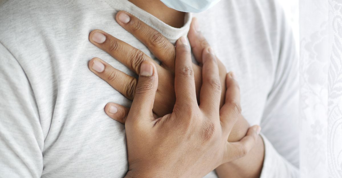
1. Introduction to Musculoskeletal Chest Pain
Musculoskeletal chest pain refers to discomfort originating from the structural components of the anterior thoracic wall, including the ribs, sternum, costal cartilages, and the overlying musculature. It is a highly prevalent condition, accounting for a significant proportion of emergency department visits and outpatient consultations. The primary clinical challenge is that the presentation of chest wall pain frequently mimics the terrifying symptoms of acute myocardial infarction or pulmonary embolism.
For the patient, any sharp, stabbing, or heavy sensation in the chest naturally triggers severe anxiety regarding cardiac health. The physician’s foremost responsibility is to rapidly systematically rule out life-threatening cardiopulmonary emergencies before assigning a benign musculoskeletal diagnosis.
Once cardiac and pulmonary etiologies are definitively excluded, the focus shifts to identifying the specific musculoskeletal structure responsible for the pain. Conditions such as costochondritis, intercostal muscle strains, and sternoclavicular joint pathology require targeted physical assessments to diagnose accurately. Effective management relies on reassuring the patient, reducing localized inflammation, and modifying physical activities to allow the injured thoracic structures to heal.
2. Anatomy of the Thoracic Wall
To pinpoint the source of musculoskeletal chest pain, one must understand the anatomy of the thoracic cage. The chest wall is a dynamic, highly flexible bony structure designed to protect vital organs while expanding continuously to facilitate respiration.
The sternum, or breastbone, sits centrally. True ribs attach directly to the sternum via flexible segments of hyaline cartilage known as costal cartilages. These costochondral junctions are highly susceptible to mechanical strain and inflammation. The spaces between the ribs are filled with layers of intercostal muscles, which assist in forced inhalation and exhalation.
The entire bony cage is enveloped by large, powerful muscle groups, including the pectoralis major, serratus anterior, and the latissimus dorsi. A network of intercostal nerves runs just beneath the lower edge of each rib, providing sensory innervation to the chest wall. Inflammation, trauma, or repetitive strain affecting any of these distinct tissues will manifest as localized chest pain.
3. Differentiating Cardiac vs. Musculoskeletal Pain
The absolute priority in clinical assessment is differentiating benign chest wall pain from acute cardiac ischemia. While overlap exists, specific characteristics strongly suggest a musculoskeletal etiology.
Musculoskeletal pain is typically somatic. It is usually sharp, highly localized to a specific spot, and frequently described as stabbing or aching. Crucially, musculoskeletal pain is almost always reproducible by physical palpation; pressing firmly on the affected area precisely recreates the patient’s specific pain. It also worsens predictably with movement, twisting of the torso, or deep inspiration.
Conversely, cardiac pain originating from ischemic heart muscle is visceral. Patients frequently describe it as a deep, diffuse, crushing pressure or a heavy weight sitting on the center of the chest. It often radiates to the jaw, neck, or left arm, and is accompanied by systemic symptoms such as profound diaphoresis, nausea, and severe shortness of breath. Cardiac pain is typically unbothered by deep breathing or chest wall palpation.
4. Costochondritis
Costochondritis is the most common specific diagnosis for musculoskeletal chest pain. It involves acute, self-limiting inflammation of the costochondral or chondrosternal junctions, where the ribs meet the sternum. The exact etiology is often idiopathic, though it frequently follows a recent viral respiratory infection, a bout of severe coughing, or an episode of intense, unfamiliar upper body exertion.
Patients present with sharp, aching pain located on the anterior chest wall, most commonly affecting the second through fifth ribs on the left side, which heavily contributes to the fear of a heart attack. The pain intensifies significantly during deep breathing, coughing, or physical activity involving the arms.
During the physical examination, the physician will elicit exquisite point tenderness when palpating directly over the affected cartilage joints. In typical costochondritis, there is no visible swelling, heat, or redness of the overlying skin, distinguishing it from other, rarer chest wall syndromes.
5. Tietze Syndrome
Tietze syndrome is frequently conflated with costochondritis, but it is a distinct clinical entity. While it also involves painful inflammation of the costal cartilages, Tietze syndrome is characterized by the presence of visible, palpable, and often erythematous swelling at the affected joint.
This condition is significantly less common than costochondritis and typically affects younger individuals, particularly adolescents and young adults. The swelling usually targets a single joint, most frequently the second or third costochondral junction.
The swelling in Tietze syndrome can be prominent and firm, sometimes persisting for months or even years after the acute pain has subsided. Because the swelling mimics a physical mass, radiological imaging is occasionally required to definitively rule out a primary bone or cartilage tumor of the chest wall.
6. Intercostal Muscle Strains
The intercostal muscles, situated between the ribs, are highly vulnerable to mechanical strain. These injuries typically occur during sudden, forceful twisting motions of the torso, heavy lifting, or violent coughing fits associated with bronchitis or asthma.
An intercostal muscle tear causes sharp, localized, and highly debilitating pain that wraps around the side of the rib cage. The pain is severely aggravated by any movement that stretches the rib cage, making deep breathing, sneezing, and laughing intensely painful.
Because the pain prevents deep pulmonary expansion, patients often take shallow, rapid breaths to guard the injured muscle. This hypoventilation can lead to atelectasis, where the lower lung segments collapse, increasing the risk of secondary pneumonia. Adequate pain control is therefore medically essential to maintain proper pulmonary function while the muscle heals.
7. Rib Fractures and Blunt Trauma
Direct blunt force trauma to the chest, sustained during motor vehicle accidents, falls, or contact sports, frequently results in rib contusions or frank fractures. Even a minor, non-displaced hairline fracture causes agonizing pain due to the continuous motion of the rib cage during respiration.
The clinical presentation involves localized, severe pain directly over the site of impact. The physician assesses for bony crepitus, a grating sensation felt when the broken ends of the bone rub together during breathing.
While simple, isolated rib fractures heal spontaneously with rest, the physician must assess for dangerous complications. Sharp bone fragments can puncture the underlying pleura or lung tissue, causing a pneumothorax. If a patient with trauma presents with a sudden, massive increase in musculoskeletal chest pain and extreme respiratory distress, an immediate chest x-ray is mandatory.
8. Sternoclavicular Joint Pathology
The sternoclavicular joint connects the medial end of the collarbone to the upper sternum. It is a highly mobile joint that absorbs immense force during upper body movements. Pain localized specifically to the very top of the breastbone is often related to sternoclavicular joint dysfunction.
This joint is susceptible to osteoarthritis, particularly in older adults, presenting as a chronic, dull ache that worsens with shoulder movement. In younger patients, pain is usually traumatic, resulting from sports injuries that cause sprains, subluxations, or full dislocations of the joint.
A posterior dislocation of the sternoclavicular joint is a medical emergency, as the collarbone is pushed backward into the superior mediastinum, potentially compressing the trachea, esophagus, or the great blood vessels exiting the heart.
9. Neuropathic Chest Wall Pain
Pain experienced on the chest wall is not always driven by muscle or bone injury; it can originate from the nerves supplying the region. Intercostal neuralgia is a neuropathic pain condition caused by irritation or damage to the intercostal nerves.
The pain is frequently described as burning, electrical, or shooting, and typically wraps horizontally around the rib cage in a band-like pattern following the path of the specific irritated nerve. A classic cause of severe intercostal neuralgia is Herpes Zoster.
Following the initial, excruciating burning pain, the characteristic vesicular rash of shingles will erupt along the specific nerve dermatome. Recognizing the neuropathic quality of the pain allows the clinician to initiate antiviral and specific nerve-pain medications rather than standard anti-inflammatories.
10. Fibromyalgia and Chronic Widespread Pain
When a patient presents with chronic, diffuse musculoskeletal chest pain that is not linked to a specific trauma or localized cartilage inflammation, the physician must consider central pain syndromes such as fibromyalgia.
Fibromyalgia involves a hypersensitization of the central nervous system, where benign sensory inputs are amplified into severe pain signals. Patients frequently present with multiple, symmetrical tender points across the upper chest, specifically around the second rib and the clavicles.
This pain is typically accompanied by profound fatigue, sleep disturbances, and cognitive clouding. Management diverges significantly from localized chest wall injuries, focusing on neuromodulating medications, graded aerobic exercise, and psychological interventions rather than localized structural therapies.
11. Clinical Diagnosis and The Emergency Assessment
Any patient presenting to a medical facility with chest pain requires rapid triaging. The foundational diagnostic step is a 12-lead electrocardiogram. This rapid, non-invasive test instantly detects electrical abnormalities characteristic of an acute myocardial infarction.
If the patient’s history or risk profile suggests cardiac involvement, serum cardiac biomarkers, specifically troponin, are drawn. A normal electrocardiogram and negative serial troponins definitively rule out acute heart muscle damage, allowing the physician to safely focus on a musculoskeletal diagnosis.
Once cardiac and pulmonary emergencies are excluded, the diagnosis of conditions like costochondritis relies heavily on the physical examination. The ability to precisely reproduce the patient’s chief complaint through firm, localized palpation of the chest wall joints is the gold standard for confirming a musculoskeletal etiology.
12. Radiological Imaging
In the majority of benign musculoskeletal chest pain cases, such as costochondritis or mild muscle strains, radiological imaging is unnecessary and will return completely normal results, as cartilage and muscle inflammation do not appear on standard X-rays.
A chest X-ray is ordered if there is a history of trauma to rule out rib fractures, or if the patient presents with a cough and fever, to rule out underlying pneumonia or pleurisy.
If severe, persistent swelling is noted, or if there is a suspicion of a sternoclavicular joint dislocation, a computed tomography scan of the chest provides highly detailed, three-dimensional views of the bony architecture, easily identifying occult fractures or suspicious structural masses missed by plain radiographs.
13. Pharmacological Relief Strategies
The primary medical intervention for musculoskeletal chest wall pain focuses on reducing localized inflammation and providing acute analgesia. Nonsteroidal anti-inflammatory drugs, such as ibuprofen or naproxen, are the first-line therapy for costochondritis and muscle strains.
Patients are instructed to take these medications on a scheduled basis, rather than just as needed, to achieve a sustained anti-inflammatory effect in the tissues. For severe muscle spasms associated with intercostal strains, short-term use of central muscle relaxants may be indicated to break the pain-spasm cycle.
If the pain is severe, sharply localized, and refractory to oral medications, a physician may perform a localized injection of corticosteroids mixed with a local anesthetic directly into the affected costochondral joint, providing rapid and often permanent relief of the inflammation.
14. Physical Rehabilitation and Posture
Musculoskeletal chest pain is heavily exacerbated by poor ergonomics and biomechanical strain. Patients who spend long hours hunched over computers frequently develop chronic shortening of the pectoral muscles and severe rounding of the shoulders. This places continuous, agonizing mechanical stress on the anterior chest wall joints.
Physical therapy is a highly effective intervention. Therapists guide the patient through targeted stretching of the anterior chest musculature and strengthening of the upper back and scapular retractors. This improves thoracic posture, opening the chest and physically removing the mechanical tension from the sternum and rib joints.
Patients are also advised on activity modification. Heavy lifting, repetitive pushing motions, and high-impact sports must be strictly avoided during the acute healing phase to prevent continuous re-injury of the fragile costal cartilage.
15. Frequently Asked Questions
1. How can a doctor be sure my chest pain isn’t a heart attack?
Doctors use a combination of tools. If your EKG and blood tests looking for heart damage are completely normal, and the doctor can exactly recreate your pain by pressing firmly on your ribs, it strongly confirms the pain is coming from the muscles or joints, not your heart.
2. What is costochondritis and why does it hurt to breathe?
Costochondritis is an inflammation of the cartilage that connects your ribs to your breastbone. Because your ribs must expand and move every single time you inhale, taking a deep breath forces that inflamed, swollen joint to move, causing sharp, stabbing pain.
3. Will an X-ray show if I have a pulled muscle in my chest?
No. Standard X-rays only show dense bone tissue. They are useful to ensure you don’t have a broken rib or a lung infection, but they cannot show inflamed muscles or cartilage. The diagnosis relies on your doctor’s physical examination.
4. How long does chest wall pain usually last?
It varies widely depending on the severity of the strain or inflammation. Mild costochondritis or a pulled intercostal muscle usually resolves in two to four weeks with rest and anti-inflammatory medication. Severe strains or cartilage inflammation can sometimes take several months to heal completely.
5. Should I wrap or tape my chest if I pulled a muscle there?
No. Taping or wrapping the chest is no longer recommended by doctors. Binding the chest prevents your lungs from expanding fully, which can lead to fluid buildup in the lower lungs and cause severe pneumonia. You must manage the pain with medication and gentle breathing.
16. Bibliography
Disclaimer: The content is for informational purposes only and does not replace medical advice. Always consult your doctor for personalized treatment.