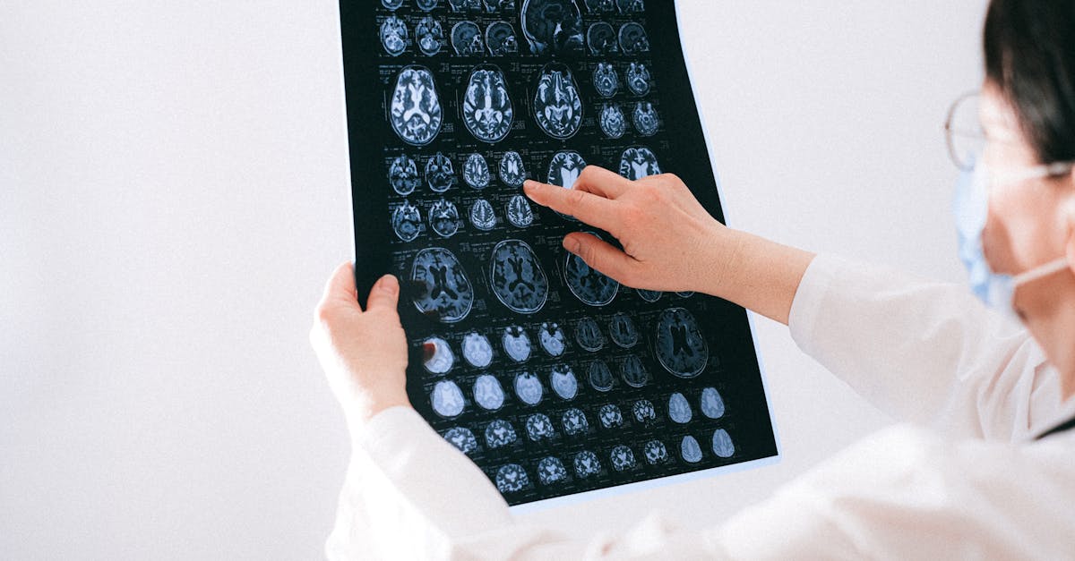
1. Introduction to the Nervous System
The human nervous system is the most complex, highly organized, and vital regulatory system within the human body. Serving as the biological command center, it is responsible for detecting environmental changes, processing sensory information, and executing precise motor responses. A normal, healthy nervous system functions with seamless efficiency, allowing individuals to navigate their environment, maintain internal homeostasis, and engage in high-level cognitive processes such as memory, emotion, and language.
At its core, the nervous system operates through the rapid transmission of electrical impulses and chemical signals across vast networks of specialized cells called neurons. This intricate communication network is responsible for everything from the conscious decision to lift a glass of water to the subconscious regulation of heart rate and digestion.
In clinical practice, determining that a patient has a “normal nervous system” requires a meticulous and structured neurological examination. When a physician confirms that the nervous system is normal, it provides profound reassurance, indicating that the structural integrity of the brain, spinal cord, and peripheral nerves is intact, and that the electrical communication pathways are functioning without pathological interruption.
2. Central and Peripheral Divisions
To understand the normal function of the nervous system, it must be viewed through its two primary anatomical divisions: the Central Nervous System (CNS) and the Peripheral Nervous System (PNS). The CNS consists of the brain and the spinal cord. Safely encased within the skull and the vertebral column, the CNS acts as the primary processing unit, integrating incoming sensory data and generating outgoing motor commands.
The Peripheral Nervous System acts as the communication relay between the CNS and the rest of the body. It consists of cranial nerves that emerge directly from the brain, and spinal nerves that branch out from the spinal cord to innervate the limbs and torso.
The PNS is further divided into two functional branches: the somatic nervous system, which governs voluntary movements and transmits conscious sensory information (like touch and temperature), and the autonomic nervous system, which operates subconsciously to regulate the internal organs, blood vessels, and glandular secretions.
3. The Neuron: The Fundamental Unit
The fundamental functional unit of the nervous system is the neuron. A healthy human brain contains approximately 86 billion neurons, each forming thousands of connections (synapses) with neighboring cells. A typical neuron consists of three main parts: the dendrites, the cell body (soma), and the axon.
Dendrites are branching structures that act as antennas, receiving chemical signals from other neurons. The cell body processes these signals. If the stimulatory signal is strong enough, the neuron fires, generating an electrical impulse known as an action potential. This electrical charge travels rapidly down the axon, a long, cable-like projection that transmits the signal to the next cell.
In a normal nervous system, the axons of motor and sensory neurons are wrapped in a protective lipid layer called the myelin sheath. Produced by specialized glial cells (Schwann cells in the PNS, oligodendrocytes in the CNS), myelin acts as biological insulation. It vastly accelerates the speed of electrical transmission, ensuring that motor commands reach the muscles instantaneously and sensory data is processed in real time.
4. Synaptic Transmission and Neurotransmitters
Neurons do not physically touch one another; they communicate across microscopic gaps called synapses. When an electrical action potential reaches the end of an axon (the terminal bouton), it triggers the release of chemical messengers known as neurotransmitters into the synaptic cleft.
These neurotransmitters drift across the gap and bind to specific receptors on the dendrites of the adjacent neuron, much like a key fitting into a lock. In a healthy nervous system, a delicate balance of excitatory and inhibitory neurotransmitters ensures smooth, coordinated neurological function.
Glutamate is the primary excitatory neurotransmitter, driving brain activity and learning. Gamma-aminobutyric acid (GABA) is the primary inhibitory neurotransmitter, keeping neuronal firing in check and preventing chaotic electrical storms (seizures). Dopamine regulates motor control and the reward pathways, while serotonin modulates mood and sleep architecture. A normal nervous system flawlessly maintains the synthesis, release, and reuptake of these vital chemicals.
5. The Blood-Brain Barrier
The central nervous system operates in a highly protected, strictly regulated microenvironment. This protection is provided by the blood-brain barrier, a highly selective semipermeable border of endothelial cells that lines the capillaries within the brain.
In a normal nervous system, the blood-brain barrier acts as a rigorous biological filter. It allows essential nutrients, such as oxygen and glucose, to pass freely into the brain tissue, while tightly blocking the entry of neurotoxins, systemic bacteria, and large inflammatory molecules circulating in the bloodstream.
This barrier is essential for maintaining the delicate ionic balance required for neuronal electrical firing. When a physician determines a nervous system is normal, it implies that this barrier is structurally sound, defending the brain from systemic infections (like meningitis or encephalitis) that would otherwise trigger catastrophic neurological dysfunction.
6. The Clinical Neurological Examination
When a patient presents with vague symptoms such as mild tingling or transient weakness, the physician relies on the clinical neurological examination to determine if the nervous system is truly normal. This exam is a highly structured, systematic assessment that tests specific neural pathways from the cerebral cortex down to the peripheral nerve endings.
The exam is divided into several distinct components: mental status, cranial nerves, motor system, sensory system, coordination, and reflexes. By systematically testing these areas, the clinician maps the functional integrity of the entire neuroanatomy.
If the patient flawlessly performs the required tasks and exhibits normal physiological responses, the clinician can confidently rule out significant structural lesions, demyelinating diseases, or degenerative neurological conditions without immediately resorting to expensive and invasive imaging studies.
7. Mental Status and Cognitive Function
The neurological exam begins with an assessment of the patient’s mental status, evaluating the function of the higher cerebral cortex. A normal mental status indicates that the frontal, temporal, and parietal lobes are functioning harmoniously.
The physician engages the patient in conversation to assess their level of alertness, orientation to time and place, and the fluency of their speech. A normal nervous system produces clear, articulate speech without word-finding difficulties (aphasia) or slurring (dysarthria).
Cognitive testing involves evaluating short-term memory, attention span, and executive function. The patient may be asked to recall three unrelated words after a few minutes or perform a serial subtraction task (e.g., counting backward from 100 by 7s). A healthy brain executes these tasks efficiently, indicating normal synaptic processing and intact memory encoding circuits.
8. Evaluating the Cranial Nerves
The twelve pairs of cranial nerves emerge directly from the brain and brainstem, supplying motor and sensory function to the head and neck. Testing these nerves provides a direct clinical window into the structural integrity of the brainstem.
A normal cranial nerve exam confirms symmetrical facial movements (testing the Facial nerve, CN VII), normal visual acuity and peripheral vision (Optic nerve, CN II), and intact extraocular eye movements without double vision or involuntary shaking (Oculomotor, Trochlear, and Abducens nerves).
The physician also checks the pupillary light reflex. Shining a light into one eye should cause both pupils to constrict briskly and equally. This normal response confirms that the complex neural loop traveling from the optic nerve through the midbrain and back via the oculomotor nerve is intact and healthy.
9. Motor Strength and Muscle Tone
Assessing the motor system evaluates the pathway from the motor cortex in the brain, down the spinal cord, and out to the peripheral muscles. A normal motor examination reveals well-developed muscle bulk without signs of atrophy (wasting) or fasciculations (involuntary twitching).
The physician tests muscle tone by passively moving the patient’s limbs. Normal muscle tone provides a slight, smooth resistance to passive stretching. Abnormalities such as rigid stiffness (spasticity) or extreme floppiness (flaccidity) indicate significant pathology in either the central or peripheral motor pathways.
Motor strength is tested by having the patient push and pull against the physician’s resistance across major muscle groups. A normal nervous system generates full, symmetrical strength (graded as 5/5 on the clinical scale) in all extremities, confirming that the descending motor tracts in the spinal cord are communicating flawlessly with the peripheral muscles.
10. The Sensory Pathways
The sensory examination maps the ascending pathways that carry information from the skin back up to the parietal lobe of the brain. The nervous system utilizes different nerve fibers and spinal tracts to carry distinct types of sensory data.
The physician tests for light touch, sharp pain (pinprick), and temperature sensation. These signals travel up the spinothalamic tract of the spinal cord. In a normal nervous system, the patient can accurately detect these sensations equally on both sides of the body, indicating no damage to the peripheral nerves or spinal pathways.
Proprioception (the sense of joint position) and vibration sense are also meticulously tested. Using a tuning fork placed on a bony prominence, the physician assesses the dorsal column pathways of the spinal cord. A healthy nervous system allows a patient to accurately identify the direction a toe is moved with their eyes closed, confirming flawless sensory transmission from the distal extremities to the brain.
11. Data: Normal Reflex Responses
Deep tendon reflexes are involuntary motor responses that test the integrity of the reflex arc at specific levels of the spinal cord. The following table highlights the standard clinical reflexes and what a normal response indicates.
| Reflex Tested | Spinal Cord Level Evaluated | Normal Clinical Response (Grade 2+) |
|---|---|---|
| Biceps Reflex | Cervical 5, 6 (C5, C6) | Brisk contraction of the biceps, slight elbow flexion. |
| Triceps Reflex | Cervical 7, 8 (C7, C8) | Brisk contraction of the triceps, slight elbow extension. |
| Patellar (Knee) Reflex | Lumbar 3, 4 (L3, L4) | Brisk contraction of the quadriceps, slight knee extension. |
| Achilles (Ankle) Reflex | Sacral 1, 2 (S1, S2) | Brisk plantar flexion (downward movement) of the foot. |
12. Coordination and the Cerebellum
The cerebellum, located at the base of the brain, does not initiate movement but is responsible for fine-tuning it, ensuring that actions are smooth, accurate, and coordinated. A normal nervous system relies heavily on a healthy cerebellum to maintain balance and posture.
The physician tests cerebellar function using the finger-to-nose test. The patient is asked to repeatedly touch their own nose and then the physician’s moving finger. A normal response is a smooth, rapid, and accurate movement without any tremor or missing the target (dysmetria).
Gait and balance are also evaluated. The patient is asked to walk normally, walk heel-to-toe (tandem gait), and perform the Romberg test (standing still with feet together and eyes closed). A normal nervous system maintains perfect balance during these tasks, utilizing proprioceptive feedback and cerebellar processing to keep the body upright against gravity, a process thoroughly assessed during any dizziness evaluation.
13. Understanding Normal Variations
It is important for patients to understand that a “normal” nervous system includes a degree of physiological variation. Minor, transient symptoms do not inherently signify a neurological disease. For example, occasional muscle twitches (benign fasciculations) in the eyelid or calf are incredibly common and usually related to stress, fatigue, or caffeine intake, rather than a motor neuron disease.
Similarly, a limb “falling asleep” with a temporary pins-and-needles sensation after sitting in an awkward position is a normal physiological response. It occurs when superficial peripheral nerves are temporarily compressed, blocking electrical transmission. Once the pressure is relieved, the nerve recovers rapidly and completely.
Aging also produces normal variations. Mild slowing of reflexes, slight decreases in vibratory sense in the toes, and a subtle decline in the speed of recall memory are expected, benign changes associated with the natural aging of the central and peripheral nervous systems, and do not equate to pathology like dementia or neuropathy.
14. When to See a Doctor
While a normal nervous system operates flawlessly in the background of daily life, specific warning signs indicate that this intricate network has been disrupted and requires immediate medical evaluation. Patients should seek urgent neurological assessment if they experience the sudden onset of weakness, numbness, or paralysis on one side of the body, as this is a classic sign of a stroke.
Immediate emergency care is also required for sudden, severe difficulty speaking, loss of vision, or an explosive, excruciating headache. These symptoms suggest a critical vascular event within the central nervous system.
Patients should consult a physician for more gradual symptoms as well. Progressive clumsiness, frequent unexplained falls, a slowly developing tremor, chronic burning pain in the hands or feet, or noticeable changes in memory and personality warrant a comprehensive neurological examination to determine if a pathological process is interfering with the normal function of the nervous system.
15. Frequently Asked Questions (FAQ)
1. What does it mean when a doctor says my neurological exam is completely normal?
It means that after testing your strength, reflexes, sensation, and coordination, the doctor found that your brain, spinal cord, and nerves are communicating perfectly. It is a highly reassuring sign that you do not have a serious structural nerve disease or brain injury.
2. Are occasional muscle twitches normal?
Yes. Occasional, brief muscle twitches (like in the eyelid or thigh) are very common and entirely normal. They are usually caused by stress, lack of sleep, or too much caffeine, rather than a disease of the nervous system.
3. Why does the doctor hit my knee with a hammer during a check-up?
The doctor is testing your deep tendon reflexes. Striking the tendon sends a rapid electrical signal to your spinal cord and back to the muscle, causing a quick kick. A normal kick proves that the nerve pathways in that specific part of your spinal cord are intact and functioning rapidly.
4. Is it normal to forget things as I get older?
Mild forgetfulness, such as occasionally misplacing keys or struggling for a moment to remember a name, is a normal part of the aging process in a healthy nervous system. It only becomes a medical concern if memory loss disrupts your daily life or makes it hard to complete familiar tasks.
5. How fast do nerve signals travel in a normal nervous system?
In healthy, myelinated nerves (nerves coated in biological insulation), electrical signals can travel at speeds of up to 268 miles per hour (120 meters per second). This incredible speed is why you pull your hand away from a hot stove instantly.
Bibliography
Disclaimer: The content is for informational purposes only and does not replace medical advice. Always consult your doctor for personalized treatment.