1. Introduction
A dental abscess can indeed cause severe, rapidly expanding swelling in the lower jaw, representing a potentially life-threatening medical emergency. A dental abscess is a localized collection of purulent fluid, or pus, generated by a bacterial infection originating within the necrotic pulp of a tooth. If the infection is not contained by the immune system or promptly treated by a dental professional, the bacteria can easily break through the protective barriers of the jawbone and invade the soft tissues of the face and neck.
The lower jaw, or mandible, is intimately connected to several critical anatomical compartments known as fascial spaces. When the aggressive bacteria from a mandibular abscess perforate the bone, they pour into these fascial spaces, inciting a massive inflammatory response. This rapid accumulation of pus and inflammatory fluid causes profound swelling that can distort facial features, restrict jaw movement, and most alarmingly, compress the airway.
Understanding the progression from a simple cavity to severe maxillofacial swelling is essential for triaging patients correctly. An untreated odontogenic infection does not simply resolve on its own. As the swelling increases, it can lead to systemic sepsis or fatal airway obstruction, known as Ludwig angina. Immediate clinical intervention, involving both surgical drainage and aggressive antibiotic therapy, is mandated to halt the progression of the disease.
2. Pathophysiology of Odontogenic Infections
The journey toward a severe jaw swelling begins microscopically within the tooth structure. The outermost layer, the enamel, protects the internal pulp chamber, which houses the nerves and blood vessels of the tooth. When advanced dental caries destroy the enamel and dentin, oral bacteria gain direct access to the sterile pulp chamber.
The bacteria rapidly colonize and infect the pulp, causing acute pulpitis. Because the pulp is encased in rigid tooth structure, the resulting inflammatory swelling quickly cuts off the blood supply, leading to pulpal necrosis. Once the nerve dies, the acute toothache may temporarily stop, creating a false sense of security for the patient while the bacteria continue to multiply in the necrotic tissue.
The infection then travels down the root canal system and exits the tip of the root, known as the apex, into the surrounding alveolar bone. Here, the immune system attempts to wall off the infection, creating a periapical abscess. As bacterial toxins and dead immune cells accumulate, the pressure builds, and the purulent fluid seeks the path of least resistance through the bone.
3. Anatomy of the Mandible and Fascial Spaces
The direction in which the severe swelling develops is dictated entirely by the anatomical relationship between the infected tooth root and the surrounding muscle attachments of the mandible. The lower jaw is surrounded by complex layers of connective tissue called fascia, which compartmentalize the head and neck into distinct fascial spaces.
These fascial spaces are normally virtual spaces, meaning they are flat and empty. However, they offer almost no resistance to expanding fluid. Once pus erodes through the cortical bone of the mandible, it pours into these spaces, ballooning them outward. The buccinator muscle on the outside of the jaw and the mylohyoid muscle on the floor of the mouth act as critical anatomical boundaries.
If the root apex of the infected tooth sits above the attachment of the buccinator muscle, the pus will swell into the vestibule of the mouth. However, if the root apex, particularly of the lower molars, sits below these muscle attachments, the pus is directed deeply into the submandibular or submental spaces, leading to massive external lower jaw and neck swelling.
4. Submandibular and Submental Space Involvement
The lower second and third molars are the most frequent culprits behind severe lower jaw swelling. The roots of these teeth commonly extend below the attachment line of the mylohyoid muscle. When an abscess from these teeth perforates the lingual, or inner, plate of the mandible, the infection spills directly into the submandibular space.
Involvement of the submandibular space presents as a massive, firm, and exquisitely tender swelling located just beneath the lower jawline. The skin overlying the swelling becomes stretched, erythematous, and warm to the touch. This presentation is a classic cellulitis, indicating that the infection is spreading diffusely through the soft tissues.
The submental space, located directly beneath the chin, can also become involved, particularly from infections originating in the lower incisors. When these spaces fill with inflammatory fluid, the swelling becomes so severe that the normal contour of the jawline is completely obliterated, leaving the patient with a swollen, rigid neck.
5. Trismus and Masticatory Muscle Spasm
A defining and alarming clinical feature of severe jaw swelling is the development of trismus. Trismus is the severe restriction of jaw movement, commonly referred to as lockjaw. It occurs when the spreading infection and severe inflammatory edema infiltrate the muscles of mastication, particularly the medial pterygoid and masseter muscles.
The inflammation causes these powerful muscles to go into a sustained, involuntary spasm. Patients with severe trismus may only be able to open their mouth a few millimeters. This physical restriction severely complicates treatment, as it prevents the clinician from examining the oral cavity, securing an airway, or easily accessing the offending tooth for extraction.
The presence of trismus is a clinical red flag. It indicates that the infection has escaped the localized confines of the alveolar bone and has penetrated deeply into the masticatory fascial spaces, representing a significant escalation in the severity of the condition.
6. Ludwig Angina: A Critical Emergency
The most feared complication of a lower jaw dental abscess is Ludwig angina. This is a rapidly progressing, bilateral cellulitis of the submandibular, sublingual, and submental spaces. It is a life-threatening medical emergency characterized by massive, board-like swelling of the floor of the mouth and the front of the neck.
As the purulent fluid fills these spaces simultaneously, it pushes the tongue upward and backward against the roof of the mouth and into the posterior pharynx. This mechanical displacement rapidly compromises the airway. Patients exhibit signs of severe respiratory distress, including stridor, drooling due to an inability to swallow, and assuming a forward-leaning posture to breathe.
Ludwig angina does not form a single, localized pocket of pus that can be easily popped. Instead, it is a diffuse, aggressive tissue infection. It requires immediate, aggressive intervention in a hospital setting, prioritizing airway management, often requiring emergency intubation or tracheostomy before the infection can be surgically addressed.
7. Osteomyelitis of the Jaw
In some instances, rather than spreading outward into the soft tissues, the bacterial infection spreads deeply through the medullary cavity of the jawbone itself, causing osteomyelitis. The mandible is particularly susceptible to osteomyelitis because its dense cortical bone has a relatively limited blood supply compared to the upper jaw.
The increasing pressure from the pus within the bone compresses the blood vessels, leading to localized ischemia and necrosis of the jawbone. Patients experience severe, deep, continuous pain in the jaw, often accompanied by numbness in the lower lip if the infection compresses the inferior alveolar nerve.
Swelling associated with osteomyelitis is typically firm and deeply fixed to the jawbone, distinguishing it from the softer, rapidly expanding swelling of fascial space cellulitis. Treatment involves long-term intravenous antibiotics and surgical debridement of the dead bone to halt the spread of the disease.
8. Signs of Systemic Inflammatory Response
A severe dental abscess is not just a localized problem; it profoundly affects the entire body. As the bacteria multiply in the jaw, they release toxins into the bloodstream, triggering a systemic inflammatory response. Recognizing these systemic signs is crucial for determining the level of care required.
| Systemic Sign | Clinical Implication | Urgency Level |
|---|---|---|
| Fever (above 38°C / 100.4°F) | Infection has overwhelmed local immune defenses | High – requires systemic antibiotics |
| Tachycardia (rapid heart rate) | Cardiovascular compensation for systemic stress | High – indicates potential early sepsis |
| Lethargy and Malaise | Significant systemic toxicity and exhaustion | High – mandates prompt intervention |
| Dysphagia (difficulty swallowing) | Swelling is compressing the pharynx or airway | Critical – immediate emergency room referral |
9. Clinical and Radiographic Diagnosis
Diagnosing the source of a severe jaw swelling requires identifying the specific infected tooth driving the pathology. The clinical examination evaluates the extent of the facial asymmetry, the firmness of the swelling, the degree of trismus, and the presence of any draining sinus tracts inside the mouth.
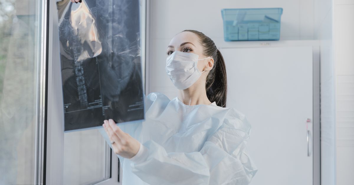
Radiographic imaging is fundamental. A panoramic radiograph provides a comprehensive view of the entire mandible. It allows the clinician to spot large areas of radiolucency, appearing as dark shadows at the roots of the teeth, which confirm the presence of large periapical abscesses and bone destruction.
In severe cases where the swelling is massive or involves the deep spaces of the neck, a Computed Tomography (CT) scan with contrast is ordered. The CT scan precisely maps the location of fluid collections, shows exactly which fascial spaces are involved, and most importantly, assesses the patency of the patient airway.
10. Antibiotic Therapy and Resistance
While surgical intervention is the definitive treatment, antibiotic therapy is a critical adjunct for managing severe facial swelling. The choice of antibiotic targets the complex, mixed flora of oral bacteria, which includes both aerobic and anaerobic species.
High-dose amoxicillin, often combined with clavulanic acid to combat resistant strains, or clindamycin for penicillin-allergic patients, are standard first-line therapies. In severe cases involving deep space infections or systemic toxicity, broad-spectrum intravenous antibiotics are administered in a hospital setting.
It is crucial to understand that antibiotics alone cannot cure a massive dental abscess. The blood supply to the necrotic center of the abscess is nonexistent, meaning systemic antibiotics cannot reach the core of the infection. They serve only to halt the spread of bacteria through the surrounding healthy tissue while awaiting surgical source control.
11. Surgical Incision and Drainage
The fundamental tenet of treating any severe abscess is prompt surgical incision and drainage. The clinician must physically open the abscess to relieve the extreme pressure, evacuate the purulent fluid, and alter the anaerobic environment that allows the bacteria to thrive.
If the swelling is localized within the mouth, a small incision is made through the gums directly over the swelling. For massive extraoral swellings, an external incision through the skin under the jawline may be required, performed under general anesthesia in an operating room.
Once the pus is evacuated, a surgical drain, such as a corrugated rubber strip, is often sutured into the incision. The drain keeps the wound open for several days, ensuring that any newly formed purulent fluid continues to flow out of the body rather than re-accumulating within the fascial spaces.
12. Endodontic Therapy vs Extraction
Removing the source of the infection is the final, non-negotiable step in resolving a severe dental abscess. The infected, necrotic tooth must be addressed, or the swelling will inevitably return. The decision lies between saving the tooth via endodontic therapy or removing it entirely through extraction.
Endodontic therapy, or a root canal, involves drilling into the tooth, physically removing all necrotic nerve tissue and bacteria from the root canals, sanitizing the interior, and sealing it. This is the preferred method if the tooth is structurally sound and restorable.
If the tooth is severely decayed, fractured below the gumline, or lacks adequate bone support due to periodontal disease, it must be extracted. Extraction provides the added benefit of creating a massive drainage pathway directly through the empty socket, which is highly beneficial in rapidly decompressing a severe jaw infection.
13. Post-Operative Management
Following surgical drainage and source control, the swelling in the lower jaw will gradually subside over a period of three to seven days. The patient must complete the entire course of prescribed antibiotics to eradicate any remaining bacteria lingering in the soft tissues.
Pain management is achieved with a combination of nonsteroidal anti-inflammatory drugs (NSAIDs) like ibuprofen to manage tissue inflammation, and acetaminophen for central pain control. Narcotic analgesics are occasionally prescribed for severe post-operative pain but are generally minimized.
Patients are instructed to perform warm saltwater rinses several times a day. The hypertonic saltwater draws out remaining fluid, keeps the intraoral incision clean, and promotes rapid healing of the oral mucosa. Close follow-up is mandatory to ensure the swelling is fully resolving and trismus is reversing.
14. Prevention of Odontogenic Infections
The catastrophic progression to a severe jaw swelling is almost entirely preventable through routine dental care. Odontogenic infections do not occur overnight; they are the end-stage result of months or years of untreated dental decay or periodontal disease.
Regular dental examinations and professional cleanings allow for the early detection and restoration of small cavities long before bacteria can penetrate the pulp chamber. Addressing mild tooth sensitivity promptly prevents the death of the nerve and the subsequent formation of a periapical abscess.
For patients with impacted wisdom teeth, proactive extraction before they become infected is a primary preventive strategy. Lower wisdom teeth are notoriously difficult to clean and frequently develop chronic infections that easily escape into the deep fascial spaces of the jaw.
15. Frequently Asked Questions (FAQ)
1. Can a swollen jaw from a tooth infection go away on its own?
No. A dental abscess lacks a blood supply at its core, meaning your immune system cannot reach or kill the bacteria. The swelling will only continue to spread and become more dangerous until a dentist physically removes the infection.
2. Is it safe to pop a dental abscess on my gum myself?
Absolutely not. Attempting to pop the swelling yourself introduces new bacteria and risks pushing the dangerous infection deeper into your bloodstream or fascial spaces, leading to life-threatening complications.
3. Why can I barely open my mouth with a jaw infection?
The infection and swelling spread into the powerful chewing muscles of your jaw. This irritation causes the muscles to lock into a continuous, painful spasm called trismus, preventing normal movement.
4. Can antibiotics cure a severely swollen jaw without pulling the tooth?
Antibiotics help stop the infection from spreading to your brain or throat, but they cannot cure the necrotic tooth. You must have a root canal or extraction to remove the source, or the swelling will return.
5. At what point should I go to the emergency room for a swollen jaw?
You should go to the nearest emergency room immediately if the swelling is rapidly expanding down your neck, if you have difficulty swallowing or breathing, if you develop a high fever, or if you cannot open your mouth.
6. Does a root canal hurt when you have a severe abscess?
Profound local anesthesia is more difficult to achieve in the presence of severe acidic infection. Dentists often prescribe antibiotics for a few days to reduce the acute inflammation before performing the root canal, ensuring a painless procedure.
16. Bibliography
- Read relevant clinical studies on PubMed
- Search for academic articles on Google Scholar
- Read relevant clinical studies on PubMed
- Search for academic articles on Google Scholar
Disclaimer: The content is for informational purposes only and does not replace medical advice. Always consult your doctor for personalized treatment.