1. Introduction
When your thumb joint locks into a bent position, it is almost exclusively the result of a condition known as trigger thumb, clinically termed stenosing tenosynovitis. This structural issue occurs when the flexor tendon, which pulls the thumb downward, develops a swollen nodule that becomes mechanically stuck beneath a tough, fibrous sheath at the base of the digit. As the tendon struggles to glide smoothly through this constricted tunnel, the thumb catches, clicks, and eventually locks, requiring forceful, painful effort—or the use of the other hand—to straighten it out.
The human hand is an intricate marvel of biomechanical engineering. The tendons operate like slick, lubricated cables passing through a series of rigid pulleys to ensure precise finger movement. When the delicate balance of this pulley system is disrupted by inflammation, repetitive strain, or underlying metabolic diseases, the resulting friction causes the tendon to swell. The thumb, bearing the brunt of daily grasping and pinching tasks, is incredibly susceptible to this specific mechanical failure.
Understanding the anatomy of the hand and the pathophysiology of tendon inflammation is essential to resolving this frustrating impairment. A locked thumb is a clear signal that the local tissue is trapped in a cycle of irritation and swelling. By diagnosing the stage of stenosing tenosynovitis and applying targeted orthopedic interventions, clinicians can reliably eliminate the mechanical block, calm the inflammation, and restore full, painless mobility to the thumb.
2. Anatomy of the Thumb Flexor Tendons
To comprehend why the thumb locks, one must visualize the mechanical anatomy of the digit. The movement of the thumb is controlled by muscles located deep within the forearm. These muscles connect to the bones of the thumb via long, string-like structures called flexor tendons. Specifically, the flexor pollicis longus tendon runs along the palm side of the thumb, attaching to the bone at the very tip.
When the forearm muscle contracts, it pulls the tendon, which flexes the thumb inward toward the palm. To prevent this long tendon from bowing away from the bone during flexion—much like a fishing line bowing away from a bent rod—the tendon is held tightly in place by a series of fibrous, tunnel-like bands called pulleys.
The tendon glides back and forth through these pulleys with every movement. To reduce friction, the tendon is encased in a delicate, fluid-filled tube called a synovial sheath. The health and smooth operation of the thumb depend entirely on this tendon gliding effortlessly through the rigid pulley system without any mechanical snagging.
3. Stenosing Tenosynovitis Explained
Trigger thumb is clinically known as stenosing tenosynovitis. “Stenosing” refers to the abnormal narrowing of a passageway, and “tenosynovitis” indicates inflammation of the tendon and its surrounding synovial sheath. The pathology begins with localized irritation at the base of the thumb, usually at the metacarpophalangeal joint, where the thumb meets the palm.
Continuous mechanical stress or systemic inflammation causes the synovial sheath to swell and thicken. As the sheath thickens, the space inside the tunnel narrows. The tendon passing through this narrowed space experiences severe friction. This constant rubbing causes the tendon itself to fray and develop a localized, swollen bump, or nodule.
Once this nodule forms, the mechanical disaster is set. Every time the individual bends their thumb, the swollen nodule is pulled down through the tight pulley. When they attempt to straighten the thumb, the nodule hits the edge of the pulley and becomes trapped. The tendon cannot glide back, leaving the thumb physically locked in a bent position.
4. The A1 Pulley Mechanism
The specific site of the blockage in trigger thumb is almost universally the A1 pulley. This is the first and thickest pulley in the flexor tendon sheath system, located right at the crease where the thumb joins the fleshy base of the palm.
The A1 pulley is subjected to the highest amount of mechanical stress during forceful gripping or pinching activities. It acts as the primary anchor point. When the flexor tendon develops a nodule, it is the rigid, unyielding edge of the A1 pulley that blocks its passage.
When a patient forces their thumb straight, the nodule is violently yanked through the narrow A1 pulley. This forceful passage creates the sudden, painful snap or popping sensation characteristic of the condition. This snapping further traumatizes both the tendon and the pulley, creating a vicious cycle of increasing inflammation and swelling.
5. Causes of Tendon Swelling
The exact cause of the initial tendon irritation is often multifactorial. Repetitive microtrauma is a leading culprit. Occupations or hobbies that require forceful, repetitive pinching, gripping, or wringing motions place continuous, heavy stress directly on the A1 pulley of the thumb.
Sustained pressure on the palm is another trigger. Using heavy power tools, shears, or even gripping a steering wheel tightly for long periods can physically crush the flexor tendon against the bony structures of the hand, initiating an inflammatory cascade.
In modern society, the ubiquitous use of smartphones has contributed to an increase in tendon irritation. Repeatedly scrolling and typing with the thumbs places the digit in an awkward, non-ergonomic position for hours a day, straining the flexor pollicis longus tendon and setting the stage for tenosynovitis.
6. Inflammatory and Metabolic Risk Factors
While mechanical overuse is a significant factor, trigger thumb frequently arises in the absence of obvious repetitive strain, pointing to underlying systemic health issues. Individuals with metabolic disorders, most notably diabetes mellitus, are at a substantially higher risk of developing stenosing tenosynovitis.
Chronic hyperglycemia alters the biochemical structure of collagen, the primary building block of tendons. The tendons of diabetic patients become stiffer, thicker, and more prone to degenerative swelling. Furthermore, diabetic patients frequently develop trigger digit in multiple fingers simultaneously, and the condition is notoriously more resistant to conservative treatment.
Systemic inflammatory joint diseases, such as rheumatoid arthritis or gout, also dramatically increase the risk. In these conditions, the immune system mistakenly attacks the synovial tissues lining the joints and tendon sheaths, causing profound, spontaneous swelling that traps the tendon without any mechanical trigger.
7. Pediatric Trigger Thumb
Trigger thumb is not exclusively an adult condition; it is frequently observed in infants and toddlers. Pediatric trigger thumb presents differently than the adult version. It is rarely associated with pain or a snapping sensation; instead, the parents simply notice that the child’s thumb is permanently stuck in a flexed position at the interphalangeal joint.
The cause of pediatric trigger thumb is believed to be congenital or developmental, resulting from a mismatch between the size of the flexor tendon and the diameter of the A1 pulley. A dense, painless nodule (known as Notta’s node) can usually be felt at the base of the infant’s thumb.
Unlike adults, children rarely resolve the condition with steroid injections. However, a significant percentage of pediatric cases resolve spontaneously as the child grows. If the thumb remains locked beyond a certain age, a minor surgical release of the pulley is performed to ensure normal development of the hand mechanics.
8. Differentiating Trigger Thumb from Osteoarthritis
Because the base of the thumb is a highly common site for osteoarthritis, clinicians must carefully distinguish between tendon entrapment and joint degeneration.
| Clinical Feature | Trigger Thumb (Tenosynovitis) | Thumb Osteoarthritis |
|---|---|---|
| Primary Symptom | Catching, popping, or physically locking | Deep, grinding ache with movement |
| Location of Discomfort | Palm side, at the base crease of the thumb | Base of the thumb near the wrist joint (CMC joint) |
| Morning Stiffness | Thumb is locked shut upon waking | Joint is stiff and achy, but moves freely |
| Physical Palpation | Palpable, tender lump that moves with the tendon | Hard, bony swelling around the joint margins |
While osteoarthritis involves the grinding away of the cartilage between the bones, trigger thumb is strictly a soft tissue failure occurring within the tendon tunnel.
9. Clinical Physical Examination
The diagnosis of trigger thumb is predominantly clinical, meaning it relies on a physical examination rather than extensive imaging. The physician will ask the patient to slowly open and close their hand, observing the mechanics of the thumb.
The clinician will palpate the palmar surface of the hand at the base of the thumb. They are feeling for a discrete, tender nodule. As the patient flexes and extends the thumb, the physician can feel this nodule move back and forth, abruptly catching or snapping as it attempts to slide under the A1 pulley.
The severity of the condition is graded based on the mechanical block. A mild case involves pain and clicking without locking. A moderate case involves locking that the patient can actively straighten with a painful snap. A severe case involves a thumb that is permanently locked and requires the patient to use their other hand to passively force the joint open.
10. The Role of Diagnostic Imaging
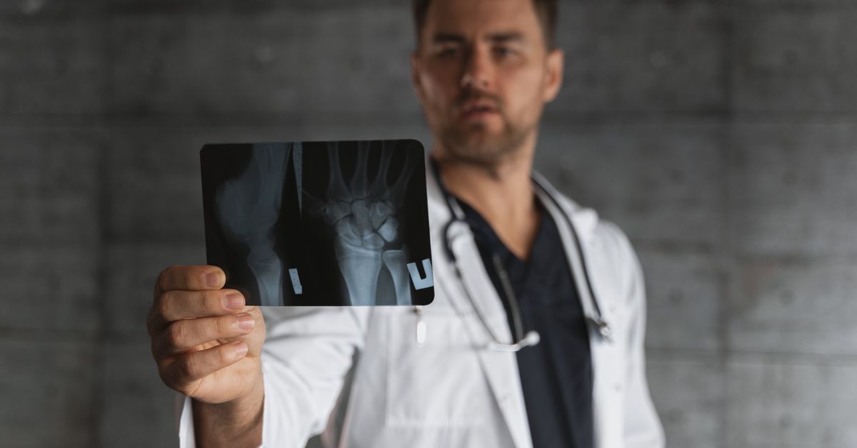
Standard X-rays are generally unhelpful for diagnosing trigger thumb, as tendons and pulleys are soft tissues that do not appear on radiographs. An X-ray is only ordered if the physician suspects concurrent osteoarthritis or a bone fracture causing the restricted movement.
If the diagnosis is unclear, a high-resolution musculoskeletal ultrasound is the imaging modality of choice. The ultrasound allows the clinician to view the tendon in real-time as the thumb moves.
The scan can precisely measure the thickened A1 pulley, visualize the swollen nodule on the flexor tendon, and detect the presence of excess fluid (tenosynovitis) within the tendon sheath, providing definitive objective confirmation of the mechanical entrapment.
11. Conservative Splinting and Rest
The initial treatment for a locking thumb focuses on reducing the mechanical friction to allow the inflammation to subside. Splinting is a highly effective conservative therapy. A rigid thumb spica splint is worn to immobilize the joint, completely preventing the thumb from bending.
By physically stopping the tendon from moving back and forth through the tight A1 pulley, the frictional trauma is halted. The swollen nodule is given a chance to shrink back to its normal size.
Patients are typically advised to wear the splint continuously for several weeks, or at the very least during sleep. Many patients find their thumb is completely locked when they wake up because the fingers naturally curl inward during sleep; wearing a splint at night keeps the tendon gliding path straight and prevents this painful morning lock.
12. Corticosteroid Injections
If rest and splinting fail to resolve the locking, a local corticosteroid injection is the next line of defense and is considered the gold standard for non-surgical treatment. A physician injects a concentrated dose of liquid steroid, often mixed with a local anesthetic, directly into the tendon sheath at the base of the thumb.
The corticosteroid acts as a potent, localized anti-inflammatory agent. It rapidly shrinks the swollen synovial tissue and reduces the size of the tendon nodule. As the swelling subsides, the tendon can once again glide smoothly through the A1 pulley without catching.
Steroid injections are highly effective, curing the condition permanently in over sixty percent of patients. However, the success rate is notably lower in patients with diabetes, and repeated injections are generally avoided due to the risk of the steroid weakening or rupturing the tendon over time.
13. Surgical Release Procedures
For severe cases where the thumb is permanently locked, or when conservative measures and injections have failed, surgical intervention provides a definitive cure. The procedure, known as a trigger thumb release, is a brief, outpatient surgery performed under local anesthesia.
The surgeon makes a small incision in the palm at the base of the thumb. They locate the tight A1 pulley and simply cut it open longitudinally. Cutting the pulley releases the roof of the narrow tunnel, providing the swollen tendon with ample space to glide freely.
Because the other pulleys in the thumb remain intact, cutting the A1 pulley does not negatively impact the mechanical strength or function of the thumb. The relief is immediate; the patient can usually flex and extend the thumb without catching while still on the operating table.
14. Post-Surgical Recovery and Rehabilitation
Recovery from a trigger thumb release is generally rapid. Patients are encouraged to begin moving their thumb gently on the same day as the surgery to prevent stiff scar tissue from forming around the healing tendon.
Some localized soreness and swelling in the palm are expected for a few weeks. The patient must keep the incision clean and dry until the sutures are removed. Full recovery and return to heavy lifting or strenuous manual labor usually take four to six weeks.
Hand therapy is rarely required after a simple release, but a physical therapist may provide targeted stretching exercises to ensure full range of motion is restored and to address any lingering stiffness in the interphalangeal joint that developed while the thumb was chronically locked.
15. When to Seek Medical Care
While trigger thumb is not a life-threatening condition, delaying treatment can result in permanent joint stiffness and a prolonged loss of hand function.
Seek a medical evaluation if your thumb clicks, catches, or is painful to straighten. It is critical to consult an orthopedic specialist or a hand surgeon if the thumb becomes completely locked in a bent position and cannot be straightened, or if attempting to straighten it causes excruciating pain.
Furthermore, if the catching sensation is accompanied by a high fever, or if the thumb feels remarkably hot to the touch and appears bright red, seek emergency medical care immediately to rule out a severe bacterial infection within the tendon sheath.
16. Frequently Asked Questions FAQ
1. Why does my thumb get stuck when I bend it?
Your thumb bends using a tendon that slides through a tight tunnel called a pulley. If the tendon becomes swollen from repetitive use or inflammation, it forms a small bump. When you bend your thumb, this bump gets yanked through the pulley, but it gets physically stuck when you try to straighten it, locking the thumb.
2. Will a trigger thumb heal on its own without a doctor?
Mild cases of clicking or catching can sometimes heal if you rest the thumb completely and avoid gripping activities. However, if the thumb is physically locking and requires you to pop it open with your other hand, it will rarely heal without medical intervention, such as a splint or a steroid injection.
3. Is it bad to keep popping my trigger thumb?
Yes, it is very bad for the tissue. Every time you force the thumb to pop past the mechanical block, you are causing severe friction and trauma to the tendon. This makes the swelling worse, increases the inflammation, and guarantees the condition will progress.
4. How do doctors fix a thumb that is permanently locked?
If steroid injections do not work, a hand surgeon will perform a quick, ten-minute procedure under local anesthesia. They make a tiny cut at the base of your thumb and snip the tight pulley band. This opens the tunnel, instantly allowing the tendon to glide freely forever.
5. Does having diabetes make trigger thumb worse?
Yes. High blood sugar alters the structure of your tendons, making them stiffer and more prone to swelling. Diabetic patients are much more likely to develop trigger thumb, often in multiple fingers, and the condition is usually more resistant to steroid injections, frequently requiring a surgical release.
17. Bibliography
Disclaimer: The content is for informational purposes only and does not replace medical advice. Always consult your doctor for personalized treatment.