1. Introduction to Thumb Pain
Pain in the thumb is a significant clinical issue that frequently arises from repetitive mechanical strain, inflammatory tendon conditions, or degenerative joint disease. Because the thumb is involved in nearly every prehensile task performed by the hand, any dysfunction directly impacts an individual’s ability to grip, pinch, and manipulate objects. The initial therapeutic approach involves identifying the precise anatomical structure causing the discomfort to ensure targeted treatment.
The thumb possesses a unique biomechanical structure that grants it exceptional mobility compared to the other digits. This extensive range of motion is what enables opposability, a fundamental trait for complex hand function. However, the reliance on the thumb for forceful gripping places immense compressive loads on its basal joint and surrounding soft tissues. Over time, these repetitive forces can lead to structural deterioration and chronic pain.
A thorough medical evaluation is required to differentiate between acute traumatic ligament injuries, nerve compression syndromes, and progressive arthritic changes. Early intervention utilizing splinting, activity modification, and pharmacological management can prevent further joint degradation and preserve optimal hand function.
2. Anatomy of the Thumb and Carpometacarpal Joint
The skeletal structure of the thumb consists of two phalangeal bones and one metacarpal bone. Unlike the other fingers which possess three phalanges, the thumb relies on a complex articulation at its base to achieve multi-directional movement. This critical joint is the carpometacarpal joint, where the first metacarpal bone meets the trapezium bone of the wrist.
The carpometacarpal joint is designed as a saddle joint. Its unique shape allows for flexion, extension, abduction, adduction, and opposition. A robust network of ligaments surrounds the joint capsule, providing vital stability during forceful pinch and grip activities. The ulnar collateral ligament at the metacarpophalangeal joint is particularly important for lateral stability.
Movement is powered by both extrinsic muscles originating in the forearm and intrinsic muscles located within the fleshy base of the thumb known as the thenar eminence. The tendons of these muscles glide through protective sheaths lubricated by synovial fluid. The median and radial nerves provide the necessary sensory and motor innervation ensuring precise control and tactile feedback.
3. Overuse Syndromes and Repetitive Strain
Repetitive strain injuries are a leading cause of thumb pain in the modern workforce. Continuous performance of specific manual tasks such as typing, texting on mobile devices, or operating hand tools places persistent mechanical stress on the musculotendinous units. The tendons become fatigued and develop microscopic tears leading to localized inflammation.
The clinical presentation of overuse injury involves a gradual onset of aching pain that exacerbates with specific repetitive activities and subsides with rest. Patients often describe a sense of fatigue or clumsiness in the hand. Swelling is typically minimal but the soft tissues at the base of the thumb may feel tender upon direct palpation.
Management of repetitive strain relies heavily on activity modification. Taking frequent breaks to rest the hands and utilizing ergonomic tools can significantly reduce the mechanical burden on the thumb. A temporary period of splinting may be recommended to enforce rest and allow the inflamed tissues to heal.
4. De Quervain Tenosynovitis
De Quervain tenosynovitis is a painful inflammatory condition affecting the tendons on the thumb side of the wrist. Specifically, it involves the tendons of the extensor pollicis brevis and abductor pollicis longus muscles. As these tendons travel from the forearm into the thumb they pass through a narrow fibrous tunnel called the first dorsal compartment.
When subjected to chronic friction from repetitive lifting or grasping the synovial sheath surrounding the tendons becomes thickened and inflamed. This swelling restricts the smooth gliding motion of the tendons within the narrow tunnel. Patients experience sharp pain at the base of the thumb and the radial side of the wrist which worsens significantly when forming a fist or grasping objects.
The classic clinical sign of De Quervain tenosynovitis is a positive Finkelstein test. This provocative maneuver involves the patient flexing their thumb into the palm closing the fingers over it and then deviating the wrist toward the ulnar side. A sharp exacerbation of pain confirms the diagnosis and directs therapy toward reducing compartment inflammation.
5. Trigger Thumb and Tendon Pathology
Trigger thumb is a form of stenosing tenosynovitis that affects the flexor pollicis longus tendon. This tendon is responsible for bending the tip of the thumb. It passes through a series of fibrous pulleys that keep the tendon closely applied to the bones. The first annular pulley located at the base of the thumb is the most common site of pathology.
Due to repeated mechanical friction a nodule may form on the tendon or the pulley itself may become thickened. As the thumb flexes and extends the swollen tendon forcefully snaps or catches as it pulls through the narrowed pulley. This mechanical catching produces a painful clicking sensation and in severe cases the thumb may become transiently locked in a flexed position.
Patients frequently report joint stiffness in the morning with the catching sensation becoming less pronounced as the day progresses. Initial treatment involves wearing a splint to restrict interphalangeal joint movement and administering localized corticosteroid injections to reduce the nodular swelling.
6. Basal Joint Arthritis and Degenerative Disease
Basal joint arthritis, affecting the carpometacarpal joint, is one of the most common forms of osteoarthritis in the upper extremity. The repetitive compressive forces generated during normal daily activities slowly erode the smooth articular cartilage covering the ends of the trapezium and the first metacarpal bone.
As the cartilage wears away bone rubs against bone triggering an inflammatory response and stimulating the formation of bony outgrowths called osteophytes. Patients with basal joint arthritis report a deep aching pain at the base of the thumb that intensifies with pinching activities such as turning a key or opening a jar.
Over time the progressive joint destruction leads to visible deformity. The base of the metacarpal may subluxate or shift out of alignment creating a prominent bony bump at the wrist. The resultant loss of mechanical advantage leads to profound weakness in grip strength. Treatment strategies focus on joint support through custom orthotics and pain control via nonsteroidal anti-inflammatory medications.
7. Acute Traumatic Injuries and Sprains
Acute trauma to the thumb frequently occurs during sports, falls, or workplace accidents. The forces applied during these events can overwhelm the tensile strength of the supportive ligaments leading to a sprain or complete rupture. Rapid forced hyperextension or lateral deviation of the digit are common mechanisms of injury.
A sprain presents with immediate sharp pain, rapid swelling, and localized ecchymosis. The patient may feel a popping sensation at the moment of injury. The thumb becomes painful to move and the surrounding soft tissues are markedly tender to touch. Clinical evaluation is necessary to grade the severity of the sprain and determine joint stability.
Initial management follows the standard protocols for acute soft tissue injury including rest, application of ice, compression, and elevation. Depending on the specific ligament involved and the degree of instability conservative management with strict immobilization may be sufficient to allow fibrous healing.
8. Skier Thumb and Ligament Tears
Skier thumb represents a specific and clinically significant injury to the ulnar collateral ligament of the metacarpophalangeal joint. The injury is traditionally associated with snow skiing when a skier falls while holding a ski pole forcibly bending the thumb away from the hand. However it is also common in other contact sports.
A complete rupture of the ulnar collateral ligament severely compromises the stability of the thumb pinch mechanism. Without this crucial ligament the proximal phalanx shifts away from the metacarpal during any attempt to grasp an object resulting in a weak and painful pinch.
If the torn ligament retracts and becomes trapped beneath the adjacent adductor aponeurosis a Stener lesion is formed. This anatomical displacement prevents the ligament ends from healing naturally. In such cases surgical repair or reconstruction of the ligament is essential to restore joint stability and prevent the early onset of post-traumatic arthritis.
9. Carpal Tunnel Syndrome and Nerve Compression
While carpal tunnel syndrome is primarily known for causing numbness and tingling in the hand it can also produce significant thumb pain. The median nerve travels through the narrow carpal tunnel at the wrist and provides sensory innervation to the thumb, index, and middle fingers. Compression of this nerve leads to neuropathic symptoms.
The pain associated with median nerve compression is often described as burning or electrical in nature. It frequently awakens the patient at night and is exacerbated by activities requiring prolonged wrist flexion such as driving or holding a phone. Over time severe compression can lead to motor weakness and atrophy of the thenar muscles at the base of the thumb.
Although the primary pathology is located at the wrist the resulting symptoms heavily impact the thumb. Diagnostic nerve conduction studies are utilized to quantify the degree of nerve impairment. Utilizing nocturnal wrist splints to maintain a neutral position relieves the pressure on the nerve and is a highly effective first-line treatment.
10. Identifying the Characteristics of the Pain
The specific presentation and location of the pain are essential diagnostic clues. A careful clinical history helps narrow down the vast differential diagnosis and directs subsequent examination and testing.
| Pain Characteristic | Common Associated Condition |
|---|---|
| Catching or clicking during bending | Trigger Thumb (Stenosing tenosynovitis) |
| Sharp pain on the wrist side during lifting | De Quervain Tenosynovitis |
| Deep ache aggravated by pinch grip | Carpometacarpal (Basal) Joint Arthritis |
| Numbness, tingling, or electrical pain | Median Nerve Compression (Carpal Tunnel) |
Noting the onset of symptoms whether sudden following trauma or insidious over several months further clarifies whether the etiology is acute or degenerative in nature.
11. Clinical Examination and Imaging Modalities
A comprehensive clinical examination of the painful thumb begins with a visual inspection for swelling, deformity, or muscle atrophy. The clinician palpates specific anatomical landmarks to identify the exact point of maximal tenderness. Range of motion is assessed both actively and passively to detect stiffness or mechanical catching.
Provocative maneuvers are a critical component of the examination. The grind test involves applying axial pressure and rotating the metacarpal bone to assess for basal joint arthritis. Ligamentous stability is tested by applying varus and valgus stress to the joints.
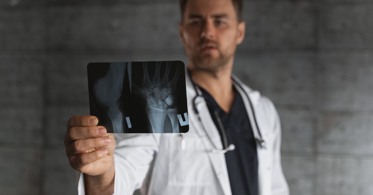
When structural bony damage or severe arthritis is suspected standard radiographs are the imaging modality of choice. X-rays reveal the extent of joint space narrowing, the presence of osteophytes, and can identify occult fractures. Magnetic resonance imaging or diagnostic ultrasound may be employed if complex soft tissue pathology or ligamentous tears require detailed visualization.
12. Non-Surgical Interventions and Splinting
Conservative management is the cornerstone of treating the vast majority of thumb pain etiologies. The immediate goal is to reduce mechanical stress and control localized inflammation. Immobilization utilizing specialized splints or braces is highly effective. A thumb spica splint stabilizes the basal and metacarpophalangeal joints allowing irritated tendons and capsules to rest.
Pharmacological management includes the use of oral nonsteroidal anti-inflammatory drugs or topical analgesic gels. For conditions that do not respond to oral medications such as severe De Quervain tenosynovitis or basal joint arthritis localized corticosteroid injections offer profound relief. These injections deliver potent anti-inflammatory medication directly to the site of pathology.
Hand therapy plays a vital role in the rehabilitation process. A certified hand therapist can instruct the patient in targeted stretching exercises to maintain tissue flexibility and strengthening protocols to enhance the dynamic stability provided by the surrounding musculature.
13. Surgical Options and Rehabilitation
Surgical intervention is considered when conservative measures fail to provide adequate pain relief or when structural instability profoundly limits hand function. The specific surgical procedure depends entirely on the underlying diagnosis. For trigger thumb a simple release of the restrictive first annular pulley restores smooth tendon gliding.
In cases of advanced basal joint arthritis surgical options include trapeziectomy where the arthritic trapezium bone is completely removed. This procedure may be combined with a ligament reconstruction and tendon interposition to stabilize the resulting space and prevent metacarpal subsidence.
Post-operative rehabilitation is a rigorous and necessary phase of recovery. Following a period of surgical immobilization graduated hand therapy is essential to prevent scar tissue contracture, restore functional range of motion, and rebuild grip strength. Complete recovery may take several months of dedicated therapeutic work.
14. When to Seek Medical Assessment
Prompt medical evaluation is crucial following any acute injury that results in visible deformity, rapid swelling, or an inability to move the thumb. These signs strongly suggest a fracture or complete ligamentous rupture that requires immediate stabilization to ensure optimal healing.
If conservative home measures such as rest and over-the-counter pain medications do not yield improvement within two weeks a clinical consultation is advised. Persistent pain that interferes with the ability to perform basic daily activities or work tasks requires professional diagnostic evaluation.
Additionally the development of neurological symptoms including progressive numbness, tingling, or noticeable weakness in the hand must not be ignored. Prolonged nerve compression can lead to irreversible nerve damage and permanent loss of motor function if left untreated.
15. Frequently Asked Questions (FAQ)
1. Can texting cause permanent damage to my thumb?
Frequent texting causes repetitive strain and inflammation in the tendons leading to pain. While rarely causing permanent damage if caught early chronic unchecked inflammation can lead to persistent tendinopathy.
2. How do I know if my thumb pain is from arthritis or a tendon issue?
Arthritis typically causes a deep aching pain localized to the base of the thumb that worsens with pinching. Tendon issues like De Quervain tenosynovitis cause sharp pain along the side of the wrist that worsens with movement.
3. Will a splint weaken my thumb muscles?
Prolonged and unnecessary splinting can lead to mild muscle atrophy. However when used appropriately under medical guidance for acute inflammation the benefits of tissue rest far outweigh the risks of temporary weakness.
4. Are corticosteroid injections safe for the thumb?
Yes, localized corticosteroid injections are safe and highly effective for treating severe inflammation. However repeated injections into the same joint or tendon sheath are limited to prevent tissue degradation.
5. Can carpal tunnel syndrome only affect the thumb?
While carpal tunnel syndrome typically affects the thumb, index, and middle fingers it is possible for the initial neuropathic symptoms to be felt predominantly in the thumb before progressing to the other digits.
Bibliography
Disclaimer: The content is for informational purposes only and does not replace medical advice. Always consult your doctor for personalized treatment.