
1. Introduction to Pelvic and Upper Leg Joint Pain
Pain localized to the joints of the pelvis and the upper leg represents a complex clinical challenge that can significantly diminish an individual’s mobility and quality of life. The pelvic region serves as the critical mechanical junction between the axial skeleton of the spine and the appendicular skeleton of the lower extremities. Because this anatomical area is responsible for transferring the entire weight of the upper body to the legs during movement, the joints and surrounding soft tissues are subjected to continuous, profound mechanical stress.
The diagnostic evaluation of pelvic and upper leg pain requires a systematic approach to differentiate between true articular pathology, surrounding soft tissue inflammation, and pain referred from the lower back. Patients typically present with localized aching, a deep sensation of stiffness, or sharp discomfort that correlates directly with specific weight-bearing activities. Establishing the precise anatomical origin of these symptoms is the essential first step in formulating an effective therapeutic intervention.
Comprehensive medical management relies on an accurate anatomical diagnosis. The therapeutic approach ranges from conservative rehabilitation aimed at restoring muscular balance and core stability to precise surgical interventions designed to repair structural damage. Early clinical assessment prevents acute inflammatory conditions from evolving into chronic, debilitating mechanical failure of the pelvic complex.
2. Anatomy of the Pelvis and Hip Region
The structural architecture of the pelvis consists of three primary bones—the ilium, ischium, and pubis—which fuse together to form a robust bony ring. This ring articulates with the sacrum at the base of the spine via the sacroiliac joints. The sacroiliac joints are robust, relatively immobile articulations secured by a dense network of powerful ligaments, designed to absorb and transfer mechanical forces during walking and running.
At the anterior aspect of the pelvis, the two pubic bones meet at the pubic symphysis, a specialized cartilaginous joint that provides subtle flexibility to the pelvic ring. On the lateral aspect of the pelvis lies the acetabulum, a deep socket that accepts the spherical head of the femur to form the hip joint. The hip joint is a profoundly stable ball-and-socket articulation enveloped by a thick fibrous capsule and supported by powerful muscular groups.
The musculature surrounding the pelvis and upper leg includes the gluteal muscles, the hip flexors, the adductors of the inner thigh, and the hamstrings. A rich network of sensory nerves, including the sciatic and femoral nerves, courses through this dense anatomical region. Any disruption in this delicate biomechanical balance alters the normal distribution of physical forces, leading to concentrated mechanical stress and subsequent joint pain.
3. Sacroiliac Joint Dysfunction
The sacroiliac joints firmly connect the sacrum to the iliac bones of the pelvis. While these joints possess very little inherent motion, they are subjected to substantial mechanical transfer forces. Dysfunction or active inflammation of these joints, clinically termed sacroiliitis, is a frequent cause of posterior pelvic and upper leg pain.
Patients with sacroiliac joint dysfunction typically describe a deep, dull, aching pain localized directly over the dimples of the lower back. This pain often refers downward into the upper buttock and occasionally into the posterior thigh, rarely extending below the knee. The discomfort is characteristically worsened by transitioning from a seated to a standing position, standing on one leg, or climbing stairs.
Sacroiliac joint pain is notably common during pregnancy due to the hormone relaxin, which increases ligamentous laxity and joint mobility to prepare the pelvis for childbirth. Management involves the use of specialized pelvic stabilization belts, targeted core strengthening exercises, and localized corticosteroid injections directly into the joint space under fluoroscopic guidance for chronic, severe cases.
4. Osteoarthritis of the Hip
Osteoarthritis is a prevalent cause of chronic, intra-articular pain in the upper leg and pelvis, particularly in older adults. It is a progressive, degenerative disease characterized by the gradual deterioration and ultimate loss of the hyaline articular cartilage covering the femoral head and the acetabulum. As this protective layer wears away, the subchondral bone is exposed, leading to increased mechanical friction and localized inflammation.
Patients suffering from hip osteoarthritis typically report a deep, aching pain located primarily in the groin or deep within the anterior upper leg. The pain is exacerbated by weight-bearing activities and initially improves with rest. As the disease advances, patients experience profound morning stiffness and a progressive, structural loss of internal rotation and extension.
To compensate for the joint instability, the body forms osteophytes along the joint margins. This progressive structural alteration inevitably leads to a pronounced antalgic gait, recognizable as a distinct limp. Management strategies focus on weight reduction, physical therapy to strengthen the surrounding muscles, and eventually total hip arthroplasty for definitive pain relief.
5. Pubic Symphysis Dysfunction
Pubic symphysis dysfunction involves inflammation or excessive mobility at the cartilaginous joint located at the front of the pelvis. This condition is most frequently observed in pregnant women and athletes involved in sports that require repetitive kicking or rapid changes of direction. The abnormal mechanical stress leads to chronic inflammation of the joint and the surrounding ligamentous attachments.
Patients experience a sharp, localized pain directly over the pubic bone in the lower central pelvis. The discomfort often radiates into the inner thighs or the groin region. Activities that involve unilateral weight-bearing, such as walking, climbing stairs, or getting in and out of a car, reliably provoke severe pain.
Clinical evaluation involves specific provocative maneuvers, including direct palpation of the pubic symphysis, which typically elicits pinpoint tenderness. Conservative management relies on utilizing a supportive pelvic belt, modifying activities to avoid unilateral leg loading, and performing targeted physical therapy to stabilize the pelvic floor and core musculature.
6. Greater Trochanteric Pain Syndrome
Pain localized to the lateral aspect of the upper leg and pelvis is most commonly attributed to greater trochanteric pain syndrome. This umbrella term encompasses trochanteric bursitis and tendinopathy of the gluteus medius or minimus muscles. The greater trochanter is the large bony prominence on the side of the upper femur, serving as the attachment site for the powerful gluteal tendons.
Repetitive mechanical friction, particularly from the overlying iliotibial band during walking or running, causes the underlying bursa or the gluteal tendons to become inflamed and degenerative. Patients present with localized, aching pain over the outside of the hip, which frequently radiates down the lateral thigh.
A hallmark clinical symptom is severe pain when lying directly on the affected side, an issue that often disrupts sleep. The pain is also worsened by prolonged standing or rising from a low chair. Treatment involves targeted physical therapy focusing on gluteal strengthening, core stabilization, and localized corticosteroid injections to rapidly reduce local tissue inflammation.
7. Femoroacetabular Impingement
Femoroacetabular impingement is a structural biomechanical disorder where abnormal bony morphology causes the femoral head and the acetabular rim to crash against each other during normal range of motion. This abnormal contact creates intense shear forces that rapidly damage the acetabular labrum and the adjacent articular cartilage of the hip joint.
There are two primary types of structural deformity. A cam impingement is characterized by an abnormally thickened femoral neck, while a pincer impingement occurs when the acetabulum creates an overly deep overhang. Many patients possess a mixed presentation of both bony deformities.
Patients, often young and active adults, experience a deep, aching groin pain that dramatically worsens during activities requiring deep hip flexion and internal rotation, such as squatting or sitting in low chairs. Early recognition and surgical correction via hip arthroscopy can effectively relieve pain and halt the progression of joint destruction.
8. Acetabular Labral Tears
The acetabular labrum is a robust fibrocartilaginous ring that lines the rim of the hip socket. It functions to deepen the joint, act as a shock absorber, and create a fluid seal that maintains joint lubrication. The labrum is susceptible to tearing from repetitive microtrauma, structural impingement, or acute twisting injuries.
A torn labrum disrupts the crucial fluid seal of the joint, leading to mechanical instability and accelerated articular cartilage wear. Patients describe a deep, sharp, or catching pain located in the anterior groin. The pain is frequently accompanied by mechanical symptoms such as clicking, locking, or a distinct feeling that the joint is giving way during pivoting movements.
Diagnosis relies on advanced imaging, specifically a magnetic resonance arthrogram, which uses intra-articular contrast dye to delineate the labral tissue. While some tears respond to conservative management, symptomatic tears in active patients often require arthroscopic surgical repair to restore the anatomical seal and prevent premature osteoarthritis.
9. Ankylosing Spondylitis and Inflammatory Arthritis
Ankylosing spondylitis is a systemic, autoimmune inflammatory disorder that predominantly affects the axial skeleton, frequently beginning in the sacroiliac joints of the pelvis. The hyperactive immune system causes profound inflammation of the joint capsules and ligamentous insertions, leading to significant pelvic and lower back pain.
Unlike mechanical joint pain, the discomfort associated with ankylosing spondylitis is typically worse during periods of rest and is particularly severe upon waking in the morning. The pain and profound joint stiffness gradually improve with physical activity and exercise. Over time, the chronic inflammation can lead to the pathological fusion of the sacroiliac joints and the vertebrae.
Accurate diagnosis requires comprehensive serological testing, specifically identifying the human leukocyte antigen B27 marker, combined with characteristic radiographic findings. Management mandates the rapid initiation of disease-modifying antirheumatic drugs and biologic therapies to suppress the systemic immune response and prevent irreversible structural fusion.
10. Referred Pain from the Lumbar Spine
Pain perceived in the posterior pelvis and upper leg frequently originates not from the pelvic joints themselves, but from pathology within the lumbar spine. A herniated intervertebral disc or spinal stenosis can compress the nerve roots that converge to form the sciatic nerve, referring pain directly through the pelvic region.
This condition, known as lumbar radiculopathy or sciatica, produces a distinct neuropathic pain that originates in the lower back and shoots directly through the deep gluteal region. The pain frequently radiates down the posterior thigh and into the lower leg. It is described as burning, electrical, or a sharp shooting sensation.
Clinical differentiation involves a thorough neurological examination. Specific maneuvers, such as the straight leg raise test, stretch the sciatic nerve and reliably reproduce the radiating pain, confirming a spinal origin. Treatment directs focus toward decompressing the affected lumbar nerve roots through physical therapy or targeted epidural injections.
11. Pelvic Ring Fractures and Acute Trauma
Acute high-energy physical trauma, such as a motor vehicle accident or a fall from a significant height, can cause severe fractures to the bones composing the pelvic ring. Because the pelvis is a robust, ring-like structure, a fracture in one area is frequently accompanied by a secondary fracture or ligamentous disruption in another area of the ring.
A pelvic fracture presents with immediate, excruciating pain, profound and rapid soft tissue swelling, extensive bruising, and a complete inability to bear any body weight. The patient may exhibit signs of mechanical instability in the lower extremities. Due to the rich vascular network surrounding the pelvis, these fractures carry a significant risk of severe internal hemorrhage.
Immediate radiographic evaluation is strictly mandatory following acute pelvic trauma. Uncomplicated, stable fractures may be managed conservatively with strict bed rest and gradual mobilization. However, displaced or mechanically unstable fractures require urgent orthopedic surgical intervention for precise internal fixation to restore perfect anatomical alignment and stability.
12. Differentiating Clinical Symptoms
A meticulous clinical analysis of the specific location and mechanical triggers of the pain directs the clinician toward an accurate anatomical diagnosis.
| Pain Location and Characteristics | Common Associated Diagnosis |
|---|---|
| Deep anterior groin pain, worse with weight-bearing | Hip Osteoarthritis or Labral Tear |
| Pain over the lower back dimples, radiating to the upper buttock | Sacroiliac Joint Dysfunction |
| Sharp pain directly over the lower central pelvic bone | Pubic Symphysis Dysfunction |
| Pain on the outer hip, severe when lying on the affected side | Greater Trochanteric Pain Syndrome |
The presence of systemic symptoms, sudden profound structural deformity, or a complete inability to bear weight warrants immediate advanced clinical evaluation to prevent permanent tissue damage or extensive joint destruction.
13. Diagnostic Imaging Modalities
The clinical examination involves precise palpation of bony landmarks, assessing the integrity of specific joints through manual stress testing, and observing the patient’s dynamic gait pattern. Standard weight-bearing plain radiographs are the universally accepted first line of imaging, rapidly identifying acute fractures, structural deformities, and the joint space narrowing indicative of osteoarthritis.
When occult stress fractures, subtle bone marrow edema, or significant soft tissue pathology are suspected, magnetic resonance imaging provides unparalleled anatomical resolution. This imaging modality is incredibly sensitive for identifying labral tears, severe tendon degeneration, and the active inflammatory changes of sacroiliitis.
Diagnostic ultrasound is increasingly utilized for the dynamic, real-time assessment of soft tissues. It is also an excellent clinical tool for accurately guiding therapeutic corticosteroid injections directly into inflamed tendon sheaths, the greater trochanteric bursa, or the sacroiliac joint, ensuring maximal medication delivery with minimal procedural discomfort.
14. Physical Therapy and Biomechanical Correction
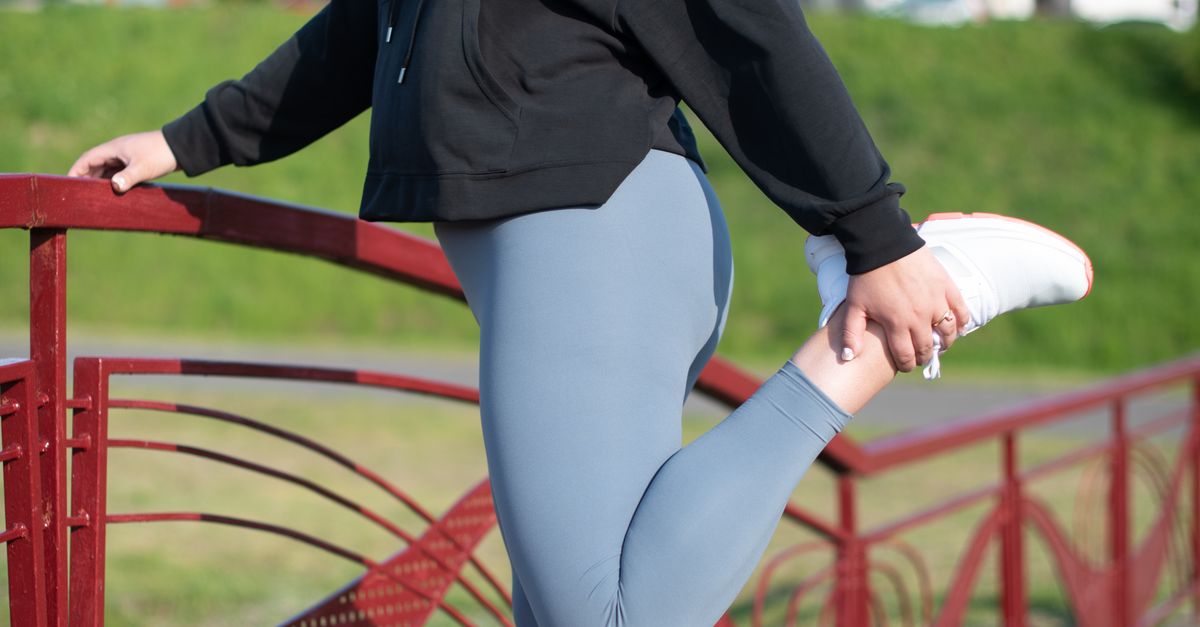
The initial management of joint pain in the pelvis and upper leg relies heavily on conservative, non-pharmacological interventions. The absolute cornerstone of long-term management is comprehensive physical therapy. A structured, progressive exercise program prevents severe muscular deconditioning and enhances dynamic pelvic stability.
Therapy focuses on restoring proper biomechanical alignment by stretching tight hip flexors and dynamically strengthening the core stabilizers, the pelvic floor, and the gluteal musculature. This specific muscular re-education effectively unloads the painful articular surfaces and reduces the non-physiological stress placed upon the healing soft tissues.
Biomechanical correction through the use of specialized supportive footwear or custom orthotics can also address underlying leg length discrepancies or abnormal foot pronation, which frequently contribute to ascending mechanical stress across the pelvic joints.
15. Pharmacological and Surgical Interventions
Pharmacological intervention utilizes oral nonsteroidal anti-inflammatory drugs to mitigate acute mechanical pain and reduce soft tissue edema. For chronic, severe intra-articular pain due to osteoarthritis or specific inflammatory conditions, targeted corticosteroid injections provide potent, localized suppression of the inflammatory response, significantly improving joint mobility.
When conservative measures fail to provide adequate pain relief, or in the presence of severe, progressive structural deformity that impairs the ability to walk, surgical intervention is indicated. Hip arthroscopy is utilized to repair labral tears and correct femoroacetabular impingement through minimally invasive techniques.
For severe, end-stage osteoarthritis or extensive avascular necrosis of the femoral head, total hip arthroplasty provides comprehensive joint replacement. The damaged articular surfaces are entirely resected and replaced with durable prosthetic components, offering profound pain relief and the restoration of functional independence.
16. Frequently Asked Questions FAQ
1. Can sitting too much cause pelvic joint pain?
Yes. Prolonged sitting tightens the hip flexor muscles and weakens the gluteal muscles. This muscular imbalance alters the biomechanics of the pelvis, placing abnormal stress on the sacroiliac joints and the lower back, frequently leading to chronic aching pain.
2. How do I know if my upper leg pain is a muscle strain or a joint issue?
A muscle strain typically occurs suddenly during physical exertion and is tender when you directly press the muscle belly. Joint pain is usually felt deeper, often in the groin or deep buttock, and typically restricts the actual range of motion of the leg.
3. Is it safe to exercise with sacroiliac joint pain?
Gentle, targeted exercise is essential for recovery. Low-impact activities such as swimming or specific core stabilization exercises help support the joint. However, activities involving heavy impact or asymmetrical leg loading, such as running or deep lunges, should be avoided until the inflammation resolves.
4. Why does my pelvis hurt during pregnancy?
During pregnancy, the body produces a hormone called relaxin, which softens the ligaments to allow the pelvis to expand for childbirth. This increased ligamentous laxity often leads to mechanical instability and severe inflammation in the sacroiliac joints or the pubic symphysis.
5. What is the recovery time for a hip replacement?
Modern total hip replacements have excellent recovery profiles. Most patients can bear weight on the new joint immediately after surgery. Substantial pain relief and functional mobility are typically achieved within four to six weeks, with complete physical rehabilitation taking a few months.
17. Bibliography
Disclaimer: The content is for informational purposes only and does not replace medical advice. Always consult your doctor for personalized treatment.
