
1. Introduction
Acute cholecystitis due to a gallbladder calculus without obstruction of the common bile duct is a severe inflammatory condition of the gallbladder wall. This condition arises when a gallstone becomes physically impacted in the neck of the gallbladder or the cystic duct. The critical clinical distinction in this diagnosis is that while the outflow of the gallbladder itself is blocked, the main common bile duct remains entirely clear. Therefore, the liver can continue to drain bile into the intestines normally, preventing the onset of systemic jaundice.
Despite the preservation of common bile duct flow, an impacted stone in the cystic duct initiates a cascade of rapid and destructive events within the localized tissues of the gallbladder. The resulting inflammation, swelling, and potential bacterial overgrowth demand immediate medical evaluation. Managing acute calculous cholecystitis requires swift diagnostic imaging to confirm the stone’s precise location, aggressive supportive medical therapy, and prompt surgical intervention to remove the diseased organ and prevent life-threatening complications such as gallbladder perforation or gangrene.
2. Anatomy of the Cystic Duct
The gallbladder is a muscular sac that stores concentrated bile. It connects to the main biliary system via a narrow, tortuous tube called the cystic duct. The interior lining of the cystic duct features the spiral valves of Heister, complex mucosal folds that help regulate the flow of bile in and out of the gallbladder.
Because the cystic duct is narrow and its internal structure is irregular, it is the most common anatomical site for a migrating gallstone to become lodged. When a stone wedges firmly into this narrow passage, it acts like a cork in a bottle. The gallbladder can no longer empty its contents, but because the cystic duct joins the common hepatic duct downstream to form the common bile duct, the liver’s primary drainage pathway remains completely unobstructed.
3. Pathophysiology of Wall Inflammation
The impaction of a stone in the cystic duct triggers a profound inflammatory response, primarily driven by chemical and mechanical factors rather than an initial bacterial infection. As the gallbladder continues to secrete mucus into the closed space, the internal pressure rises significantly.
This rising pressure physically stretches the muscular wall of the gallbladder, causing intense visceral pain. Simultaneously, the concentrated bile salts trapped inside act as severe chemical irritants, destroying the protective mucosal lining of the gallbladder wall. The damaged tissue releases inflammatory mediators, such as prostaglandins, which further exacerbate the local swelling, edema, and localized tissue destruction.
4. Vascular Compromise and Ischemia
As the inflammatory process accelerates, the swelling of the gallbladder wall becomes a critical threat to its own survival. The wall contains a delicate network of microscopic blood vessels supplied by the cystic artery.
When the internal pressure of the distended gallbladder exceeds the pressure within these small blood vessels, capillary blood flow is choked off. This leads to localized tissue ischemia, or oxygen starvation. If the impaction is not relieved, the oxygen-deprived tissue begins to die. This necrotic progression transforms a simple inflamed gallbladder into a gangrenous gallbladder, making the tissue extremely fragile and highly susceptible to rupture.
5. Secondary Bacterial Infection
While acute cholecystitis begins as a sterile, chemical inflammation, the stagnant, pressurized environment quickly becomes an ideal breeding ground for bacteria. The damaged mucosal barrier allows bacteria, which normally reside in small numbers within the biliary tract or ascend from the intestine, to proliferate rapidly.
Common pathogens include Escherichia coli, Klebsiella species, and Enterococcus. The secondary bacterial infection accelerates the tissue destruction and pushes the localized inflammation into a systemic infectious response. The patient transitions from experiencing isolated abdominal pain to developing a fever, chills, and an elevated white blood cell count.
6. Clinical Presentation and Murphy’s Sign
A patient with acute calculous cholecystitis typically presents with severe, unrelenting pain in the right upper quadrant of the abdomen. Unlike the transient pain of simple biliary colic, which resolves after a few hours, the pain of acute cholecystitis persists continuously for days and often worsens with deep breathing or movement.
During the physical examination, the physician will test for Murphy’s sign, a classic clinical indicator of a severely inflamed gallbladder. The physician places their hand firmly under the patient’s right rib cage and asks the patient to take a deep breath. As the lungs expand, the diaphragm pushes the inflamed gallbladder down into the physician’s hand. If the patient abruptly stops breathing in due to a sudden, sharp catch of pain, Murphy’s sign is positive, strongly suggesting acute cholecystitis.
7. Distinguishing from Choledocholithiasis
It is imperative for the clinical team to confirm that the obstruction is confined to the cystic duct and has not migrated into the common bile duct, a condition known as choledocholithiasis.
| Clinical Indicator | Acute Cholecystitis (Cystic Duct Only) | Choledocholithiasis (Common Bile Duct) |
|---|---|---|
| Jaundice (Yellow skin/eyes) | Absent | Present and prominent |
| Urine Color | Normal | Dark, tea-colored due to bilirubin |
| Liver Enzymes | Generally normal or mildly elevated | Significantly elevated |
8. Diagnostic Ultrasound Imaging
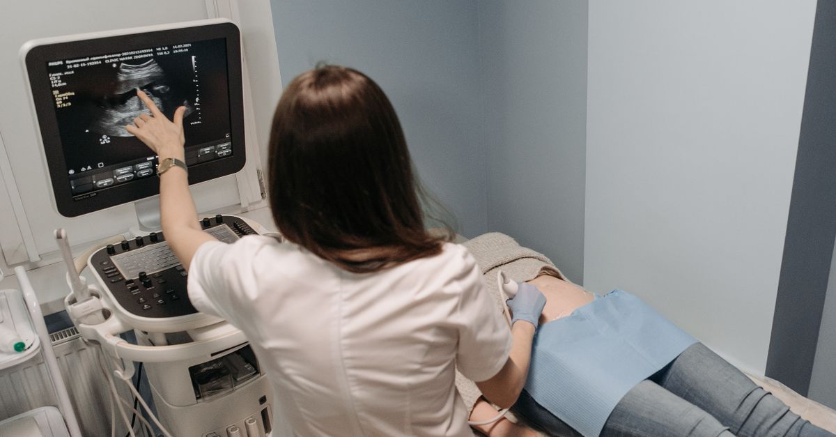
Transabdominal ultrasound is the primary, most sensitive imaging modality utilized in the emergency setting. The sonographer evaluates the right upper quadrant to visualize the gallbladder and the surrounding structures.
The classic ultrasound findings for acute cholecystitis include the direct visualization of gallstones, a thickened gallbladder wall (typically measuring greater than three millimeters), and pericholecystic fluid, which is a thin layer of inflammatory fluid pooling directly outside the gallbladder. The sonographer may also elicit a “sonographic Murphy’s sign” by pressing the ultrasound probe directly over the visualized gallbladder to assess localized tenderness.
9. Hepatobiliary Iminodiacetic Acid HIDA Scan
If the ultrasound findings are equivocal, but the clinical suspicion for acute cholecystitis remains high, a HIDA scan is performed. This is a nuclear medicine imaging test that evaluates the functional flow of bile.
A harmless radioactive tracer is injected into the patient’s vein, taken up by the liver, and excreted into the bile. A specialized camera tracks the tracer as it moves through the biliary system. In a healthy patient, the tracer will flow into both the gallbladder and the small intestine. In a patient with an impacted stone in the cystic duct, the tracer will flow clearly into the intestine (confirming no common duct obstruction) but will completely fail to enter the gallbladder, definitively proving the diagnosis of acute cystic duct obstruction.
10. Initial Medical Stabilization
Once the diagnosis is confirmed, the patient is admitted to the hospital for immediate medical stabilization. The patient is made strictly nil per os (NPO), meaning they take nothing by mouth. This rests the digestive system and prevents any further hormonal stimulation of the inflamed gallbladder.
Intravenous fluids are initiated to correct dehydration caused by vomiting and poor oral intake. Potent intravenous analgesics, such as non-steroidal anti-inflammatory drugs or targeted narcotics, are administered to control the severe visceral pain. The clinical team monitors vital signs closely to ensure the patient remains hemodynamically stable.
11. Intravenous Antibiotic Therapy
Given the high probability of secondary bacterial infection in the stagnant bile, empiric intravenous antibiotics are a mandatory component of acute management.
The selected antibiotics must provide broad coverage against common biliary and intestinal pathogens. A typical regimen might involve a cephalosporin combined with metronidazole, or a broad-spectrum penicillin derivative. The antibiotics serve to contain the infection within the gallbladder, preventing the bacteria from crossing into the bloodstream and causing life-threatening systemic sepsis while the patient is prepared for surgery.
12. Surgical Management: Laparoscopic Cholecystectomy
The definitive and curative treatment for acute calculous cholecystitis is the surgical removal of the diseased gallbladder, a procedure known as a cholecystectomy.
Current surgical guidelines strongly advocate for early laparoscopic cholecystectomy, ideally performed within the first forty-eight to seventy-two hours of symptom onset. Operating early, before the inflammation causes dense, fibrous scar tissue to form around the gallbladder and surrounding structures, significantly reduces the complexity of the surgery and lowers the risk of surgical complications. The surgeon carefully dissects the cystic duct, ensuring the main bile duct remains untouched and perfectly intact.
13. Percutaneous Cholecystostomy
In specific scenarios, a patient may present with acute cholecystitis but be deemed too critically ill or physiologically frail to survive general anesthesia and surgery. For these high-risk patients, a percutaneous cholecystostomy is performed as a life-saving, temporizing measure.
Guided by ultrasound or CT imaging, an interventional radiologist inserts a small drainage tube through the abdominal wall directly into the inflamed gallbladder. This immediately evacuates the pressurized, infected bile to the outside of the body, relieving the tension and allowing the inflammation to subside. Once the patient recovers and is medically optimized, a definitive surgical cholecystectomy can be planned for a later date.
14. Postoperative Recovery and Prognosis
The prognosis for patients undergoing prompt surgical removal of the gallbladder for acute cholecystitis is exceptionally good. Following a laparoscopic approach, most patients are discharged from the hospital within one to two days.
The body adapts seamlessly to the absence of the gallbladder. The liver continues to produce bile, which now flows continuously and directly down the unobstructed common bile duct into the intestine. Patients can generally resume a normal, healthy diet without long-term digestive consequences, entirely free from the risk of future gallbladder attacks.
15. When to Seek Immediate Medical Attention
Acute cholecystitis is a medical emergency that cannot be managed at home. You must proceed to an emergency department immediately if you experience severe, continuous pain in your upper right abdomen that prevents you from sitting still or finding a comfortable position. Furthermore, the development of a high fever, shaking chills, persistent vomiting that prevents you from keeping liquids down, or severe tenderness when the abdomen is touched requires urgent clinical evaluation and intervention.
16. Frequently Asked Questions FAQ
1. Why does a stone in the cystic duct cause so much more damage than a stone sitting in the main gallbladder?
A stone floating freely in the gallbladder body rarely causes damage. However, when a stone wedges tightly into the narrow cystic duct, it seals the exit. The gallbladder continues to produce mucus, building immense, destructive pressure inside the closed sac.
2. Can antibiotics cure acute cholecystitis without surgery?
Antibiotics can kill the secondary bacteria, but they cannot remove the physical stone blocking the duct. If the gallbladder is not removed surgically, the blockage remains, and the severe inflammation will inevitably return or cause the gallbladder to rupture.
3. Will my liver be damaged by this condition?
Because this specific condition involves no obstruction of the common bile duct, the bile from your liver can still drain safely into your intestines. Therefore, your liver is generally protected from direct back-pressure damage.
4. Why do doctors want to operate so quickly?
Operating within the first few days of the attack is safer. After a few days, the severe inflammation turns into thick, sticky scar tissue that cements the gallbladder to nearby vital organs, making laparoscopic surgery much more difficult and dangerous.
5. Is the surgery performed through a large incision?
Usually, no. The vast majority of these operations are performed laparoscopically through four tiny keyhole incisions using a camera. A large, open incision is only required if severe scarring makes the keyhole approach unsafe.
Bibliography
Disclaimer: The content is for informational purposes only and does not replace medical advice. Always consult your doctor for personalized treatment.
