1. Introduction
A stabbing pain in the left wrist when twisting a door knob is predominantly caused by a structural injury to the triangular fibrocartilage complex or inflammation of the extensor carpi ulnaris tendon. The mechanical action of twisting the wrist places profound rotational sheer stress on the ulnar side of the joint, forcefully irritating compromised ligaments or inflamed tendon sheaths. Clinical resolution relies on implementing rigid splinting, utilizing targeted nonsteroidal anti-inflammatory medications, and occasionally pursuing arthroscopic surgical repair for complete structural tears.
2. Biomechanics of Wrist Rotation
The human wrist is an incredibly complex articulation allowing for a vast array of movements. The specific action used to turn a door knob involves rotation of the forearm, clinically defined as supination (turning the palm upward) and pronation (turning the palm downward).
During these rotational movements, the radius bone physically crosses over the stationary ulna bone. This dynamic crossover places immense biomechanical torque on the ulnar side of the wrist, which is the side aligned with the pinky finger. The structural stability required to perform this twisting motion smoothly relies entirely on a delicate network of ligaments and cartilage that bind the radius and ulna together at the distal radioulnar joint.
3. The Triangular Fibrocartilage Complex
The primary stabilizer of the ulnar side of the wrist is the triangular fibrocartilage complex, commonly abbreviated as TFCC. The TFCC is a sophisticated, hammock-like structure composed of thick ligaments and a central cartilaginous disc suspended between the radius, ulna, and the small carpal bones.
This complex structure acts as the primary shock absorber for the wrist and provides the essential tethering force that keeps the forearm bones aligned during rotational stress. Because the central disc of the TFCC possesses a notoriously poor blood supply, any mechanical injury to this tissue heals exceptionally slowly and is prone to chronic dysfunction.
4. Pathology of TFCC Tears
A tear in the triangular fibrocartilage complex is the leading culprit for sharp, twisting pain. These tears can occur acutely, such as from falling onto an outstretched hand or abruptly twisting the wrist under a heavy load, or they can develop degeneratively over years of repetitive strain.
When the TFCC is torn, the structural hammock loses its tension. Attempting to twist a door knob forces the wrist bones to grind against the damaged, frayed edges of the cartilage. This mechanical impingement triggers a sharp, localized, stabbing sensation deep within the ulnar aspect of the wrist, frequently accompanied by a distinct clicking or catching sound.
5. Extensor Carpi Ulnaris Tendonitis
If the cartilage remains intact, the stabbing pain may originate from the surrounding musculotendinous structures. The extensor carpi ulnaris is a prominent tendon running directly over the ulnar side of the wrist, responsible for extending and stabilizing the joint.
Repetitive rotational movements can cause the sheath surrounding this tendon to become severely inflamed, a condition known as tenosynovitis. Additionally, the subsheath that holds the tendon in its groove can tear, allowing the tendon to painfully snap in and out of place during twisting motions. The resulting friction produces intense, localized pain that mimics a deep structural joint injury. Addressing this inflammation is a critical component of treating localized joint pain.
6. Ulnar Impaction Syndrome
Ulnar impaction syndrome is a degenerative anatomical condition that drastically increases the mechanical load on the TFCC. In some individuals, the ulna bone is genetically slightly longer than the adjacent radius bone, a structural variance termed positive ulnar variance.
When the wrist is twisted or tilted toward the pinky finger, this abnormally long ulna repeatedly smashes into the delicate carpal bones and the TFCC. Over time, this chronic, repetitive impaction wears holes completely through the cartilage and causes deep bruising within the carpal bones, manifesting as sharp, stabbing pain during any loaded rotational activity.
7. Distal Radioulnar Joint Instability
The distal radioulnar joint is the specific articulation where the radius and ulna pivot against each other near the wrist. The ligaments of the TFCC normally tightly secure this joint. If these stabilizing ligaments are severely sprained or ruptured, the joint becomes physically unstable.
During the action of twisting a door knob, the lack of ligamentous restraint allows the radius and ulna to partially separate or subluxate. This abnormal shifting of the bones strains the remaining intact soft tissues to their breaking point, resulting in a sudden, defensive muscle spasm and a sharp spike of joint pain.
8. The Role of Daily Repetitive Strain
Many patients cannot recall a specific trauma that initiated their wrist pain. In these instances, the structural degradation is the result of insidious, cumulative micro-trauma. Occupations or hobbies that require continuous, forceful twisting—such as using screwdrivers, playing tennis, or heavy manual labor—subject the ulnar wrist to thousands of high-torque cycles.
This relentless mechanical demand surpasses the biological recovery rate of the cartilage and tendons. The tissues slowly degrade, fray, and inflame. The stabbing pain eventually presents suddenly during a mundane task, like opening a door, acting as the final threshold where the compromised tissues fail to bear the load.
9. Differentiating Tendonitis from Cartilage Tears
Accurate clinical differentiation between tendon pathology and internal cartilage damage is crucial for treatment planning. Tendonitis of the extensor carpi ulnaris typically presents with visible swelling along the back of the wrist and is highly tender to direct palpation over the tendon itself.
Conversely, a TFCC tear produces deep, internal pain that is difficult for the patient to pinpoint exactly. The pain is usually most severe when the wrist is simultaneously compressed and twisted. Tendonitis generally responds faster to conservative anti-inflammatory therapies, whereas a true cartilage tear requires prolonged immobilization or surgical repair.
10. Clinical Provocative Testing
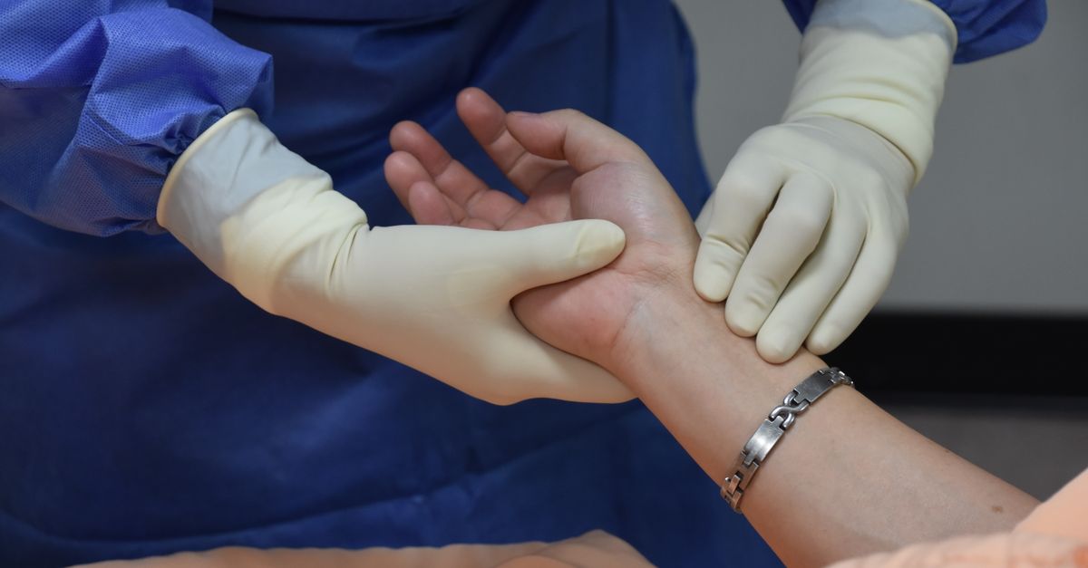
An orthopedic specialist utilizes specific provocative maneuvers to isolate the pain source. The fovea sign is a highly sensitive test for TFCC tears; the physician applies direct pressure to a specific anatomical depression between the ulnar styloid and the flexor carpi ulnaris tendon. Severe tenderness here strongly indicates a TFCC lesion.
The TFCC compression test involves the clinician holding the patient’s forearm, grasping their hand, bending the wrist toward the ulnar side, and applying an axial compression force while rotating the wrist. Reproducing the sharp, stabbing pain or eliciting a mechanical click definitively confirms pathology within the triangular fibrocartilage complex.
11. Diagnostic Imaging and Arthrography
While standard X-rays are obtained to evaluate bone length and rule out occult fractures, they cannot visualize cartilage or tendons. To accurately assess soft tissue damage, magnetic resonance imaging is the standard of care.
For optimal visualization of the tiny ligaments within the wrist, an MR arthrogram is frequently preferred. This involves injecting a contrast dye directly into the wrist joint prior to the MRI scan. The dye flows into any microscopic tears or defects within the TFCC, highlighting the exact location and severity of the structural failure for the treating physician.
12. Splinting and Mechanical Rest
Initial conservative management mandates strict mechanical rest. The patient is prescribed a specialized, rigid wrist splint or a Munster cast. Unlike standard wrist braces, an effective splint for ulnar-sided pain must completely restrict the rotational movement of the forearm (pronation and supination).
Immobilizing the joint halts the continuous mechanical sheer force grinding the torn cartilage or inflamed tendons. The splint is typically worn consistently for four to six weeks, providing the compromised tissues with the necessary stable environment to initiate the biological healing process.
13. Corticosteroid Injections
If severe inflammation prevents healing despite immobilization, pharmacological intervention is employed. The physician may administer a targeted corticosteroid injection directly into the ulnar side of the wrist joint or into the specific inflamed tendon sheath.
Corticosteroids deliver profound, localized suppression of the inflammatory cascade. While the injection does not physically repair a torn ligament, it drastically reduces the painful swelling within the joint capsule. This creates a pain-free window, allowing the patient to participate comfortably in rehabilitative physical therapy and regain functional range of motion.
14. Data: Causes of Ulnar-Sided Wrist Pain
| Underlying Pathology | Anatomical Structure | Primary Provocative Motion |
|---|---|---|
| TFCC Tear | Cartilage disc and ligaments. | Twisting under a heavy load; deep clicking. |
| ECU Tendonitis | Tendon on the outer wrist. | Bending the wrist backward; visible swelling. |
| Ulnar Impaction Syndrome | Elongated ulna bone crashing into carpal bones. | Tilting the wrist toward the pinky finger. |
| DRUJ Instability | Joint between the radius and ulna. | Severe pain when turning the palm upward. |
15. Surgical Intervention Options
When conservative therapies, prolonged splinting, and injections fail to resolve the stabbing pain, surgical intervention becomes necessary. Wrist arthroscopy is a minimally invasive surgical procedure where a miniature camera and instruments are inserted into the joint.
An orthopedic surgeon can visualize the torn TFCC directly. If the tear is in the center of the cartilage, which lacks a blood supply, the frayed edges are carefully trimmed away (debridement) to eliminate the mechanical catching. If the tear is located at the outer rim, where a blood supply exists, the surgeon will physically suture the ligaments back onto the bone, restoring the structural integrity of the wrist.
16. Frequently Asked Questions
1. Will doing wrist curls and strengthening exercises fix the pain?
No. If the pain is caused by a torn TFCC or severely inflamed tendon, aggressive strengthening exercises will only generate more mechanical friction and worsen the damage. Strict immobilization is required first to allow the tissues to heal before any strengthening begins.
2. Why does the pain happen with door knobs but not when I type on a computer?
Typing primarily involves moving the fingers while the wrist remains in a relatively static, flat position. Turning a door knob requires supination and pronation, which twists the forearm bones around each other, placing maximum rotational sheer force precisely on the damaged ulnar tissues.
3. Can an old sports injury cause this pain years later?
Yes. An untreated sprain or minor tear sustained years ago can alter the biomechanics of your wrist. Over time, this subtle instability leads to premature wear and tear on the cartilage, eventually presenting as sudden, sharp pain during a mundane activity.
4. Is ulnar-sided wrist pain a sign of arthritis?
While severe, bone-on-bone arthritis can cause wrist pain, it usually presents as a constant, dull ache accompanied by profound stiffness in the morning. Sharp, stabbing pain isolated to a specific twisting movement is much more indicative of a localized cartilage tear or tendon entrapment.
5. How long does a TFCC tear take to heal with a brace?
Because the central portion of the TFCC has a notoriously poor blood supply, healing is exceptionally slow. It typically requires a minimum of four to six weeks of strict, continuous immobilization in a brace, followed by several weeks of gradual physical therapy, to achieve clinical improvement.
17. Bibliography
Disclaimer: The content is for informational purposes only and does not replace medical advice. Always consult your doctor for personalized treatment.