
1. Introduction
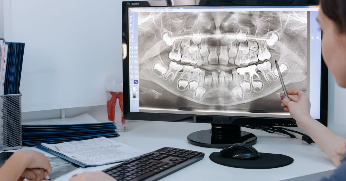
In dental and maxillofacial radiography, the term radiolucency refers to areas on an X-ray film that appear dark or black, indicating that X-ray photons have passed through the tissue with minimal resistance. When a radiolucency is associated with a tooth or its surrounding bony architecture, it serves as a critical diagnostic marker for tissue destruction, bone loss, or structural demineralization. Healthy teeth, composed of exceptionally dense enamel and dentin, heavily absorb X-rays and appear radiopaque, or bright white. A localized dark shadow within this dense structure or at the root apex immediately alerts the clinician to an underlying pathological process.
The clinical implications of a dental radiolucency are vast. Most commonly, these dark areas represent active bacterial infections, such as deep dental caries dissolving the enamel, or severe periapical abscesses where infection has traveled through the tooth root to destroy the surrounding jawbone. However, radiolucencies can also indicate non-infectious pathologies, including benign dental cysts, aggressive odontogenic tumors, or systemic metabolic bone diseases.
Accurate interpretation of these radiographic shadows is the cornerstone of modern endodontics and oral surgery. Dentists must meticulously analyze the size, shape, borders, and exact anatomical location of the radiolucent area. By combining this precise visual data with clinical vitality testing and patient history, the dental team can differentiate a simple cavity from a destructive jaw lesion, formulating a targeted treatment plan to eradicate the infection, preserve the tooth, and restore the integrity of the maxillofacial skeleton.
2. Principles of Radiopacity and Radiolucency
Understanding how dental X-rays function is essential for interpreting clinical findings. X-rays pass through the body and strike a digital sensor or physical film. The density and atomic weight of the tissue determine how many X-ray photons reach the sensor.
Dense, heavily mineralized tissues like tooth enamel, dentin, and cortical jawbone block the X-rays. Because the photons are absorbed by the tissue and do not strike the sensor, these areas appear bright white, a characteristic defined as radiopaque. Metallic dental fillings and crowns appear starkly radiopaque due to their extreme density.
Conversely, tissues with low density, such as the soft dental pulp inside the tooth, the periodontal ligament space, or areas filled with air, fluid, or pus, allow X-rays to pass through easily. These photons strike the sensor and darken the image, creating a radiolucency. When a disease process destroys dense bone or dissolves enamel, the affected area becomes less dense and transitions from radiopaque to radiolucent on the resulting image.
3. Dental Caries (Tooth Decay)
The most frequent cause of a radiolucency strictly within the crown of a tooth is dental caries, commonly known as a cavity. Caries are caused by acidogenic bacteria in the oral cavity that metabolize dietary sugars and produce lactic acid. This acid actively demineralizes the highly calcified enamel and the underlying dentin.
As the minerals are stripped away, the affected portion of the tooth loses its density. On a bitewing X-ray, this demineralization appears as a distinct, dark, wedge-shaped or localized radiolucency cutting into the bright white enamel.
Detecting caries radiographically before they become visible to the naked eye allows the dentist to intervene early. By removing the decayed, radiolucent tissue and replacing it with a dense, radiopaque composite filling, the clinician halts the bacterial progression before it can breach the inner pulp chamber of the tooth.
4. Periapical Abscesses and Lesions
When a deep carious lesion or severe dental trauma kills the living pulp tissue inside a tooth, the necrotic debris becomes a breeding ground for bacteria. These bacteria travel down the root canal and exit through the tiny opening at the tip of the root, known as the apex.
Once the infection reaches the apex, the host immune system responds aggressively, generating inflammation and pus. This inflammatory response actively destroys the dense alveolar bone surrounding the root tip to create space for the accumulating pus.
On a periapical X-ray, this bone destruction presents as a classic, round, dark radiolucency hovering directly at the tip of the tooth root. This periapical radiolucency is the definitive radiological hallmark of a dead, infected tooth, necessitating prompt endodontic therapy (a root canal) or complete tooth extraction to drain the infection and prevent its spread into the fascial spaces of the neck or face.
5. Radicular Cysts and Granulomas
A periapical radiolucency does not always represent an acute, pus-filled abscess; it frequently indicates a chronic, slow-growing inflammatory lesion known as a periapical granuloma or a radicular cyst.
A periapical granuloma is a mass of chronically inflamed granulation tissue at the root apex, representing the body’s attempt to wall off the infection coming from the dead tooth. Over time, the epithelial cells within this granuloma can proliferate and form a fluid-filled sac lined with tissue, creating a radicular cyst.
Radiographically, both granulomas and cysts present as dark, radiolucent circles at the root tip. While a cyst may exhibit a more distinct, smooth, sclerotic white border compared to an acute abscess, it is nearly impossible to differentiate them definitively on an X-ray alone. Regardless of the exact histological makeup, the presence of this radiolucency confirms that the tooth is non-vital and requires comprehensive endodontic intervention.
6. Periodontal Disease and Bone Loss
Radiolucencies are also the primary indicator of severe periodontal disease, which is a chronic bacterial infection of the gums that destroys the bone supporting the teeth. Healthy alveolar bone should appear as a dense, flat, radiopaque line extending almost to the neck of the tooth.
In periodontitis, the chronic inflammation causes the bone to recede away from the teeth. On an X-ray, this bone loss appears as a widespread radiolucency between the teeth. The clinician evaluates whether the bone loss is horizontal (an even drop in bone level across multiple teeth) or vertical (a deep, localized trench of dark radiolucency running down the side of a specific root).
Severe vertical bone loss creates deep periodontal pockets that harbor aggressive bacteria. Recognizing these radiolucent patterns directs the periodontist to perform deep scaling and root planing or surgical bone grafting procedures to halt the progression of the disease before tooth mobility and eventual tooth loss occur.
7. External and Internal Root Resorption
Root resorption is an insidious condition where the body’s own immune cells, specifically odontoclasts, inappropriately attack and dissolve the hard structure of the tooth root. Internal resorption begins inside the pulp chamber and hollows out the tooth from the inside. On an X-ray, this appears as a localized, balloon-like radiolucency expanding the normally narrow root canal space.
External root resorption occurs on the outer surface of the root, frequently triggered by severe orthodontic forces (braces), physical trauma, or pressure from an adjacent impacted tooth. The X-ray will show a ragged, irregular radiolucency eating away at the side or the apex of the root, making the tooth appear blunted or shortened.
Managing resorption is exceptionally challenging. If caught early, advanced endodontic therapies utilizing specialized calcium silicate cements can halt internal resorption. However, severe external resorption often renders the tooth structurally unsalvageable, leading to extraction.
8. Odontogenic Tumors and Jaw Lesions
When a large radiolucency is discovered in the jawbone that is not centered on the apex of a specific tooth, the clinician must consider odontogenic tumors and developmental cysts. Ameloblastoma is a benign, locally aggressive tumor originating from the cells that form tooth enamel.
On a panoramic X-ray, an ameloblastoma typically presents as a large, multilocular radiolucency. It is often described as having a “soap bubble” or “honeycomb” appearance due to the distinct, bony partitions dividing the dark, cystic spaces.
These tumors are expansive, capable of silently hollowing out the mandible and pushing healthy teeth out of alignment. While benign, they require aggressive surgical resection to remove the entire radiolucent margin, as they possess a notoriously high recurrence rate if any cellular remnants are left behind in the bone.
9. Dentigerous Cysts
A dentigerous cyst, or follicular cyst, is a specific developmental abnormality that occurs around the crown of an unerupted or impacted tooth. The most common site for this pathology is the impacted lower third molar (wisdom tooth).
The cyst forms due to fluid accumulation between the developing tooth crown and the surrounding enamel organ. On an X-ray, it presents as a distinct, well-circumscribed, dark radiolucency wrapping symmetrically around the crown of the impacted tooth and attaching at the cementoenamel junction.
While dentigerous cysts are benign, they can grow to a substantial size, causing silent, progressive destruction of the surrounding jawbone and increasing the risk of a pathological jaw fracture. Surgical removal of the cyst, typically performed concurrently with the extraction of the impacted wisdom tooth, is the standard, curative treatment.
10. Evaluating Lesion Margins
When a dentist or oral radiologist identifies a radiolucency, they meticulously evaluate the borders of the dark shadow to predict the biological behavior of the lesion.
| Margin Characteristic | Visual Appearance | Clinical Implication |
|---|---|---|
| Well-Defined with Corticated Border | Sharp border with a bright white rim | Slow-growing, benign process (e.g., simple cyst) |
| Punched-Out | Sharp border without a white rim | Intermediate growth (e.g., multiple myeloma, specific cysts) |
| Ill-Defined or Ragged | Fuzzy, blurry border blending into bone | Aggressive spreading infection or malignant tumor |
| Moth-Eaten | Multiple tiny, irregular dark holes | Severe, rapid bone destruction (e.g., osteomyelitis) |
11. Clinical Vitality Testing
A radiograph provides excellent structural data, but it cannot determine the physiological status of the dental pulp. When a periapical radiolucency is identified, the dentist must perform pulp vitality testing before initiating treatment.
Thermal testing, using extreme cold applied to the tooth surface, determines if the nerve inside the tooth is alive and capable of transmitting a sensation. Electric pulp testing delivers a mild, graded electrical current to assess nerve vitality.
If the tooth does not respond to thermal or electrical stimuli, it is confirmed as necrotic (dead). The combination of a dead nerve and a periapical radiolucency provides absolute diagnostic proof that an endodontic infection has destroyed the bone, officially directing the clinician to proceed with a root canal or extraction.
12. Advanced Imaging: Cone Beam Computed Tomography
Standard two-dimensional dental X-rays are exceptionally useful but possess inherent limitations. They compress a three-dimensional jaw into a flat image, leading to overlapping anatomical structures that can obscure a subtle radiolucency or misrepresent the true extent of a lesion.
Cone Beam Computed Tomography has revolutionized maxillofacial diagnostics. This specialized 3D imaging technology allows the clinician to rotate and slice the jawbone digitally. It provides unparalleled visualization of the complex root canal anatomy, exposing hidden infected roots and subtle periapical radiolucencies that are invisible on traditional films.
Furthermore, Cone Beam Computed Tomography accurately maps the proximity of large radiolucent cysts or tumors to critical anatomical landmarks, such as the inferior alveolar nerve or the maxillary sinus floor, ensuring meticulous safety during surgical excisions.
13. Systemic Bone Diseases
Occasionally, a radiolucency in the jawbone is the first clinical manifestation of a systemic medical condition rather than a localized dental infection. Multiple myeloma, a malignant cancer of plasma cells, classically presents with multiple, sharply defined “punched-out” radiolucencies scattered throughout the skull and mandible.
Hyperparathyroidism, a metabolic disorder causing excess parathyroid hormone, triggers the rapid removal of calcium from bones. On a dental X-ray, this condition can cause the dense lamina dura—the bright white bone outlining the tooth socket—to disappear completely, while the overall jawbone takes on a generalized, washed-out, radiolucent appearance.
Identifying these atypical radiolucent patterns requires the dentist to refer the patient to a medical specialist, facilitating the early diagnosis and treatment of severe systemic metabolic or oncological diseases.
14. Treatment Protocols for Infectious Radiolucencies
The clinical resolution of an infectious periapical radiolucency requires complete eradication of the bacterial source. Root canal therapy achieves this by physically removing the necrotic nerve tissue, disinfecting the hollow root canal space with potent antimicrobial irrigants, and sealing the space with a dense, radiopaque rubber material called gutta-percha.
Once the internal bacterial reservoir is eliminated, the host immune system ceases its inflammatory attack on the jawbone. Over the subsequent six to twelve months, specialized bone-building cells (osteoblasts) migrate into the void.
Follow-up X-rays will show the dark radiolucency slowly filling in with bright, radiopaque bone, radiographically proving that the treatment was successful and the skeletal structure has healed completely. If the tooth structure is too severely compromised to support a root canal, extraction removes the infection source, allowing the bone to heal prior to dental implant placement.
15. The Role of Dental Restorations
Patients must understand that radiolucencies not only indicate bone loss but are the primary method for detecting secondary decay hidden beneath existing dental work. A clinical exam cannot see underneath a dense silver or composite filling.
Routine bitewing X-rays allow the dentist to check the margins where the filling meets the natural tooth. If the seal has failed, bacteria leak underneath and dissolve the dentin. This presents as a dark radiolucency actively undermining the bright white filling material.
Early detection of this hidden radiolucency allows the dentist to remove the failing restoration, clean the new decay, and place a fresh filling before the bacteria reach the pulp chamber, saving the patient from a painful toothache and complex endodontic therapy. For insights into related facial pain management, review our clinical guide on facial weakness.
16. Frequently Asked Questions (FAQ)
1. What does a “dark spot” on my dental X-ray mean?
In dental X-rays, dark spots are called radiolucencies. Because X-rays pass easily through soft areas, a dark spot usually means that dense tooth enamel or jawbone has been dissolved away, most commonly by a bacterial cavity or an infection at the root tip.
2. Does a dark spot at the root always mean I need a root canal?
Usually, yes. A dark circle at the tip of the root indicates that the nerve inside the tooth has died, and bacteria have leaked into the jawbone, dissolving it to create a pocket of pus or inflammation. A root canal removes the dead nerve and cures the infection.
3. Will the bone grow back after the infection is treated?
Yes. Once the root canal or extraction removes the source of the bacteria, your immune system stops attacking the area. Over several months, your body will naturally refill the dark hole with strong, healthy white bone.
4. Can a dark spot on a jaw X-ray be cancer?
While the vast majority of dark spots are simple dental infections or benign cysts, aggressive or strange-looking radiolucencies can sometimes indicate jaw tumors or systemic diseases. Your dentist uses the shape and borders of the spot to decide if a surgical biopsy is needed.
5. Why do I need X-rays if my teeth do not hurt?
Many destructive dental conditions, including deep cavities between the teeth, chronic bone loss from gum disease, and slow-growing cysts, do not cause pain until they are extremely advanced. X-rays detect these silent radiolucencies early, saving your teeth and preventing extensive surgery.
17. Bibliography
Disclaimer: The content is for informational purposes only and does not replace medical advice. Always consult your doctor for personalized treatment.
