1. Introduction
A reduced sensation of skin, clinically known as hypoesthesia, indicates an interruption or damage to the neural pathways that transmit sensory information from the body’s surface to the brain. The bottom line is that skin numbness is not a dermatological issue, but a neurological symptom pointing to peripheral nerve damage, metabolic disorders, or central nervous system pathology, making a precise neurological evaluation mandatory to prevent irreversible nerve death and severe secondary injuries. Diagnosing the exact location of the nerve impairment dictates the course of medical or surgical intervention.
The human skin is heavily innervated with specialized sensory receptors that detect touch, temperature, and pain. These receptors send continuous electrical signals along peripheral nerves, up the spinal cord, and into the somatosensory cortex of the brain. When a patient experiences a loss of sensation, it signifies a roadblock anywhere along this complex transmission highway.
Ignoring numbness is clinically dangerous. Without protective sensation, patients are highly susceptible to severe burns, unnoticed lacerations, and chronic ulcerations. Identifying the root cause—whether it is chronic high blood sugar, a compressed spinal disc, or an autoimmune attack—allows clinicians to halt the damage and, in some cases, restore sensory function.
2. Understanding Cutaneous Sensation and Dermatomes
To pinpoint the source of reduced sensation, clinicians rely on the anatomical map of dermatomes. A dermatome is a specific area of skin that is primarily innervated by a single spinal nerve root. By carefully mapping exactly where the numbness occurs, a physician can often trace the symptom back to the precise damaged nerve.
For example, numbness isolated strictly to the thumb and index finger correlates with the median nerve, while numbness running down the outside of the thigh corresponds to specific lumbar nerve roots in the lower back. This mapping distinguishes localized nerve compression from systemic nerve damage.
Sensory nerves are composed of different types of fibers. Large, myelinated fibers transmit light touch and vibration, while smaller, unmyelinated fibers transmit pain and temperature. A patient may lose the ability to feel a light brush against the skin but retain the ability to feel a sharp pinprick, depending on which specific nerve fibers are sustaining damage.
3. Peripheral Neuropathy: The Systemic Threat
Peripheral neuropathy is a broad clinical term describing damage to the peripheral nervous system, which connects the brain and spinal cord to the rest of the body. When the long sensory nerves that reach down into the hands and feet are damaged, patients experience a symmetric “stocking-glove” pattern of reduced sensation.
The numbness typically begins in the longest nerves first, starting at the tips of the toes and slowly marching up the legs over months or years. Eventually, the fingertips become involved. Patients often describe the sensation as feeling like they are walking on cotton or wearing thick gloves.
This type of systemic nerve damage is frequently caused by metabolic toxins, chronic inflammation, or vascular insufficiency that starves the distal nerves of oxygen and nutrients, leading to gradual axonal degeneration.
4. The Impact of Diabetic Neuropathy
Diabetes mellitus is the leading global cause of peripheral neuropathy. Chronically elevated blood glucose levels are highly toxic to neural tissue. The excess glucose damages the microscopic blood vessels, known as the vasa nervorum, that supply oxygen and nutrients directly to the peripheral nerves.
As the nerves suffocate and undergo oxidative stress, they lose their ability to transmit sensory signals. Patients with diabetic neuropathy suffer a profound loss of protective sensation in their feet. Because they cannot feel pain, a small blister from tight shoes can rapidly evolve into a massive, infected diabetic ulcer.
Strict glycemic control is the absolute cornerstone of managing this condition. While severely damaged nerves may not fully recover, maintaining normal blood sugar levels definitively halts the progression of the neuropathy and prevents the onset of total sensory loss.
5. Nerve Compression and Entrapment Syndromes
Unlike the symmetric numbness of peripheral neuropathy, nerve entrapment syndromes cause reduced sensation in highly specific, localized areas due to mechanical compression of a single nerve. Carpal tunnel syndrome is the most prevalent example, where the median nerve is compressed as it passes through the narrow fibrous tunnel in the wrist.
Patients with carpal tunnel syndrome experience profound numbness, tingling, and weakness in the thumb, index, and middle fingers, often waking up at night needing to shake their hands out. Similarly, compression of the ulnar nerve at the elbow (cubital tunnel syndrome) causes numbness strictly in the ring and pinky fingers.
Radiculopathy occurs when a nerve root is compressed as it exits the spinal column, usually due to a herniated disc or spinal stenosis. This causes sharp, shooting pain and numbness that radiates down the entire length of the arm or leg along the specific dermatome path.
6. Central Nervous System Pathologies
If the peripheral nerves are intact, reduced skin sensation can originate from damage within the central nervous system—the brain or the spinal cord. A cerebrovascular accident, or stroke, can damage the somatosensory cortex. In this scenario, the patient’s skin and peripheral nerves function perfectly, but the brain cannot receive or interpret the signals, resulting in sudden, unilateral numbness of the face, arm, or leg.
Multiple sclerosis is an autoimmune disease where the body attacks the protective myelin sheath surrounding nerve fibers in the brain and spinal cord. The resulting lesions disrupt signal transmission, frequently presenting as migratory patches of numbness or a “tight band” sensation across the torso.
Central causes of numbness are considered medical emergencies, especially if the onset is sudden and accompanied by weakness, visual changes, or difficulty speaking, requiring immediate neurological imaging and intervention.
7. Vitamin Deficiencies and Toxic Neuropathies
Nerve tissue requires specific micronutrients to maintain its structural integrity and synthesize neurotransmitters. Vitamin B12 is crucial for maintaining the myelin sheath. A severe deficiency, common in pernicious anemia, older adults, or strict vegans, causes subacute combined degeneration of the spinal cord, leading to profound loss of sensation and proprioception (the awareness of body position).
Toxic neuropathies occur when external chemicals damage the nerve fibers. Chronic, excessive alcohol consumption is highly neurotoxic and a leading cause of nutritional neuropathy. Furthermore, many chemotherapeutic agents used in cancer treatment cause chemotherapy-induced peripheral neuropathy, presenting as severe numbness and tingling in the extremities.
Identifying and removing the toxic agent, or aggressively supplementing the deficient vitamin, allows the peripheral nervous system the opportunity to regenerate, though recovery is often a slow process spanning many months.
8. Structural Data: Distinguishing Causes of Numbness
Categorizing the pattern of sensory loss is a critical diagnostic step.
| Pattern of Numbness | Clinical Presentation | Likely Underlying Cause |
|---|---|---|
| Stocking-Glove Distribution | Symmetric numbness starting in toes, moving up to calves and fingertips. | Diabetic Neuropathy, Toxic Neuropathy, B12 Deficiency |
| Single Nerve Distribution | Numbness isolated to specific fingers (e.g., thumb and index only). | Carpal Tunnel Syndrome, Nerve Entrapment |
| Dermatomal (Radiating) | Numbness radiating from the neck down an arm, or lower back down a leg. | Spinal Disc Herniation, Radiculopathy |
| Unilateral (One-Sided) | Sudden numbness affecting exactly one half of the face, arm, and leg. | Stroke, Transient Ischemic Attack (TIA) |
| Patchy / Migratory | Unpredictable patches of numbness that come and go across the body. | Multiple Sclerosis, Autoimmune Demyelination |
9. Infectious and Autoimmune Etiologies
Several infectious diseases directly target the peripheral nerves. Lyme disease, caused by a tick-borne spirochete, can cause cranial nerve palsies and peripheral radiculoneuropathy, resulting in localized numbness. The Varicella-Zoster virus, responsible for shingles, lies dormant in nerve roots and, upon reactivation, causes severe pain and altered sensation along a single dermatome.
Autoimmune conditions, such as Guillain-Barré syndrome, involve a rapid, post-infectious immune attack on the peripheral nerves. This presents as ascending weakness and sensory loss, starting in the feet and rapidly progressing upward, requiring urgent hospital admission to monitor respiratory function.
Conditions like lupus and rheumatoid arthritis can also cause vasculitis—inflammation of the blood vessels—which subsequently chokes off the blood supply to the nerves, leading to a loss of skin sensation.
10. The Neurological Diagnostic Examination
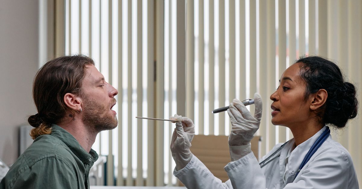
A clinical evaluation for reduced sensation begins with a detailed neurological exam. The physician tests different sensory modalities separately. Light touch is assessed using a fine piece of cotton. Pain sensation is tested with a sterile pinprick, and temperature sensation is evaluated using cold metal instruments.
Proprioception, the ability to sense where a limb is in space without looking, is tested by moving the patient’s toes up or down and asking them to identify the position with their eyes closed. Vibration sense is highly sensitive to early nerve damage and is tested using a tuning fork applied to the bony prominences of the ankles and wrists.
Reflexes are also checked; a loss of the Achilles tendon reflex is often one of the earliest objective signs of severe peripheral neuropathy.
11. Electromyography and Nerve Conduction Studies
When the clinical examination suggests peripheral nerve damage, the gold standard diagnostic test is an electromyography and nerve conduction study. A nerve conduction study involves placing surface electrodes on the skin and delivering small electrical shocks to the nerve. The machine measures the speed and strength of the electrical signal as it travels down the nerve.
If the myelin sheath is damaged, the signal speed will be significantly slowed. If the axon (the core of the nerve) is damaged, the signal amplitude will be weak. Electromyography involves inserting a tiny needle electrode directly into the muscle to evaluate the electrical activity during rest and contraction.
These tests allow neurologists to pinpoint the exact location of a nerve compression or confirm the presence and severity of a systemic polyneuropathy, guiding precise treatment decisions.
12. Pharmacological Symptom Management
While medications cannot directly repair dead nerve tissue, they are highly effective in managing the abnormal sensations that often accompany numbness, such as burning, tingling, or neuropathic pain. Standard pain relievers like ibuprofen or acetaminophen are entirely ineffective for nerve pain.
Clinicians rely on neuroactive medications. Anticonvulsants, such as gabapentin and pregabalin, stabilize the hyperactive electrical membranes of damaged nerves, significantly dampening the painful signals. Certain antidepressants, particularly serotonin-norepinephrine reuptake inhibitors like duloxetine, modify the brain’s perception of pain and are heavily utilized in diabetic neuropathy.
Topical treatments, including lidocaine patches or capsaicin cream, can provide localized relief for specific areas of altered sensation without causing systemic side effects.
13. Physical Therapy and Surgical Decompression
For nerve compression syndromes, conservative management includes physical therapy and splinting. Wearing a rigid wrist splint at night keeps the wrist in a neutral position, maximizing the space in the carpal tunnel and relieving pressure on the median nerve, which often resolves the numbness.
Physical therapists teach nerve gliding exercises, designed to help the compressed nerves slide smoothly through the surrounding inflamed tissues without snagging. They also focus on postural correction to relieve pressure on spinal nerve roots.
If conservative measures fail, and the electromyography shows severe nerve block or impending muscle atrophy, surgical decompression is necessary. Procedures like a carpal tunnel release or a spinal laminectomy physically cut the restricting ligaments or bone, removing the pressure and allowing the nerve the opportunity to heal and restore sensation.
14. Preventative Care for the Insensate Foot
Patients with permanent reduced sensation in their feet, particularly those with diabetes, must practice rigorous preventative foot care. The loss of the biological pain alarm means the patient must rely entirely on visual inspection to prevent amputations.
Patients are instructed to examine the bottom of their feet daily using a mirror, looking for any redness, calluses, or cuts. Walking barefoot is strictly prohibited, even indoors. Shoes must be thoroughly inspected before wearing to ensure no foreign objects are inside.
Podiatrists play a crucial role in managing thick calluses and trimming nails safely, as self-care with sharp instruments often leads to accidental injury when the patient cannot feel the blade cutting into the skin.
15. Frequently Asked Questions (FAQ)
1. Is numbness in my fingers a sign of a stroke?
If numbness occurs suddenly and is accompanied by weakness, confusion, trouble speaking, or facial drooping, it is a medical emergency and could be a stroke. If the numbness has developed slowly over months and only affects specific fingers, it is more likely a local nerve compression like carpal tunnel syndrome.
2. Can peripheral neuropathy be cured?
It depends on the cause. Neuropathy caused by a vitamin deficiency or a compressed nerve can often be reversed. However, neuropathy caused by years of uncontrolled diabetes is typically permanent, though treatments can halt its progression and manage the symptoms.
3. Why do my numb feet feel like they are burning at night?
Damaged nerves often misfire. Even though you cannot feel light touch, the damaged pain fibers send spontaneous, chaotic signals to the brain, which are interpreted as severe burning or electrical pain, a condition known as painful diabetic neuropathy.
4. What is a nerve conduction study like?
It involves mild electrical impulses applied to your skin, which can feel like a brief, sharp static shock. It is generally well-tolerated and provides vital information about the health and speed of your nerve pathways.
5. How long does a compressed nerve take to heal after surgery?
Nerves heal very slowly, at a rate of approximately one millimeter per day. Depending on how long the nerve was compressed and how severe the damage was, it can take several months to a year for sensation to fully return.
16. Bibliography
Disclaimer: The content is for informational purposes only and does not replace medical advice. Always consult your doctor for personalized treatment.