
1. Introduction
Myoepithelial carcinoma is an uncommon and notably aggressive malignancy arising primarily in the salivary glands, composed exclusively of neoplastic myoepithelial cells displaying destructive, infiltrative growth patterns. The bottom line is that owing to its incredibly diverse microscopic appearance and potential for early regional metastasis, securing a precise immunohistochemical diagnosis is essential for guiding radical surgical resection and postoperative radiation therapy to achieve durable locoregional control. Preventing injury to crucial cranial nerves while securing negative oncological margins presents a profound surgical challenge.
These tumors originate from the contractile cells that normally surround the secretory units of the salivary glands. Unlike mixed tumors that contain both ductal and myoepithelial cells, a pure myoepithelial carcinoma demonstrates monoclonal proliferation of only the myoepithelial lineage. This biological purity, paradoxically, presents with vast morphological diversity, frequently mimicking other sarcomas or carcinomas under the microscope.
Clinical management is heavily concentrated within specialized head and neck oncology centers. The anatomical complexity of the parotid and submandibular glands requires a multidisciplinary strategy, ensuring complete extirpation of the malignancy while executing precise nerve preservation techniques to protect the patient’s facial function and quality of life.
2. Anatomy of the Salivary Glands
To comprehend the pathology of myoepithelial carcinoma, one must understand the anatomical distribution of the salivary system. The major salivary glands include the paired parotid glands (located in front of the ears), the submandibular glands (beneath the jaw), and the sublingual glands (under the tongue). Thousands of minor salivary glands are scattered throughout the mucosal lining of the lips, palate, and pharynx.
The parotid gland is the most frequent site of origin for this malignancy. The anatomy of the parotid gland is uniquely complex because the facial nerve (Cranial Nerve VII), which controls all the muscles of facial expression, courses directly through the center of the gland, dividing it into a superficial and deep lobe.
Any rapidly growing tumor within the parotid gland threatens this vital nerve. The surgical oncologist must navigate this dense neurovascular web, attempting to resect the tumor with wide margins without severing the facial nerve, a balance that defines the primary surgical challenge of this disease.
3. The Role of the Normal Myoepithelial Cell
In healthy salivary gland architecture, the secretory units (acini) and the small ducts are lined by a dual-cell layer. The inner luminal cells secrete saliva. The outer basal layer is composed of myoepithelial cells. These specialized cells contain contractile proteins similar to smooth muscle.
When stimulated, healthy myoepithelial cells contract, forcefully expelling the secreted saliva from the acini into the ductal system and into the oral cavity. They also provide a structural basement membrane, establishing a protective barrier between the glandular tissue and the surrounding stroma.
In myoepithelial carcinoma, this protective barrier is lost. The mutated myoepithelial cells proliferate autonomously, abandoning their functional contractile role. They adopt an aggressive, invasive behavior, digesting the surrounding extracellular matrix and infiltrating deep into the adjacent parotid parenchyma, muscle, and nerve sheaths.
4. Pathophysiology of Malignant Transformation
Myoepithelial carcinomas can arise de novo, meaning they develop spontaneously as primary malignancies from previously healthy tissue. Alternatively, they can develop secondary to the malignant transformation of a pre-existing benign pleomorphic adenoma (a benign mixed tumor).
When a patient has harbored a painless, slow-growing salivary gland nodule for many years, a sudden, rapid acceleration in growth strongly suggests that the benign myoepithelial component has undergone a malignant transformation. The mutated cells acquire the capacity for uninhibited division and local tissue destruction.
Genetic analysis frequently reveals substantial chromosomal instability and profound mutations in tumor suppressor genes, particularly TP53. This genetic chaos fuels the tumor’s ability to invade local lymphatic channels, setting the stage for regional metastasis to the cervical lymph nodes in the neck.
5. Clinical Presentation and Warning Signs
The hallmark presentation of a myoepithelial carcinoma is a firm, rapidly enlarging mass in the parotid or submandibular region. Unlike benign salivary tumors which are soft, mobile, and grow over years, the malignant variant feels hard, fixed to the underlying deep tissues, and often causes localized pain due to tissue distension and nerve irritation.
When the tumor arises in the minor salivary glands of the hard palate, the patient may notice a painless, ulcerated, or bleeding mass on the roof of their mouth, occasionally interfering with swallowing or the proper fit of dental prosthetics.
Advanced local disease can invade the overlying skin, causing the skin to become tethered, discolored, or ulcerated. The rapid onset of a noticeable neck mass indicates that the tumor has already metastasized to the regional cervical lymph nodes, representing a clinically advanced stage of the disease.
6. Cranial Nerve Involvement
A stark clinical red flag for malignancy in a parotid mass is the onset of facial nerve palsy. If a patient presents with a parotid lump and simultaneous weakness or paralysis on that side of the face (an inability to close the eye, smile, or raise the eyebrow), it strongly indicates that the tumor is aggressively infiltrating the facial nerve.
Benign tumors may push or stretch the nerve, but they rarely cause paralysis. Malignant myoepithelial cells exhibit a biological propensity for perineural invasion, meaning they grow along and aggressively invade the nerve sheaths.
This neurotropic behavior destroys the nerve fibers, resulting in immediate functional loss. Identifying cranial nerve involvement prior to surgery drastically alters the surgical plan, as it frequently necessitates the deliberate resection of the involved nerve to achieve clear oncological margins.
7. Diagnostic Imaging of the Head and Neck
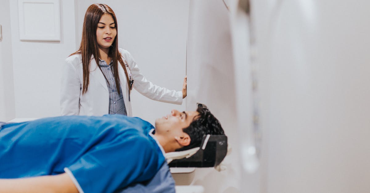
Comprehensive radiological imaging is vital for defining the tumor’s exact location, size, and relationship to critical anatomical structures. Magnetic resonance imaging with intravenous contrast is the premier modality for evaluating soft tissue tumors of the head and neck.
Magnetic resonance imaging provides exquisite detail of the facial nerve’s predicted pathway, assesses the depth of invasion into the deep lobe of the parotid gland or the parapharyngeal space, and detects any abnormal enlargement of the cervical lymph nodes.
Computed tomography scans of the neck and chest are also required for complete staging. The chest scan rules out pulmonary metastases, while the neck scan helps evaluate the bony structures, determining if the tumor has eroded into the mandible or the base of the skull, which would require complex composite surgical resections.
8. Fine-Needle Aspiration and Core Biopsy
Securing a preoperative tissue diagnosis guides the surgical approach. Ultrasound-guided fine-needle aspiration is routinely performed. A thin needle extracts cells from the mass for cytological evaluation. While safe and rapid, fine-needle aspiration may fail to definitively diagnose myoepithelial carcinoma due to the tumor’s immense cellular diversity.
If the aspiration is inconclusive, an ultrasound-guided core needle biopsy is performed to obtain a larger, intact cylinder of tissue. This provides the pathologist with the necessary architectural context to observe the tumor’s infiltrative borders.
Incrision biopsies (cutting directly into the tumor) of parotid masses are strictly avoided. Breaching the tumor capsule in the neck poses an unacceptably high risk of seeding malignant cells into the surgical field, which significantly increases the risk of a disastrous local recurrence.
9. Histological Diversity and Pleomorphism
Myoepithelial carcinoma is notorious among pathologists for its remarkable morphological heterogeneity. The tumor cells can adopt multiple distinct appearances, frequently co-existing within the same tumor mass. The cells may appear spindle-shaped (resembling a sarcoma), plasmacytoid (resembling plasma cells), epithelioid, or clear.
The cells are often suspended in a rich, myxoid (mucus-like) or hyalinized connective tissue stroma. To confirm malignancy, the pathologist must identify aggressive features: destructive invasion into surrounding normal salivary tissue, a high mitotic rate (numerous dividing cells), significant nuclear atypia, and extensive areas of tumor necrosis.
Because the tumor can look like a spindle cell sarcoma, a melanoma, or a metastatic carcinoma from another organ, accurate classification relies heavily on advanced molecular and protein staining techniques.
10. Immunohistochemistry for Definitive Diagnosis
Immunohistochemistry is the absolute diagnostic key for confirming myoepithelial lineage. The pathologist applies specific stains to the tissue sample to detect the presence of particular cellular proteins.
A true myoepithelial carcinoma will typically exhibit dual positivity for epithelial markers (such as broad-spectrum cytokeratins) and specific myoepithelial/muscle markers (such as S100 protein, calponin, p63, and smooth muscle actin).
This specific combination of positive stains confirms the tumor’s origin from the contractile cells of the salivary gland. Testing negative for other specific markers helps reliably exclude other salivary malignancies, such as adenoid cystic carcinoma or mucoepidermoid carcinoma, ensuring the patient receives the correct prognostic information.
11. Structured Data: Salivary Gland Tumors
Distinguishing this aggressive carcinoma from other salivary neoplasms is a critical diagnostic objective.
| Tumor Type | Cellular Composition | Growth Pattern | Clinical Behavior |
|---|---|---|---|
| Pleomorphic Adenoma | Mixed ductal and myoepithelial. | Encapsulated, non-invasive. | Benign, but can transform if left untreated. |
| Myoepithelial Carcinoma | Pure myoepithelial cells. | Highly infiltrative, destructive. | Aggressive, high risk of regional and distant metastasis. |
| Adenoid Cystic Carcinoma | Ductal and myoepithelial in specific patterns. | Prominent perineural invasion. | Slow-growing but relentless; prone to late metastasis. |
| Mucoepidermoid Carcinoma | Mucous, epidermoid, and intermediate cells. | Infiltrative; cystic areas common. | Behavior depends heavily on histological grade. |
12. Principles of Radical Surgical Resection
The primary and most effective treatment for myoepithelial carcinoma is radical surgical resection. The goal is to achieve an R0 resection, ensuring microscopically clear margins. For tumors located in the parotid gland, a superficial or total parotidectomy is performed.
The head and neck surgeon meticulously dissects the branches of the facial nerve away from the tumor. If the tumor is intimately wrapped around or invading the nerve, the involved segment of the nerve must be sacrificed to achieve clear oncological margins. Leaving gross tumor behind to save the nerve guarantees a fatal recurrence.
If the facial nerve is sacrificed, immediate nerve grafting or complex facial reanimation surgery is frequently performed concurrently by plastic reconstructive surgeons to restore facial tone and protect the eye from exposure keratitis.
13. Neck Dissection and Lymph Node Management
Because myoepithelial carcinoma has a notable propensity for lymphatic spread, managing the cervical lymph nodes is a crucial component of the surgical plan. If preoperative imaging or clinical examination reveals enlarged, suspicious lymph nodes in the neck, a therapeutic comprehensive neck dissection is mandated.
The surgeon removes the fatty tissue and all lymph node basins on the affected side of the neck, carefully preserving the spinal accessory nerve, internal jugular vein, and sternocleidomastoid muscle if they are not directly invaded by the tumor.
If the neck appears clinically negative on imaging, the surgeon may still perform an elective selective neck dissection to remove the first echelon of draining lymph nodes. This serves as a vital staging procedure, as microscopic nodal metastasis is common and substantially alters the need for post-operative radiation.
14. Adjuvant Radiation Therapy
Given the aggressive nature of the disease and the anatomical difficulty of achieving wide margins in the head and neck, adjuvant radiation therapy is routinely recommended following surgical recovery.
Radiation utilizes high-energy X-rays to target the surgical bed and the regional lymph node basins. It aims to eradicate any residual microscopic disease, significantly lowering the risk of locoregional recurrence. It is highly indicated if the surgical margins were close or positive, if the tumor exhibited perineural invasion, or if metastatic lymph nodes were identified.
Modern techniques, such as intensity-modulated radiation therapy, allow the radiation oncologist to precisely sculpt the radiation dose, minimizing severe damage to adjacent vital structures like the spinal cord, jawbone, and the contralateral healthy salivary glands.
15. Managing Systemic Spread
Myoepithelial carcinoma possesses the biological capacity for distant hematogenous metastasis, most frequently targeting the lungs, liver, and skeletal system. When systemic spread occurs, the disease is generally considered incurable, and the clinical focus shifts to palliation and disease control.
Systemic chemotherapy regimens, typically utilizing platinum-based agents or taxanes, may be offered, though response rates vary widely due to the rarity of the tumor and its inherent resistance to traditional cytotoxic drugs.
Comprehensive molecular profiling of the metastatic tissue is highly recommended. Identifying specific actionable genetic mutations may open pathways for utilizing targeted biological inhibitors or immunotherapy in specialized clinical trials, providing patients with advanced disease access to novel, life-extending therapies.
16. Frequently Asked Questions (FAQ)
1. What exactly is a myoepithelial cell?
Myoepithelial cells are normal, muscle-like cells that surround the saliva-producing glands. They contract to squeeze saliva into your mouth. In myoepithelial carcinoma, these specific cells mutate, stop functioning normally, and multiply aggressively to form a cancerous tumor.
2. Why is surgery on the parotid gland so difficult?
The facial nerve, which controls your ability to smile, blink, and move your face, runs directly through the middle of the parotid gland. The surgeon must carefully navigate around this nerve to remove the tumor without paralyzing your face.
3. Will I lose my facial movement after the surgery?
If the tumor is resting against or invading the facial nerve, the surgeon may have to remove that section of the nerve to get all the cancer. If this happens, reconstructive surgeons can often perform nerve grafts to restore movement, though recovery takes many months.
4. Why do I need radiation if the surgeon removed the whole lump?
This cancer is highly invasive and often sends microscopic roots into surrounding tissues that cannot be seen with the naked eye. Radiation acts as an insurance policy, cleaning up the surgical area to ensure no hidden cells grow back.
5. How is this cancer different from a benign salivary gland tumor?
Benign tumors grow very slowly, have smooth borders, and do not spread. Myoepithelial carcinoma grows rapidly, aggressively destroys surrounding tissues and nerves, and has a high risk of spreading to the lymph nodes and lungs.
17. Bibliography
Disclaimer: The content is for informational purposes only and does not replace medical advice. Always consult your doctor for personalized treatment.


