
1. Introduction to Tumourlet NOS
Tumourlet NOS, or Not Otherwise Specified, refers to a specific, microscopic proliferation of cells that displays some neoplastic features but does not meet the size criteria or biological behavior thresholds required for a formal cancer diagnosis. In medical pathology, the term tumourlet most frequently describes tiny, localized clusters of neuroendocrine cells found within the pulmonary parenchyma. These minute lesions are typically discovered entirely by chance during the evaluation of other respiratory conditions.
Despite their microscopic size and technically benign behavior, the presence of tumourlets warrants careful clinical evaluation. They represent an abnormal cellular hyperplasia that provides important clues regarding the underlying health of the surrounding tissue. In the lungs, their presence is intimately linked to chronic inflammation, airway scarring, and long-standing hypoxic conditions.
Understanding the nature of a tumourlet requires distinguishing it from early-stage, aggressive malignancies. While the cells themselves share histological similarities with low-grade neuroendocrine tumors, their confined nature and lack of progressive growth place them in a distinct clinical category. Management focuses primarily on addressing any underlying chronic tissue damage rather than aggressively targeting the tumourlet itself.
2. Defining the Not Otherwise Specified Classification
The designation NOS is frequently utilized in pathology when a lesion exhibits general characteristics of a specific cell lineage but lacks further defining features that would allow it to be classified into a more precise, narrow subcategory. It acts as a standardized medical identifier for lesions that are biologically straightforward but morphologically ambiguous.
In the context of a tumourlet, the NOS designation implies that while the cellular cluster is clearly identified as a benign or low-grade proliferation, it does not display the complex architectural features necessary to categorize it as a specific mature tumor type. It is essentially a descriptive diagnosis indicating a very small, non-invasive growth.
This classification prevents the over-diagnosis of cancer. By strictly defining these tiny clusters as tumourlets rather than early carcinomas, pathologists guide clinicians to pursue conservative management strategies, protecting patients from unnecessary surgical resections and the severe psychological burden of a cancer diagnosis.
3. Pulmonary Neuroendocrine Tumourlets
The most clinically relevant and frequently encountered type of tumourlet occurs in the lungs, specifically involving the pulmonary neuroendocrine cells. These specialized cells are naturally present in the epithelial lining of the respiratory tract, where they monitor oxygen levels and secrete peptides that regulate airway tone and blood flow.
A pulmonary neuroendocrine tumourlet is defined as a nodular proliferation of these neuroendocrine cells that measures less than five millimeters in maximum diameter. They often extend slightly beyond the basement membrane of the bronchioles but remain tightly localized, forming small nests within the adjacent fibrotic lung tissue.
When these clusters exceed five millimeters in diameter, the diagnostic classification automatically changes to a typical carcinoid tumor. This strict size threshold is vital because it formally separates a benign, reactive process from a true neuroendocrine neoplasm with theoretical metastatic potential.
4. Pathophysiology of Cellular Proliferation
The development of a pulmonary tumourlet is fundamentally a reactive physiological process. It is rarely a primary neoplastic event. Instead, it is considered a secondary hyperplastic response to severe, chronic injury of the respiratory epithelium.
When lung tissue undergoes chronic inflammation and subsequent scarring, the normal architecture of the bronchioles is distorted. In response to this fibrotic damage and the associated localized hypoxia, the neuroendocrine cells are stimulated to multiply. They undergo neuroendocrine hyperplasia, increasing in number in an attempt to regulate the damaged airway environment.
Over time, this hyperplasia can become nodular, forming the distinct clusters recognized as tumourlets. The cells within these clusters appear monotonous and regular. They do not harbor the severe genetic mutations, such as TP53 deletions or high mitotic rates, that drive the uncontrolled growth seen in true pulmonary malignancies.
5. Relationship with Chronic Lung Disease
There is a profound clinical correlation between the presence of pulmonary tumourlets and underlying chronic pulmonary diseases. Pathologists routinely identify these lesions in lung tissue specimens resected for severe bronchiectasis, chronic obstructive pulmonary disease, or idiopathic pulmonary fibrosis.
The chronic scarring characteristic of these conditions creates the exact microenvironment necessary for tumourlet formation. The fibrotic tissue acts as a matrix, supporting the nodular growth of the neuroendocrine cells. Therefore, finding a tumourlet is often a secondary marker indicating that the patient lung tissue has endured long-standing inflammatory stress.
In rare instances, patients may develop numerous tumourlets scattered throughout both lungs, a condition known as diffuse idiopathic pulmonary neuroendocrine cell hyperplasia. This specific condition can lead to airway narrowing and progressive shortness of breath, requiring specialized pulmonary management.
6. Tumourlet versus Carcinoid Tumor
The clinical differentiation between a tumourlet and a carcinoid tumor is one of the most critical distinctions in pulmonary pathology. Both lesions originate from the same neuroendocrine cell lineage and share identical cellular morphology under the microscope. The cells are small, uniform, and possess granular chromatin without significant atypia.
The primary distinguishing factor is strictly dimensional. Lesions smaller than five millimeters are classified as tumourlets, while those five millimeters or larger are classified as typical carcinoid tumors. This size threshold is supported by extensive clinical data demonstrating that lesions under five millimeters possess virtually zero risk of distant metastasis.
While a carcinoid tumor requires formal surgical resection and oncological staging, a tumourlet is typically considered an incidental finding requiring no specific oncological intervention. Accurate measurement by the pathologist is therefore the definitive step in determining the patient clinical pathway.
7. Clinical Manifestations and Respiratory Symptoms
Isolated tumourlets are entirely asymptomatic. Due to their microscopic size, they do not obstruct airways, cause bleeding, or produce enough neuroendocrine peptides to induce systemic symptoms. The clinical manifestations experienced by the patient are almost always attributable to the underlying chronic lung disease.
Patients often present with a chronic, productive cough, exertional dyspnea, and recurrent respiratory infections. These symptoms are driven by bronchiectasis or pulmonary fibrosis, not by the tumourlet itself. The tiny cellular clusters remain silent inhabitants within the scarred tissue matrix.
If a patient exhibits systemic flushing, diarrhea, or wheezing, it suggests the presence of a functioning neuroendocrine tumor, known as carcinoid syndrome. Tumourlets do not cause carcinoid syndrome. The presence of such systemic symptoms must prompt a rigorous search for a larger, biochemically active neuroendocrine malignancy elsewhere in the body. You can read more about evaluating chronic respiratory issues in our clinical guide on chronic cough.
8. Incidental Discovery on Imaging
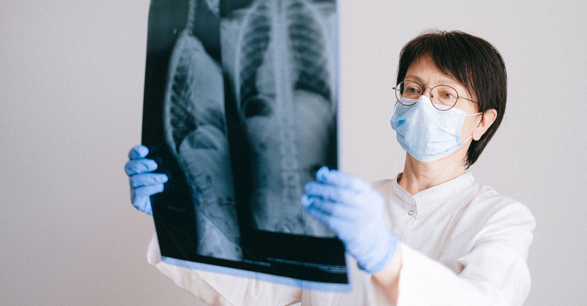
Because they are asymptomatic, tumourlets are rarely the primary target of diagnostic investigations. They are overwhelmingly discovered incidentally. Patients undergoing high-resolution computed tomography for the evaluation of chronic lung disease or during lung cancer screening programs frequently have these tiny nodules detected.
On computed tomography, a tumourlet appears as a minute, solid pulmonary nodule, often located in the peripheral lung fields or intimately associated with scarred and distorted bronchioles. Due to their size, they fall into the category of sub-centimeter pulmonary nodules, which generate significant clinical anxiety and necessitate structured follow-up protocols.
Radiologists must carefully evaluate the nodule characteristics. While a tumourlet is benign, its radiological appearance can be indistinguishable from early-stage primary lung cancer or a small metastatic deposit, leading to further diagnostic procedures.
9. High-Resolution Computed Tomography Findings
High-resolution computed tomography is the optimal modality for visualizing the subtle parenchymal changes associated with tumourlets. The scans provide detailed cross-sectional images that define the exact size, density, and location of the micro-nodules.
Radiologists look for specific patterns. Tumourlets are frequently seen clustered together in areas of established lung fibrosis, known as honeycomb change, or adjacent to dilated airways in bronchiectasis. The nodules are typically well-circumscribed and exhibit uniform tissue density without central cavitation.
Sequential imaging is the standard of care for these sub-centimeter nodules. By comparing current scans with previous imaging, clinicians can track the growth rate. Tumourlets remain remarkably stable over time, showing no significant increase in diameter over months or years, which strongly supports their benign biological nature.
10. Histopathological Features and Biopsy
When a nodule is biopsied or when lung tissue is surgically resected for other reasons, the definitive diagnosis of a tumourlet is made microscopically. The pathologist observes small nests or cords of uniform cells situated within the fibrotic submucosa of the terminal bronchioles.
The cells exhibit characteristic neuroendocrine nuclear features. The chromatin is finely stippled, often described as having a “salt and pepper” appearance. The cytoplasm is moderate in volume and slightly eosinophilic. Mitotic figures, areas of necrosis, and severe cellular pleomorphism are strictly absent.
The architectural growth is expansive but not destructively invasive. The cell nests push into the surrounding fibrotic tissue but do not destroy adjacent normal structures or invade vascular channels, confirming the lack of aggressive malignant potential.
11. Immunohistochemistry Markers
Immunohistochemical staining is utilized to definitively confirm the neuroendocrine lineage of the cells, differentiating them from small clusters of lymphocytes or reactive epithelial cells.
| Protein Marker | Cellular Function Indicated | Result in Tumourlet NOS |
|---|---|---|
| Synaptophysin | Neuroendocrine differentiation | Strongly Positive |
| Chromogranin A | Presence of secretory granules | Positive |
| CD56 (NCAM) | Neural cell adhesion | Positive |
| Ki-67 Index | Cellular proliferation rate | Very Low (typically < 2%) |
12. Differential Diagnosis and Diagnostic Challenges
The primary diagnostic challenge for the pathologist is differentiating a tumourlet from a minute focus of metastatic carcinoma or an early primary small cell lung cancer. Small cell lung cancer is also a neuroendocrine tumor, but it is substantially aggressive and requires immediate systemic treatment.
Small cell lung carcinoma cells exhibit severe nuclear molding, a very high mitotic rate, and extensive necrosis, features that are completely absent in a benign tumourlet. Additionally, the clinical context is vital; tumourlets are found in stable, scarred lungs, whereas small cell carcinoma presents as a rapidly growing, invasive mass.
Differentiating a tumourlet from a typical carcinoid tumor relies exclusively on the five-millimeter size threshold rule. Precise measurement using a calibrated microscope reticle is mandatory to ensure the lesion is classified correctly and the patient avoids unnecessary oncological staging.
13. Clinical Management and Surveillance Strategies
The diagnosis of Tumourlet NOS does not require specialized oncological treatment. Because these lesions represent a localized, benign hyperplasia, the clinical focus must shift entirely toward managing the patient underlying chronic pulmonary disease.
Optimization of respiratory function is the primary goal. Patients are treated with inhaled bronchodilators, corticosteroids to reduce airway inflammation, and targeted antibiotics for recurrent pulmonary infections associated with bronchiectasis. Smoking cessation is an absolute medical requirement to prevent further epithelial damage.
Radiological surveillance is the standard protocol for isolated tumourlets discovered on imaging. Patients undergo serial low-dose computed tomography scans at intervals determined by established pulmonary nodule guidelines, ensuring that the nodules remain dimensionally stable over time.
14. Surgical Interventions
Surgical resection is generally contraindicated for the primary treatment of an isolated tumourlet. The surgical risks associated with thoracotomy or video-assisted thoracoscopic surgery far outweigh any theoretical benefit of removing a microscopic, benign lesion.
Surgery is only indicated if the nodule demonstrates unexpected, rapid growth on surveillance imaging, raising the suspicion of a primary lung carcinoma. In such cases, a wedge resection may be performed to secure a definitive histological diagnosis.
Frequently, tumourlets are discovered incidentally in lung tissue that was surgically resected to treat severe localized bronchiectasis or as part of a lobectomy for an independent, separate lung cancer. In these instances, the finding of the tumourlet is noted in the pathology report but requires no further specific surgical action.
15. Prognosis and Long-Term Outcomes
The prognosis for a patient with an isolated Tumourlet NOS is excellent regarding the lesion itself. Tumourlets do not metastasize, they do not transform into aggressive high-grade carcinomas, and they do not cause disease-specific mortality.
The overall patient outcome is dictated entirely by the severity and progression of the concurrent chronic lung disease. Patients with severe, progressive pulmonary fibrosis or end-stage chronic obstructive pulmonary disease face significant respiratory challenges, independent of the microscopic tumourlets present within their lung parenchyma.
Long-term follow-up ensures that the patient respiratory status is optimized and that any new, unrelated pulmonary nodules are evaluated accurately. The incidental discovery of a tumourlet should be viewed by clinicians as a biological marker of chronic airway injury, prompting diligent, supportive pulmonary care.
16. Frequently Asked Questions (FAQ)
1. Is a Tumourlet NOS considered cancer?
No. It is a microscopic, benign cluster of cells. While the cells share some features with certain tumors, they do not grow uncontrollably, destroy tissue, or spread to other organs.
2. What causes a pulmonary tumourlet to form?
They generally form as a reaction to chronic lung damage. Conditions that cause scarring and inflammation in the lungs, like chronic bronchitis or fibrosis, stimulate these specific cells to multiply in small clusters.
3. Will a tumourlet cause coughing or shortness of breath?
The tumourlet itself is too small to cause symptoms. Any coughing or shortness of breath a patient experiences is caused by the underlying chronic lung disease that created the scarred environment in the first place.
4. Do I need surgery or chemotherapy for this?
No. Because tumourlets are benign and do not spread, surgery or chemotherapy is not required. Treatment is focused on managing your underlying lung health and using inhalers if needed.
5. How are tumourlets monitored over time?
Your doctor will likely recommend routine CT scans of your chest to monitor the size of the nodule. As long as it remains stable and under five millimeters, no specific intervention is needed.
17. Bibliography
Disclaimer: The content is for informational purposes only and does not replace medical advice. Always consult your doctor for personalized treatment.

