1. Introduction
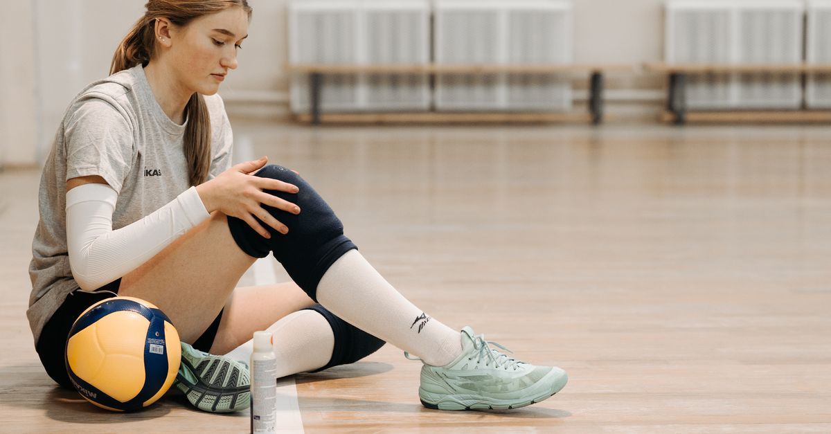
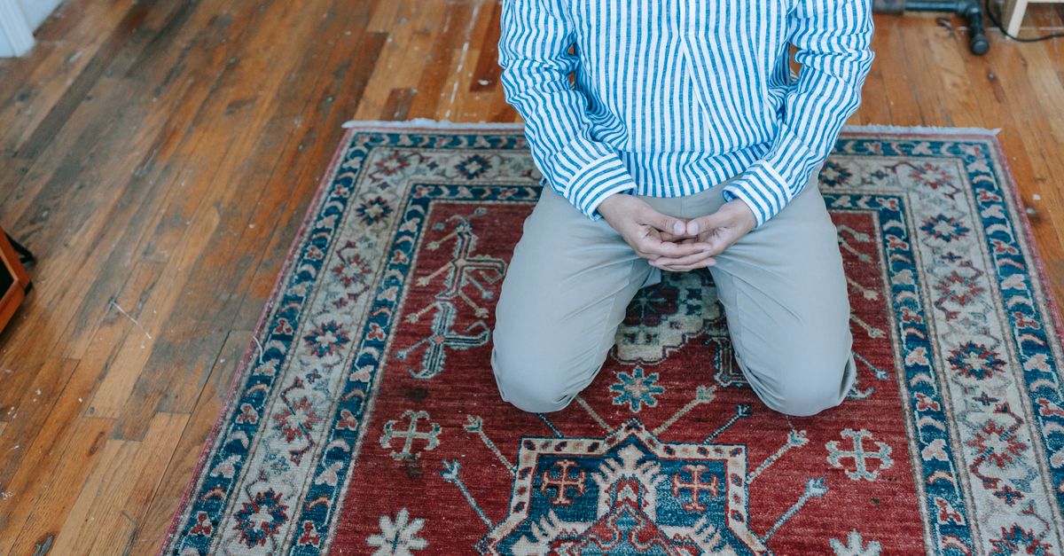
Sore and tender knees after kneeling on a hardwood floor are primarily caused by the intense mechanical compression of the prepatellar bursa and the acute restriction of blood flow to the anterior soft tissues of the joint. The rigid, unyielding surface of the wood forces the entire weight of the upper body onto the small, delicate structures located directly over the kneecap. This severe physical pressure initiates localized tissue ischemia and provokes a rapid inflammatory response within the synovial bursa, leading directly to the deep, aching tenderness experienced upon standing.
2. Anatomy of the Anterior Knee
The front of the knee is structurally designed to facilitate the gliding motion of the extensor mechanism, but it is poorly equipped for sustained weight-bearing. The prominent bone at the front is the patella, or kneecap, which sits within the tendon of the quadriceps muscle.
Overlying the patella is a thin layer of subcutaneous fat and skin. Directly between the skin and the hard bone of the patella lies a vital anatomical structure known as the prepatellar bursa.
Unlike the bottom of the foot, which is protected by a thick, honeycombed fat pad designed for impact, the anterior knee lacks dense structural cushioning, leaving the underlying bone and bursa highly exposed to external pressure.
3. The Role of the Prepatellar Bursa
A bursa is a small, fluid-filled sac lined with a synovial membrane. The human body strategically places bursae in areas where high friction occurs between bones, tendons, and skin.
The prepatellar bursa functions as a biological gliding surface. It allows the skin to move smoothly over the kneecap during the massive degree of flexion and extension required for walking and running.
While excellent at reducing shear friction, the bursa is not designed to bear heavy, static, compressive loads. When forced to act as a primary weight-bearing cushion against a rigid surface, the bursa is subjected to immense physiological stress.
4. Mechanical Compression and Ischemia
Kneeling on a hardwood floor concentrates a substantial percentage of your total body weight onto a surface area of just a few square inches. This immense focal pressure crushes the soft tissues of the anterior knee against the underlying bone.
This physical crushing immediately collapses the microscopic blood vessels supplying the skin, the fat pad, and the bursa. The occlusion of these capillaries halts local blood flow, plunging the tissues into a state of acute ischemia, or oxygen starvation.
Without oxygenated blood, the cells cannot maintain normal metabolic function. The localized ischemic stress irritates the surrounding nociceptors—pain-sensing nerve endings—generating a deep, dull, and burning ache as the kneeling posture is maintained.
5. Patellofemoral Joint Loading
The pain of kneeling extends beyond the superficial bursa; it deeply impacts the underlying joint mechanics. The patellofemoral joint is the articulation where the back of the kneecap rests against the femur.
When you kneel at a ninety-degree angle, the quadriceps tendon stretches tightly over the joint. This tension pulls the patella forcefully backward, compressing it against the femoral groove.
The articular cartilage lining the back of the patella is subjected to this massive compressive force. While cartilage is resilient, sustained static pressure squeezes out the lubricating synovial fluid, causing a deep, structural ache that originates inside the joint space rather than just on the skin surface.
6. Hoffa Fat Pad Irritation
Just below the kneecap and behind the patellar tendon sits Hoffa’s fat pad, a highly innervated and vascularized mass of adipose tissue. It acts as a protective spacer and secretes fluid to aid joint lubrication.
Depending on the exact angle of the kneel, this fat pad can be pinched between the hard floor, the patellar tendon, and the tibia.
Because Hoffa’s fat pad contains an exceptionally dense network of pain receptors, even mild mechanical compression or pinching generates severe, sharp tenderness located directly below the kneecap that worsens immediately upon standing or extending the leg.
7. Synovial Fluid Displacement
Synovial fluid behaves as a non-Newtonian fluid, meaning its viscosity changes based on pressure and movement. For the knee joint to remain healthy and pain-free, this fluid must circulate constantly over the cartilage surfaces.
Prolonged static kneeling forces the synovial fluid out from between the compressed joint surfaces, pushing it into the recesses of the joint capsule.
When you stand up after kneeling, the dry, unlubricated cartilage surfaces must suddenly articulate under the load of your body weight. This temporary lack of lubrication creates significant mechanical friction, resulting in a stiff, aching, and sometimes grinding sensation.
8. Inflammatory Mediators and Tissue Edema
The physical trauma of crushing the prepatellar bursa triggers the body’s immune system to initiate an inflammatory repair cascade. The synovial lining of the bursa becomes reactive and inflamed.
In response to the mechanical insult, the cells secrete inflammatory mediators like prostaglandins, which increase vascular permeability. This allows excess fluid to leak into the bursal sac, causing it to swell.
This condition, known as prepatellar bursitis or “housemaid’s knee,” presents clinically as a visible, soft, fluid-filled pouch situated directly over the kneecap. The distension of the bursa stretches the surrounding tissues, causing profound, localized tenderness.
9. Cartilage Stress on Rigid Surfaces
The density of the surface dictates the severity of the tissue trauma. A soft surface, like a thick carpet or a garden kneeling pad, yields to the shape of the knee, distributing the body weight over a larger anatomical area and reducing focal pressure.
A hardwood floor offers zero compliance. It forces the convex shape of the patella to bear the entire load against a rigid, unyielding plane.
This lack of shock absorption means that one hundred percent of the compressive force is reflected back into the biological tissues, rapidly accelerating the onset of ischemia, bursal inflammation, and cartilage stress.
10. Cutaneous Nerve Compression
The skin overlying the knee is supplied by branches of the saphenous nerve and other small cutaneous nerves. These delicate nerve branches travel superficially just beneath the skin.
Kneeling on a hard surface physically flattens these nerves against the bone. Prolonged nerve compression disrupts electrical conduction and causes transient neural ischemia.
This sensory disruption is why the skin over the knee may feel numb, tingly, or exquisitely sensitive to a light touch for several minutes after you finally stand up and relieve the pressure.
11. Meniscal Squeeze During Deep Flexion
If the kneeling posture involves sitting back on the heels, the knee is forced into maximum hyperflexion. In this extreme angle, the posterior horns of the menisci—the C-shaped shock absorbers inside the knee—are aggressively pinched between the femur and the tibia.
This deep meniscal squeeze places immense shear force on the cartilage. While healthy menisci can tolerate this briefly, prolonged hyperflexion disrupts the internal mechanics of the joint.
The pain from meniscal compression is distinctly felt deep within the back of the knee or along the joint line, differing significantly from the superficial pain of bursal compression.
12. Cumulative Trauma and Occupational Risks
Occasional kneeling produces transient soreness that resolves quickly. However, repetitive daily kneeling, common in occupations like flooring installation, carpentry, or plumbing, constitutes cumulative microtrauma.
Repeatedly crushing the prepatellar bursa leads to chronic inflammation. The synovial walls of the bursa thicken, and fibrotic scar tissue develops within the sac.
A chronically inflamed bursa loses its ability to function smoothly. The knee becomes permanently tender, swollen, and prone to severe flare-ups with even minimal contact against a hard surface.
13. Clinical Evaluation of Anterior Knee Pain
When knee soreness following kneeling becomes persistent or is accompanied by significant swelling, a clinical evaluation is necessary to rule out infection or structural joint damage.
A clinician relies on palpation and range of motion testing to isolate the involved tissues.
| Clinical Finding | Characteristics | Probable Anatomical Source |
|---|---|---|
| Soft, localized swelling directly over the kneecap. | Tender to touch, normal joint movement. | Prepatellar Bursitis. |
| Deep aching behind the kneecap. | Worsens when using stairs or squatting. | Patellofemoral joint stress / Cartilage irritation. |
| Sharp pain just below the kneecap. | Tender when pressing the soft tissue beside the tendon. | Hoffa’s Fat Pad impingement. |
14. Conservative Management and Joint Protection
The definitive prevention for this specific pain is mechanical protection. Utilizing high-density foam knee pads or a thick kneeling mat introduces an external layer of compliance. This distributes the body weight over a larger surface area, drastically reducing the focal compression on the bursa and the superficial nerves.
If soreness occurs, applying an ice pack to the anterior knee for fifteen minutes induces localized vasoconstriction, checking the inflammatory fluid accumulation within the bursa and numbing the sensitized nerve endings.
Avoiding prolonged static kneeling by shifting positions frequently, standing up, and stretching the quadriceps allows blood flow to return to the ischemic tissues, preventing severe metabolic fatigue.
15. Frequently Asked Questions FAQ
1. Is the swelling on my knee filled with water or blood?
In most cases of benign prepatellar bursitis, the swelling is filled with excess synovial fluid, which is clear and viscous. If the swelling occurs immediately after a traumatic, hard fall directly onto the kneecap, it may contain blood, requiring medical evaluation.
2. Will the fluid in the bursa go away on its own?
Yes, mild to moderate bursal swelling usually reabsorbs naturally over a week or two if you completely avoid kneeling and protect the joint from further mechanical trauma. Applying ice and keeping the leg elevated assists the lymphatic clearance of the fluid.
3. Do I need to have the fluid drained by a doctor?
Aspiration (draining with a needle) is generally reserved for severe, tense swelling that restricts movement, or if the clinician suspects the bursa has become infected. Routine, painless swelling is best left to heal conservatively to avoid introducing bacteria into the sterile sac.
4. Why does my knee hurt more when I transition from kneeling to standing?
When you kneel, the fluid is squeezed out of the joint, and the tissues are compressed. Standing suddenly requires the dry cartilage surfaces to bear weight and the stiff, ischemic muscles to contract rapidly, creating an immediate, sharp mechanical ache before blood flow normalizes.
5. Can chronic kneeling cause arthritis?
Chronic kneeling primarily damages the superficial bursa (bursitis), rather than the deep articular cartilage (arthritis). However, if kneeling forces the kneecap to track improperly over time, it can accelerate wear and tear on the patellofemoral joint cartilage.
16. Bibliography
Disclaimer: The content is for informational purposes only and does not replace medical advice. Always consult your doctor for personalized treatment.