1. Introduction to Fibroma of Tendon Sheath
A fibroma of the tendon sheath is a rare, solitary, and entirely benign soft tissue tumor that arises from the connective tissue lining the tendons. These slow-growing nodules are most frequently discovered in the upper extremities, particularly affecting the fingers, hands, and wrists. While completely non-cancerous, their physical presence can cause mechanical interference with daily activities, leading patients to seek medical evaluation.
The appearance of a localized mass on the hand naturally generates concern. However, healthcare professionals can assure patients that a fibroma of the tendon sheath exhibits no malignant behavior and will not spread to other areas of the body. The lesion consists of dense fibrous tissue that firmly attaches to the tendon sheath, the protective sleeve covering the tendon.
Accurate clinical and histological diagnosis is essential to differentiate this lesion from other common hand tumors. The primary objective of treatment is surgical excision, which not only provides tissue for a definitive pathological diagnosis but also restores normal mechanical function to the affected digit or joint, ensuring the patient can return to their daily routines without discomfort.
2. Anatomical Locations and Pathophysiology
The pathophysiology of a fibroma of the tendon sheath involves the localized, excessive proliferation of fibroblasts and myofibroblasts. These specialized connective tissue cells generate an overabundance of dense collagen, which forms the bulk of the solitary nodule. The tumor originates directly from the tenosynovial membrane, which is the specialized tissue responsible for lubricating tendons as they glide over joints.
Anatomically, the vast majority of these fibromas develop in the upper extremity. The fingers and the volar aspect, or palm side, of the hand are the most commonly affected sites. The thumb, index, and middle fingers are particularly susceptible. Less frequently, these benign tumors can manifest in the lower extremities, presenting around the toes, feet, or ankles.
The mass usually grows in a lobular pattern and is firmly tethered to the underlying tendon or its sheath. Because of this rigid attachment, the fibroma moves in tandem with the tendon during active flexion or extension of the affected digit, a classic clinical sign that aids physicians in initial anatomical assessment.
3. Macroscopic and Microscopic Features
When examined macroscopically following surgical removal, a fibroma of the tendon sheath appears as a well-circumscribed, firm, and nodular mass. The tissue is typically grayish-white and exhibits a rubbery texture, reflecting its dense fibrous composition. The size of the tumor is generally small, usually ranging from one to three centimeters in maximum diameter.
Microscopically, the tumor is characterized by a prominent lobular architecture separated by deep clefts. The predominant cellular component consists of bland, spindle-shaped fibroblasts and myofibroblasts embedded in a densely collagenous stroma. The cells are widely spaced and exhibit no malignant features, such as nuclear atypia or increased mitotic figures.
A unique and defining histological feature of this tumor is the presence of numerous narrow, slit-like vascular channels scattered throughout the dense collagen. These compressed blood vessels are integral to the diagnosis, allowing pathologists to accurately identify the lesion and differentiate it from other fibrous growths.
4. Genetic and Molecular Insights
Current medical research indicates that the fibroma of the tendon sheath is a neoplastic process rather than a simple reactive hyperplasia. Recent molecular studies have identified specific genetic abnormalities in a subset of these tumors. A recurrent chromosomal translocation involving the USP6 gene has been documented in some cases.
The discovery of the USP6 gene rearrangement provides fascinating insight into the biology of this benign tumor. It suggests a shared genetic mechanism with other benign fibrous lesions, such as nodular fasciitis. However, despite these molecular alterations, the clinical behavior of the fibroma remains strictly benign and non-aggressive.
While genetic testing is not routinely required for standard diagnosis and treatment, these molecular insights help scientific researchers classify soft tissue tumors more accurately and understand the underlying cellular pathways that drive fibrous tissue proliferation.
5. Clinical Presentation and Physical Signs
Patients with a fibroma of the tendon sheath generally present to their physician complaining of a slow-growing, solitary lump on their hand or finger. The mass is remarkably firm to the touch and typically painless during its initial growth phase. Patients often report that the lump has been present for several months or even years before they decided to seek a medical opinion.
Because the tumor is physically anchored to the tendon sheath, a key clinical sign is its mobility. The mass will frequently move longitudinally when the patient actively flexes or extends the corresponding finger, but it exhibits restricted mobility in the transverse, or side-to-side, direction.
As the fibroma enlarges within the confined anatomical spaces of the hand, it may begin to exert pressure on adjacent structures. This compression can lead to localized tenderness, a decreased range of motion in the affected joint, or an uncomfortable catching sensation similar to a trigger finger. If the mass compresses a nearby digital nerve, the patient might experience mild numbness or a tingling sensation in the finger.
6. Potential Causes and Demographics
The precise etiology of a fibroma of the tendon sheath remains unknown. Unlike some connective tissue disorders, there is no definitive link to repetitive occupational strain, dietary factors, or environmental exposures.
However, a significant subset of patients report a history of antecedent local trauma to the affected area. This has led to a clinical hypothesis that an initial, minor mechanical injury to the tenosynovium might trigger an abnormal, localized fibrotic healing response in susceptible individuals, eventually culminating in tumor formation.
Demographically, this benign condition most frequently affects adults between the ages of twenty and fifty. There is a documented male predominance, with men being diagnosed approximately twice as often as women. The incidence of this tumor in pediatric populations or the elderly is notably rare.
7. Diagnostic Imaging and Ultrasonography
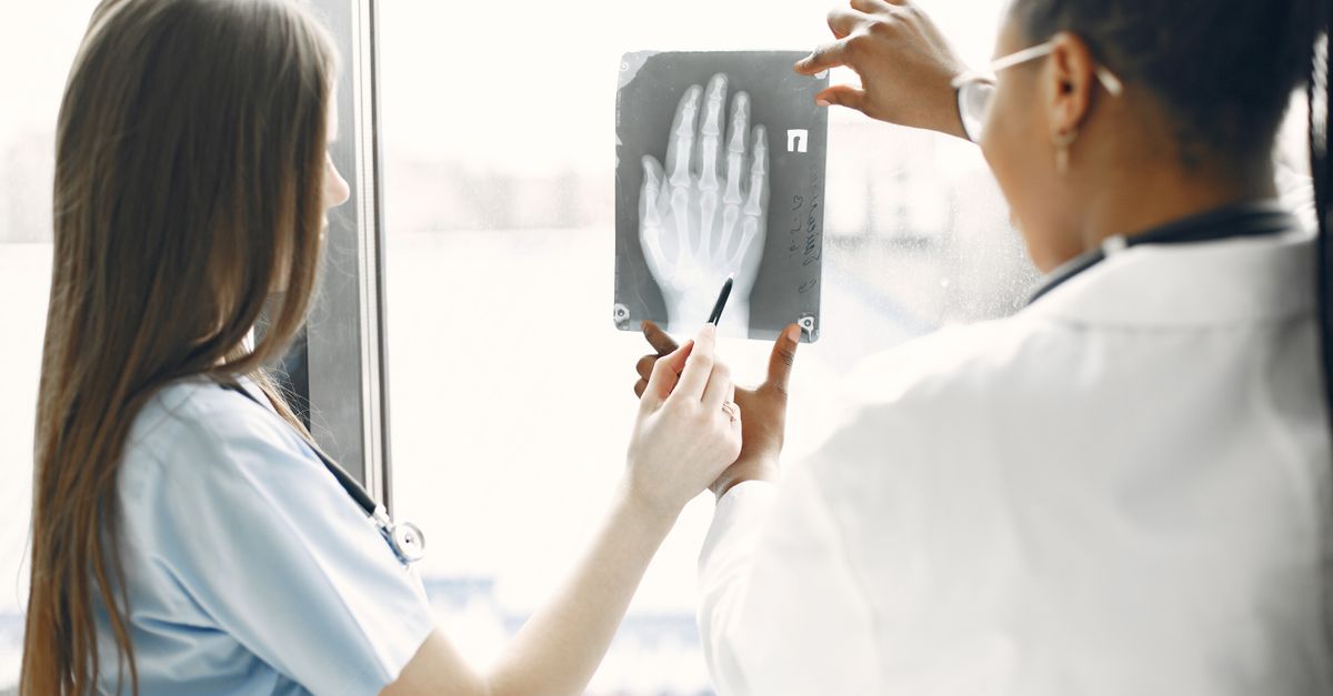
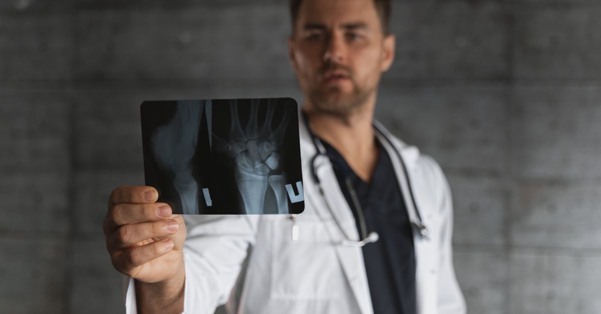
When evaluating a hand mass, diagnostic imaging provides crucial anatomical information prior to any surgical intervention. High-resolution ultrasonography is an excellent initial modality. It is non-invasive, cost-effective, and highly detailed for superficial soft tissue structures.
On an ultrasound, a fibroma of the tendon sheath typically presents as a well-defined, solid, hypoechoic mass closely associated with a tendon. The sonographer can ask the patient to move their finger during the examination, dynamically demonstrating the mass gliding along with the tendon sheath, which strongly supports the clinical diagnosis.
Furthermore, Doppler ultrasound can assess the vascularity of the lesion. These fibromas generally show minimal to no internal blood flow on Doppler imaging, a reassuring finding that helps differentiate them from highly vascularized malignant tumors.
8. Magnetic Resonance Imaging Characteristics
For deeper lesions or for precise preoperative planning in complex anatomical areas of the hand, magnetic resonance imaging is the gold standard. Magnetic resonance imaging offers superior soft tissue resolution and clearly delineates the margins of the tumor from adjacent nerves and blood vessels.
Because a fibroma of the tendon sheath is composed of dense, acellular collagen, its magnetic resonance imaging signature is highly characteristic. The mass typically demonstrates uniformly low signal intensity on both T1-weighted and T2-weighted sequences. This “dark” appearance on T2-weighted images is a classic indicator of dense fibrous tissue.
Following the administration of an intravenous contrast agent, the tumor usually exhibits mild, heterogeneous enhancement. This imaging profile helps the surgeon confirm the benign, fibrous nature of the mass and plan a surgical approach that safely preserves hand function.
9. Differential Diagnosis
The clinical and radiographic presentation of a fibroma of the tendon sheath closely mimics several other common soft tissue lesions of the hand.
| Condition | Differentiating Characteristics |
|---|---|
| Giant Cell Tumor of Tendon Sheath | Often more lobulated, may show variable signal on MRI due to hemosiderin, histologically cellular with multinucleated giant cells. |
| Ganglion Cyst | Fluid-filled, transilluminates with light, displays extremely high (bright) signal on T2-weighted magnetic resonance imaging. |
| Epidermal Inclusion Cyst | Located more superficially in the skin, often associated with prior penetrating trauma, contains keratinous material. |
| Nodular Fasciitis | Grows much more rapidly, may be tender, histologically shows plump fibroblasts in a myxoid stroma. |
A definitive distinction among these conditions relies on careful histological examination by a pathologist following surgical removal.
10. Excisional Biopsy and Pathology
Because fine-needle aspiration of dense fibrous lesions often yields insufficient cellular material, an excisional biopsy is the standard diagnostic and therapeutic approach. This means the surgeon removes the entire mass during the procedure, which is then sent to the pathology laboratory for comprehensive evaluation.
The pathologist assesses the tissue architecture, cellular morphology, and the distinctive slit-like vascular channels. In ambiguous cases, immunohistochemistry may be utilized. The spindle cells in a fibroma of the tendon sheath generally test positive for vimentin and smooth muscle actin, while testing negative for S100 and desmin.
This rigorous pathological analysis is the only way to provide a conclusive diagnosis, ensuring that the patient and the clinical team can confidently rule out any form of malignancy.
11. Surgical Treatment Modalities
The primary and most effective treatment for a fibroma of the tendon sheath is marginal surgical excision. The procedure is typically performed by an orthopedic or plastic hand surgeon under regional or local anesthesia, frequently on an outpatient basis.
During the surgery, the physician makes a careful incision over the mass. Because the tumor is intimately attached to the tendon sheath, the surgeon must meticulously dissect the dense fibrous tissue away from the underlying tendon. The objective is to remove the tumor entirely while preserving the structural integrity and gliding mechanism of the tendon.
If the tumor has encased small digital nerves or vessels, micro-surgical techniques may be employed to carefully free these vital structures. Complete excision is crucial not only for symptomatic relief but also for minimizing the risk of the tumor returning.
12. Rehabilitation and Hand Therapy
Following surgical excision, proper postoperative care is vital to restore full function to the hand. The hand is initially placed in a soft dressing or a light splint to protect the surgical site and minimize localized swelling.
Early, controlled mobilization is a critical component of recovery. To prevent the formation of internal scar tissue, which can cause tendons to adhere to surrounding structures and limit movement, patients are encouraged to begin gentle range-of-motion exercises within a few days after surgery.
In some cases, referral to a specialized occupational or hand therapist is highly beneficial. The therapist guides the patient through specific exercises, scar massage, and strengthening routines, ensuring optimal functional recovery and a swift return to normal daily activities.
13. Complications and Recurrence
Complications following the surgical excision of a fibroma of the tendon sheath are rare but can include standard surgical risks such as localized infection, delayed wound healing, or transient numbness if a digital nerve was manipulated during dissection.
The most significant clinical consideration post-surgery is the potential for local recurrence. Studies indicate a recurrence rate of approximately ten to twenty-five percent. Recurrence is generally attributed to the incomplete removal of the fibrous tissue, particularly when the tumor is tightly adhered to complex tenosynovial structures.
If a recurrence does occur, it typically manifests as a slow-growing nodule at the original surgical site. Repeated surgical excision is the standard management for recurrent cases and is highly successful.
14. When to Consult a Specialist
Individuals should schedule an evaluation with a healthcare provider if they notice a new, persistent lump on their finger, hand, or wrist. Early assessment is particularly important if the mass begins to restrict joint movement, causes a triggering sensation, or induces pain.
Prompt medical attention is also necessary if a known lump rapidly changes in size, becomes acutely painful, or if there are associated symptoms such as numbness, tingling, or weakness in the fingers.
Consulting a specialist, such as a hand surgeon, ensures that the patient receives an accurate diagnosis through physical examination and appropriate imaging, followed by an expert surgical intervention tailored to preserve hand mechanics and alleviate anxiety.
15. Frequently Asked Questions (FAQ)
1. Is a fibroma of the tendon sheath cancerous?
No. It is a completely benign soft tissue tumor. It does not have the ability to spread to other parts of the body or transform into cancer.
2. Why did I get a fibroma on my finger?
The exact cause is unknown. However, some cases are linked to prior minor injuries or repetitive mechanical trauma to the finger that triggered an abnormal, localized healing response.
3. Do I have to have surgery to remove it?
If the fibroma is small, painless, and does not interfere with hand function, careful observation is an option. However, surgery is the only way to completely remove the mass and obtain a definitive diagnosis.
4. Will the lump grow back after surgery?
There is a modest risk of recurrence, estimated between ten and twenty-five percent, particularly if microscopic fragments of the fibrous tissue remain attached to the tendon sheath.
5. How long is the recovery after surgery?
Most patients recover quickly. Stitches are typically removed after two weeks, and full functional recovery, often aided by hand therapy, is usually achieved within a few weeks to a couple of months.
16. Bibliography
Disclaimer: The content is for informational purposes only and does not replace medical advice. Always consult your doctor for personalized treatment.