1. Introduction to Calcifying Aponeurotic Fibroma
A calcifying aponeurotic fibroma, historically recognized in medical literature as a Keasbey tumor, is a rare, slow-growing soft tissue neoplasm that characteristically develops in the distal extremities of children and young adults. This fibrous tumor originates from the deep connective tissues, specifically the aponeuroses, tendons, and fascia, most frequently affecting the hands and the feet.
The discovery of a firm, deep-seated mass in a child hand naturally elicits significant concern for parents and healthcare providers, as it mandates the exclusion of malignant soft tissue sarcomas. Clinicians can provide profound reassurance that a calcifying aponeurotic fibroma is an entirely benign entity. It does not harbor the biological ability to undergo malignant transformation, nor does it metastasize to distant anatomical sites.
Despite its benign classification, this tumor is well-known in orthopedic oncology for its locally aggressive growth pattern and its exceptionally high propensity for local recurrence following surgical removal. Clinical management therefore requires a delicate balance: achieving adequate surgical excision to prevent the tumor from returning, while meticulously preserving the complex, vital anatomical structures and mechanical function of the affected hand or foot.
2. Fibroblastic Proliferation and Pathophysiology
The pathophysiology of a calcifying aponeurotic fibroma involves the localized, abnormal proliferation of fibroblasts, which are the primary cells responsible for synthesizing connective tissue and collagen. In this condition, these fibroblasts multiply in a highly infiltrative manner along the planes of the deep fascia, tendons, and aponeuroses.
The tumor exhibits a distinct biphasic growth pattern that is closely tied to the patient age and skeletal maturity. In its early, initial phase, often observed in very young children, the tumor is highly cellular, rapidly growing, and infiltrative. During this phase, it frequently lacks the characteristic calcifications for which the tumor is named, presenting instead as a dense, fibrous mass.
As the patient ages and the tumor matures into its late phase, the cellular proliferation slows significantly. The tissue undergoes extensive cartilaginous metaplasia, a process where the fibrous connective tissue transforms into cartilage-like tissue. Subsequently, dense focal calcifications develop within these areas. This unique evolution from a cellular, infiltrative process to a heavily calcified, mature mass is the defining pathophysiological hallmark of the disease.
3. Histological Hallmarks and Calcification
Definitive diagnosis requires a meticulous microscopic evaluation by a specialized soft tissue pathologist. The histological appearance varies dramatically depending on the developmental phase of the tumor at the time of surgical excision.
A classic, mature calcifying aponeurotic fibroma presents a highly distinctive architectural landscape. The pathologist observes dense, cellular sheets of plump, spindle-shaped fibroblasts interspersed with abundant collagen fibers. These fibroblasts aggressively infiltrate the surrounding normal adipose tissue and skeletal muscle, contributing to the tumor high recurrence rate.
Embedded within this dense fibrous stroma are distinct, nodular areas of cartilaginous tissue that frequently surround central zones of dense, chunky calcification. These calcified nodules are often bordered by a layer of multinucleated giant cells and plump epithelioid fibroblasts. Crucially, despite the aggressive local infiltration, the cells display no malignant nuclear atypia, and mitotic activity remains reassuringly low.
4. Clinical Presentation and Symptomatology
Patients typically present to a pediatric or orthopedic clinic with a solitary, painless mass located on the palmar surface of the hand or the plantar surface of the foot. Because the tumor originates deep within the aponeurotic tissues, it often feels remarkably firm, immobile, and intimately tethered to the underlying tendons or skeletal structures.
The growth rate of the mass is generally slow and insidious. Parents may report that the lump has been present for several months or even years, gradually increasing in size. The overlying skin typically remains entirely normal, lacking any redness, warmth, or signs of acute inflammation.
While the mass itself is inherently painless, its physical presence within the confined, anatomically complex spaces of the hand or foot can induce secondary mechanical symptoms. As the tumor enlarges, it may restrict the normal gliding motion of the tendons, leading to a decreased range of motion in the affected fingers or toes. Rarely, it may compress adjacent peripheral nerves, causing mild, localized numbness or tingling.
5. Anatomical Focus on Hands and Feet
The anatomical distribution of a calcifying aponeurotic fibroma is one of its most defining clinical characteristics. The overwhelming majority of these tumors—over seventy percent of all documented cases—arise in the distal extremities.
The volar aspect of the hand, specifically the palms and the fingers, is the absolute most frequent site of occurrence. Following the hands, the plantar aspect of the feet and the toes are the next most common locations. The tumor originates directly from the tough, fibrous bands of the palmar or plantar aponeuroses.
While significantly less common, medical literature documents rare occurrences in larger anatomical sites such as the wrist, forearm, ankle, knee, and even the paraspinal muscles of the back. However, when evaluating a deep, firm mass in the palm of a child hand, this specific tumor must always remain high on the clinician list of differential diagnoses.
6. Demographics and Pediatric Incidence
Calcifying aponeurotic fibroma is predominantly a pediatric and adolescent disease. The vast majority of diagnoses are made in patients under the age of twenty, with a significant peak incidence occurring in children and young teenagers between the ages of eight and fourteen.
While less frequent, the tumor can occasionally present in infants or, conversely, manifest for the first time in adults over the age of thirty. When occurring in adults, the tumors tend to be in the late, mature phase, exhibiting extensive calcification and much slower growth rates compared to the cellular variants seen in young children.
Regarding gender distribution, clinical studies consistently demonstrate a strong male predominance. Boys and young men are affected approximately twice as often as female patients. The condition is sporadic and does not demonstrate any known hereditary or familial inheritance patterns.
7. Potential Causes and Genetic Mutations
The exact underlying etiology of calcifying aponeurotic fibroma remains incompletely understood. Historically, it was debated whether the lesion represented a reactive inflammatory fibromatosis secondary to unrecognized minor trauma or a true neoplastic tumor.
Recent advancements in molecular biology strongly support a neoplastic origin. Cytogenetic research has identified a specific, recurrent genetic mutation in a significant subset of these tumors. Researchers frequently observe an alteration involving the FN1 gene, often manifesting as a genetic rearrangement or fusion.
This specific genetic anomaly provides compelling evidence that the tumor is a true biological neoplasm driven by a genetic error in the fibroblastic cells, rather than a simple reactive healing process. However, this mutation is strictly localized to the tumor cells and is not inherited from the parents, confirming the sporadic nature of the disease.
8. Diagnostic Imaging and Radiography
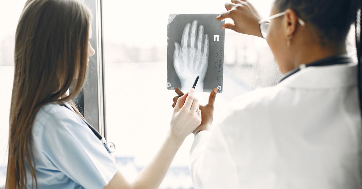
Radiological imaging plays a crucial role in the initial diagnostic workup and preoperative planning. Standard plain radiographs, or X-rays, of the affected hand or foot are typically the first imaging modality utilized.
The appearance of the tumor on an X-ray is highly dependent on its developmental phase. In the early, cellular phase typically seen in young children, the X-ray may only show a faint, non-specific soft tissue swelling, as the tumor has not yet developed mineralized components.
However, in the mature phase, the radiograph reveals the classic, pathognomonic feature: fine, stippled, or chunky radiopaque calcifications clustered within the soft tissue mass. These calcifications do not typically involve the adjacent bone. The underlying bones generally appear entirely normal, without signs of destructive erosion or periosteal reaction, which is a reassuring finding that helps rule out malignant bone tumors.
9. Magnetic Resonance Imaging Findings
For precise preoperative anatomical mapping, magnetic resonance imaging is the gold standard. Magnetic resonance imaging is essential for delineating the exact extent of the tumor, particularly due to its infiltrative growth pattern along tendon sheaths and fascial planes in the complex anatomy of the hand.
On magnetic resonance imaging, a calcifying aponeurotic fibroma typically presents as a poorly defined, infiltrative mass. On T1-weighted images, the tumor generally displays low to intermediate signal intensity. On T2-weighted images, the signal intensity is highly heterogeneous.
Areas of dense cellularity may appear hyperintense, while areas of dense collagen and the pathognomonic calcifications demonstrate pronounced, low-signal intensity voids. Following the administration of intravenous contrast, the tumor typically shows moderate, heterogeneous enhancement. This detailed imaging helps the orthopedic surgeon plan an approach that maximizes tumor removal while safeguarding vital nerves and blood vessels.
10. Differential Diagnosis
Distinguishing a calcifying aponeurotic fibroma from other soft tissue lesions of the distal extremities is a critical aspect of clinical practice.
| Soft Tissue Mass | Key Differentiating Features |
|---|---|
| Calcifying Aponeurotic Fibroma | Child/adolescent, hand/foot location, characteristic stippled calcifications on X-ray. |
| Giant Cell Tumor of Tendon Sheath | Common in adults, usually lacks significant calcification, lobulated appearance on MRI. |
| Synovial Sarcoma | Malignant, highly aggressive, often painful, features the t(X;18) genetic translocation. |
| Plantar/Palmar Fibromatosis | Common in older adults, forms firm nodules in the fascia, completely lacks calcifications. |
A precise diagnosis relies on the integration of clinical history, radiological findings, and expert pathological review. For related concerns regarding joint and tendon discomfort, patients might review joint pain guidelines.
11. The Necessity of Surgical Biopsy
Due to the deep, infiltrative nature of the mass and its potential to mimic malignant sarcomas clinically and radiologically, a definitive tissue diagnosis is absolutely mandatory before initiating definitive treatment.
An open incisional biopsy or a core needle biopsy is typically performed. An open biopsy provides a larger volume of tissue, ensuring the pathologist can accurately identify the complex biphasic architecture and the characteristic zones of calcification and cartilaginous metaplasia.
Fine needle aspiration is wholly inadequate for diagnosing this condition, as it fails to capture the essential structural tissue patterns required to differentiate this benign, locally aggressive tumor from a malignant soft tissue sarcoma.
12. Surgical Excision Strategies
The primary and only definitive treatment for a calcifying aponeurotic fibroma is surgical excision. However, the surgical strategy requires meticulous planning by an experienced orthopedic hand or foot surgeon.
Because the tumor is benign and will never metastasize, amputation or highly destructive radical surgeries are never indicated. The surgical objective is to perform a wide local excision, removing the entire tumor mass along with a margin of healthy tissue, whenever anatomically feasible.
The significant challenge lies in the tumor highly infiltrative growth pattern. The fibrous tissue frequently entwines itself around delicate digital nerves, critical blood vessels, and essential flexor or extensor tendons. The surgeon must carefully balance the desire for complete tumor removal against the absolute necessity of preserving the mechanical function and sensation of the patient hand or foot.
13. Addressing High Recurrence Rates
The most frustrating clinical aspect of managing a calcifying aponeurotic fibroma is its exceptionally high rate of local recurrence. Medical literature indicates that up to fifty percent of these tumors will recur at the original surgical site following the initial excision.
This high recurrence rate is directly attributed to the tumor ill-defined, infiltrative margins. Microscopic clusters of fibroblastic cells inevitably remain embedded in the complex fascial planes of the hand, even after an apparently thorough surgical excision. Notably, the risk of recurrence is highest in very young children, whose tumors are typically in the highly cellular, rapidly growing phase.
If a recurrence occurs, the standard clinical protocol is a repeat surgical excision. Despite multiple local recurrences, the tumor remains steadfastly benign and never undergoes malignant transformation.
14. Postoperative Rehabilitation
Following surgery, particularly in the hand, structured postoperative rehabilitation is crucial for ensuring a successful clinical outcome. The hand is initially immobilized in a splint to protect the surgical incisions and allow the deep tissues to heal securely.
Once initial healing has occurred, typically within ten to fourteen days, the patient is referred to a specialized occupational or hand therapist. The therapist guides the patient through a rigorous regimen of range-of-motion exercises, scar tissue massage, and tendon gliding protocols.
Because the surgery involves deep dissection around the tendons, there is a significant risk of internal scar tissue formation that can permanently restrict joint movement. Early, aggressive, and compliant participation in hand therapy is essential to prevent these adhesions and restore full, normal dexterity to the affected digits.
15. When to Consult a Specialist
Parents should arrange an immediate medical evaluation if they discover any new, firm, or deep-seated lump on their child hand, foot, or elsewhere on the body. An accurate, professional diagnosis is imperative for any pediatric soft tissue mass.
Prompt consultation with a pediatric orthopedic surgeon or an orthopedic oncologist is highly recommended. These specialists possess the advanced expertise required to interpret complex imaging studies, perform accurate biopsies, and execute intricate surgical excisions in functionally critical areas like the hand.
Early specialized intervention ensures that the child receives the most appropriate, function-preserving treatment while providing the family with definitive reassurance regarding the benign nature of the tumor.
16. Frequently Asked Questions (FAQ)
1. Is a calcifying aponeurotic fibroma a type of cancer?
No, it is a completely benign tumor. Although it can grow aggressively into nearby tissues in the hand or foot, it cannot spread to other organs or cause systemic cancer.
2. Why do these tumors have calcium in them?
As the tumor matures over time, the fibrous tissue naturally changes into a cartilage-like tissue, which then collects calcium deposits. This is a characteristic feature of the tumor biology and is not related to the child diet or calcium intake.
3. Will my child lose function in their hand after surgery?
The goal of the specialized hand surgeon is to completely preserve function. While the surgery is delicate, most children regain excellent dexterity, especially if they follow a dedicated postoperative hand therapy and exercise program.
4. Why is the lump so likely to come back?
The tumor does not have a smooth, contained border; it sends microscopic roots into the surrounding tendons and tissues. Even with careful surgery, a few microscopic cells may be left behind, which can slowly regrow into a new lump over several years.
5. What happens if the tumor comes back?
If the tumor recurs, the standard treatment is simply another surgical procedure to remove the new growth. Importantly, even if it comes back multiple times, it never turns into a malignant cancer.
17. Bibliography
Disclaimer: The content is for informational purposes only and does not replace medical advice. Always consult your doctor for personalized treatment.