1. Introduction to Atypical Fibrous Histiocytoma
Atypical fibrous histiocytoma is a rare and distinct cutaneous neoplasm that represents an unusual variant of the common benign dermatofibroma. It generally presents as a solitary, elevated nodule on the skin surface. The discovery of a new or changing skin lesion frequently causes profound anxiety for patients, primarily due to the fear of aggressive skin cancers such as melanoma or sarcoma.
Clinicians approach this specific lesion with a high degree of diagnostic rigor. The primary objective is to accurately identify the tumor through microscopic analysis and differentiate it from true malignancies. While the term atypical implies an abnormal cellular appearance, the biological behavior of this tumor remains overwhelmingly benign. It lacks the biological machinery to invade deep organ structures or spread through the bloodstream.
Accurate identification provides patients with immediate and factual reassurance. Because the physical appearance can closely mimic other concerning dermatological conditions, a definitive tissue biopsy is strictly required. Treatment generally consists of complete surgical excision, which effectively cures the condition and provides a permanent cosmetic and physical resolution with an exceptionally low risk of adverse outcomes.
2. Pathophysiology and Fibroblastic Origin
The pathophysiology of an atypical fibrous histiocytoma centers on the localized proliferation of dermal fibroblasts and histiocytes within the skin. Fibroblasts are the fundamental structural cells responsible for producing collagen and maintaining the connective tissue matrix. Histiocytes act as localized immune cells that participate in tissue repair and defense.
In this condition, an unknown stimulus prompts these cells to multiply and form a distinct, restricted nodule within the dermis layer. Unlike standard dermatofibromas, which frequently arise as a reactive inflammatory response to minor injuries like insect bites, the atypical variant is often considered a true benign neoplasm driven by localized cellular errors.
As the fibroblasts proliferate, they secrete dense bundles of collagen, creating the firm texture characteristic of the nodule. The cellular expansion respects the boundaries of the subcutaneous fat layer beneath the dermis, confirming its non-invasive nature. The organized, albeit cellularly altered, structure firmly separates this process from chaotic malignant tumor growth.
3. Understanding Cellular Atypia
The defining biological characteristic of this tumor lies in its cellular appearance under a microscope. Atypia refers to cells that appear structurally abnormal compared to standard, healthy cells. In many medical contexts, severe cellular atypia is an indicator of malignancy.
However, in an atypical fibrous histiocytoma, this atypia is essentially a diagnostic paradox. The fibroblasts within the tumor develop enlarged, hyperchromatic nuclei, appearing unusually dark and irregular. The cells may vary significantly in size and shape, a phenomenon known as pleomorphism.
Pathologists recognize this specific pattern as degenerative atypia or ancient change. This means the cellular abnormalities are the result of a slow, prolonged aging process within the tumor cells rather than a dangerous, rapid genetic mutation. Despite their frightening microscopic appearance, these cells divide very slowly and remain entirely benign in their biological behavior.
4. Histological Features
Definitive diagnosis requires meticulous microscopic examination by a specialized dermatopathologist. The histological architecture provides the crucial evidence needed to classify the lesion accurately.
Under the microscope, the tumor presents as a well-circumscribed but unencapsulated mass located primarily in the dermis. The defining architectural hallmark is a storiform or cartwheel pattern, where the spindle-shaped fibroblasts arrange themselves in short, intersecting, swirling bundles.
Scattered throughout this highly organized matrix are the atypical, pleomorphic giant cells containing large, irregular nuclei. A critical diagnostic feature is the evaluation of mitotic figures, which indicate active cell division. While a few normal mitotic figures may be present, there is a complete absence of atypical mitoses and an absolute lack of spontaneous tissue necrosis, decisively confirming the benign classification.
5. The Role of Immunohistochemistry
Because the highly atypical cells can visually mimic several dangerous skin cancers, immunohistochemistry is a mandatory diagnostic step. This advanced laboratory technique utilizes targeted antibodies to identify specific proteins on the surface of the tumor cells, verifying their precise cellular lineage.
The spindle cells and atypical giant cells consistently demonstrate strong positive staining for vimentin and Factor XIIIa, confirming their fibrohistiocytic origin. They also frequently test positive for CD68, a marker common in tissue macrophages and histiocytes.
Crucially, the pathologist utilizes these stains to rule out severe malignancies. The cells consistently test negative for S100 protein, Melan-A, desmin, and cytokeratins. Testing negative for these specific markers effectively excludes invasive melanoma, malignant smooth muscle tumors, and cutaneous carcinomas, ensuring a precise and safe diagnosis.
6. Clinical Presentation
Patients presenting with an atypical fibrous histiocytoma typically report the slow, insidious development of a single lump on their skin. The lesion generally presents as a well-demarcated, dome-shaped papule or a firm nodule. The size of the mass usually ranges from five millimeters to two centimeters in diameter.
The physical texture of the nodule is profoundly firm, reflecting its dense collagenous composition. The overlying skin typically appears smooth and intact, occasionally exhibiting a shiny surface if the nodule creates tension on the epidermis. The color of the lesion frequently ranges from reddish-brown to dark brown or tan, often possessing a slightly darker rim at its periphery.
These lesions are virtually always completely asymptomatic. They do not cause spontaneous pain, itching, or bleeding unless subjected to severe mechanical friction from clothing. Because the nodule grows slowly and lacks bothersome symptoms, patients frequently delay seeking a dermatological evaluation.
7. Common Anatomical Locations
An atypical fibrous histiocytoma can develop on almost any area of the cutaneous surface. However, extensive clinical case reviews reveal a distinct anatomical predilection for the lower extremities.
The thighs, knees, and anterior lower legs are the absolute most frequently documented sites for these firm, pigmented nodules. Following the lower limbs, the upper extremities, specifically the shoulders and outer arms, are highly common locations.
Occurrence on the trunk, head, neck, or face is considered exceptionally rare. This specific anatomical distribution overlaps significantly with standard dermatofibromas. The anatomical site does not influence the benign biological behavior of the lesion, but it does heavily influence the surgical approach to minimize cosmetic scarring.
8. Patient Demographics
The demographic profile of patients diagnosed with an atypical fibrous histiocytoma indicates a broad age distribution. The condition can be diagnosed in individuals ranging from young adults to the elderly.
The peak incidence occurs in young to middle-aged adults, typically those in their third to fifth decades of life. The presentation of this specific lesion in pediatric populations or infants is exceedingly uncommon and requires careful specialist review.
Regarding gender distribution, clinical literature consistently suggests a distinct female predominance, mirroring the demographic patterns of standard dermatofibromas. The condition is entirely sporadic, meaning it occurs randomly across all racial and ethnic groups without any known geographical or genetic population clustering.
9. Potential Causes and Trauma Link
The exact biological trigger that initiates the dysregulated proliferation of fibroblasts in this atypical variant remains incompletely understood within dermatopathology. Currently, it is classified largely as an idiopathic process.
While standard fibrous skin nodules are frequently linked to localized minor trauma, such as insect bites, splinter punctures, or shaving cuts, the association between physical injury and the atypical variant is less definitive. Some patients report a history of a preceding injury at the exact site, suggesting an exaggerated healing response.
However, many patients deny any prior trauma or inflammation. There is no clinical evidence to suggest that the condition is related to sun exposure, dietary factors, underlying autoimmune diseases, or systemic infections. It occurs as an isolated, localized event in a patient life.
10. Dermoscopy Evaluation
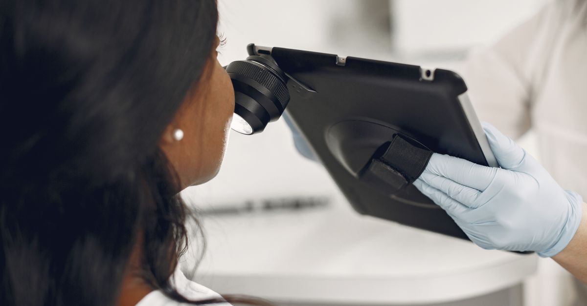
Dermoscopy is an invaluable, non-invasive clinical tool utilized by dermatologists to examine the subsurface architecture and vascular patterns of skin lesions. It provides vital diagnostic clues before a surgical biopsy is performed.
The dermoscopic features of an atypical fibrous histiocytoma are helpful for clinical assessment. The clinician frequently observes a delicate, fine pigment network at the extreme periphery of the lesion. The central area typically displays a homogeneous, structureless pinkish or pale central patch, which correlates directly with the dense dermal collagen matrix.
While dermoscopy aids in the clinical evaluation and helps rule out heavily pigmented melanocytic lesions, the presence of atypical features can sometimes create ambiguous visual patterns. Because dermoscopy cannot definitively differentiate this condition from early stage melanoma, a tissue biopsy remains an absolute requirement.
11. Differential Diagnosis
Accurately distinguishing an atypical fibrous histiocytoma from other pigmented or nodular skin lesions is an essential component of dermatological practice.
| Skin Nodule | Key Differentiating Features |
|---|---|
| Atypical Fibrous Histiocytoma | Benign, prominent nuclear atypia but zero atypical mitoses, Factor XIIIa positive. |
| Nodular Melanoma | Highly malignant, irregular borders, severe cellular chaos, S100 and Melan-A positive. |
| Atypical Fibroxanthoma | Usually found on sun-damaged head and neck skin, high mitotic rate, fast-growing. |
| Dermatofibrosarcoma Protuberans | Locally aggressive, deeply invades subcutaneous fat, strongly positive for CD34. |
A definitive distinction relies entirely on the microscopic evaluation of the tissue. For patients seeking broader information regarding unexpected skin changes, exploring dermatological abnormality guidelines provides valuable clinical context on evaluating new lesions.
12. Importance of Skin Biopsy
Because visual inspection and dermoscopy cannot reliably differentiate a benign atypical fibrous histiocytoma from malignant skin cancers, a diagnostic tissue biopsy is a strict medical requirement.
A simple shave biopsy, which only removes the top layer of the skin, is frequently inadequate because it fails to capture the deep dermal architecture necessary for a complete evaluation. To ensure an accurate diagnosis, the dermatologist must perform a full-thickness punch biopsy or a complete excisional biopsy.
These techniques obtain a complete cylinder of tissue that includes the entire dermal depth of the tumor. This allows the dermatopathologist to accurately assess the cellular architecture, verify the absence of destructive deep fat invasion, and perform the essential immunohistochemical staining, completely securing the benign diagnosis.
13. Surgical Excision Procedures
Once a deep biopsy definitively confirms the diagnosis of a benign atypical fibrous histiocytoma, the standard and highly recommended management strategy is complete surgical excision.
While the lesion is benign, complete removal is advised to prevent any potential for local recurrence and to provide absolute diagnostic certainty that no malignant components were missed in the initial biopsy sample. The dermatologist performs a standard local excision, carefully removing the remaining nodule along with a narrow margin of normal surrounding skin.
Superficial destructive treatments, such as freezing with liquid nitrogen or laser ablation, are strongly discouraged. Because the nodule resides deep within the dermis, superficial therapies frequently result in incomplete removal, leaving residual atypical cells that can subsequently regenerate into a new nodule.
14. Healing and Scar Management
The postoperative recovery following the simple local excision of this skin nodule is generally rapid and uncomplicated. Patients are advised to keep the surgical site clean, dry, and protected with a sterile dressing during the initial healing phase.
Postoperative pain is typically minimal and managed effectively with standard over-the-counter analgesic medications. Proper wound care, including avoiding excessive tension or stretching on the sutures, is crucial, especially on the lower legs where skin tension is naturally high.
Patients must be counseled prior to surgery that removing a deep dermal nodule will inevitably result in a permanent, linear surgical scar. With meticulous surgical technique and proper postoperative care, the resulting scar generally fades significantly over time, offering a highly acceptable cosmetic resolution compared to the persistent presence of an enlarging nodule.
15. Prognosis and Recurrence
The long-term prognosis for an individual diagnosed with a solitary atypical fibrous histiocytoma is exceptionally excellent. The condition is entirely benign and carries absolutely no risk of transforming into a malignant cancer or spreading systemically to distant organs.
Following a complete surgical excision with clear, negative margins, the patient is considered completely cured of the specific lesion. The condition does not predispose the individual to any future connective tissue disorders or malignant skin cancers.
Local recurrence at the surgical site is exceedingly rare. A recurrence is typically attributed solely to an incomplete initial surgical removal of the deep dermal cells. If a small nodule does return at the surgical site, a repeat local excision is highly effective and remains the standard, curative management protocol.
16. When to Consult a Dermatologist
Patients should promptly schedule a clinical evaluation with a board-certified dermatologist if they discover any new, firm, persistent, or changing lump on their skin. Accurate professional diagnosis is essential for any new skin growth to definitively rule out malignancy.
Immediate medical consultation is strongly advised if a previously stable skin nodule suddenly begins to grow rapidly, becomes ulcerated, or starts bleeding spontaneously without significant trauma. These clinical presentations are critical warning signs that demand rapid investigation for aggressive skin cancers.
Early assessment by a specialized dermatologist ensures that the appropriate diagnostic steps, including a full-thickness excisional biopsy, are executed correctly, providing the patient with factual reassurance and facilitating safe, evidence-based management decisions.
17. Frequently Asked Questions (FAQ)
1. Does the word atypical mean I have a form of skin cancer?
No. While the term atypical means the cells look unusual or irregular under a high-powered microscope, extensive medical research confirms that this specific tumor is entirely benign and harmless. It cannot spread to other organs.
2. Why do the cells look so abnormal if it is not cancer?
Pathologists refer to this as degenerative atypia. The cells have slowly aged and changed shape over a long period without undergoing the dangerous, rapid division seen in true malignant cancers.
3. Will this dark lump fade away if I leave it alone?
No, this specific type of fibrous nodule is a permanent structural mass. It will not spontaneously dissolve, shrink, or fade away on its own over time.
4. Do I absolutely have to have it surgically removed?
Complete surgical removal is highly recommended. Because the cells look highly unusual, removing the entire lump allows the pathologist to examine the whole structure, ensuring absolute certainty that no microscopic cancer cells are hidden anywhere within the tissue.
5. Is there a high chance of the tumor coming back after surgery?
No. Once the nodule is completely removed surgically by a dermatologist, the chances of it returning at the exact same site are extremely low, providing a permanent cure.
18. Bibliography
Disclaimer: The content is for informational purposes only and does not replace medical advice. Always consult your doctor for personalized treatment.