1. Introduction
Angiomatoid fibrous histiocytoma is a rare, slow-growing soft tissue neoplasm that primarily manifests in the deep dermal layers or subcutaneous tissues. It most commonly arises on the extremities, such as the arms and legs, and predominantly affects children, adolescents, and young adults. This unique tumor is characterized by blood-filled cystic spaces, a dense fibrous outer capsule, and a prominent surrounding zone of inflammatory immune cells.
Receiving a soft tissue tumor diagnosis invariably generates significant anxiety for patients and their families. It is important to emphasize that this specific condition is classified as a low-grade malignancy. While it demonstrates a propensity to recur locally if incompletely excised, its potential to spread to distant organs is exceedingly rare, occurring in less than five percent of cases. The clinical strategy is firmly focused on precise surgical eradication to achieve a definitive cure.
Addressing this rare condition requires a specialized approach involving advanced molecular pathology and expert surgical oncology. By understanding the distinct genetic and cellular characteristics of the growth, clinicians can provide personalized interventions. A comprehensive review of the disease empowers patients with the clarity needed to navigate treatment and adhere to essential follow-up care.
2. Pathological Architecture
The microscopic structure of this tumor is highly distinctive, distinguishing it from standard connective tissue growths. Under pathological examination, the tumor is composed of a nodular arrangement of spindle-shaped or round histiocyte-like cells. A defining feature is the presence of multiple pseudovascular spaces. These spaces are filled with blood but lack the true endothelial cell lining found in normal blood vessels.
Surrounding this cellular core is a thick, dense capsule composed of fibrous connective tissue. This pseudocapsule gives the tumor its firm, discrete clinical feel. Furthermore, the tumor tissue often exhibits areas of internal hemorrhage and the subsequent accumulation of haemosiderin, an iron-rich pigment that gives the tissue a dark, bruised appearance upon surgical removal.
Pathologists meticulously evaluate the cellular margins to assess the mitotic rate, which is the speed at which the cells are dividing. In an angiomatoid fibrous histiocytoma, the mitotic activity is generally low, confirming its classification as a low-grade, indolent neoplasm rather than a highly aggressive sarcoma.
3. The Inflammatory Component
A remarkable biological feature of this specific tumor is the intense inflammatory response it provokes in the surrounding host tissue. The fibrous pseudocapsule is typically encircled by a massive infiltrate of lymphocytes and plasma cells, which are key components of the human immune system.
This heavy inflammatory cuff often mimics the microscopic appearance of a normal lymph node. In fact, this striking resemblance frequently leads to initial diagnostic confusion, with the mass mistakenly suspected to be an inflamed or metastatic lymph node rather than a primary connective tissue tumor.
The precise biological reason why this specific tumor elicits such a profound immune response remains an active area of medical research. Scientists believe the tumor cells secrete specific cytokines, or chemical messengers, that actively recruit immune cells to the tumor boundary, creating this unique localized inflammatory environment.
4. Prevalence and Demographics
This condition is classified as a rare soft tissue tumor, accounting for less than one percent of all soft tissue neoplasms worldwide. Epidemiological data indicates a strong demographic predilection for younger populations. The vast majority of cases are diagnosed in individuals under the age of thirty, with a significant peak occurring during the second decade of life.
Statistical analysis reveals a relatively equal incidence rate between male and female patients, with no significant gender disparity. Furthermore, the disease is documented across diverse geographic and ethnic populations globally, showing no specific regional clustering.
Because of its scarcity and its visual resemblance to common benign cysts or swollen lymph nodes, the condition is frequently misdiagnosed during initial clinical evaluations. Diagnosis relies heavily on the expertise of specialized sarcoma pathology centers.
5. Primary Clinical Symptoms
The classic clinical presentation involves the development of a slow-growing, painless mass located deep within the skin or subcutaneous fat. The upper and lower extremities are the most frequent sites, though lesions can occasionally arise on the trunk or head and neck region.
Patients generally notice a firm, well-defined lump that gradually enlarges over a period of months or even years. The overlying skin typically remains intact and normal in color, though in some instances, it may take on a slight bluish hue due to the internal blood-filled cystic spaces of the tumor.
Because the mass grows indolently and is usually encased within a fibrous capsule, it rarely causes mechanical pain or functional impairment unless it compresses an underlying nerve. The subtle and benign appearance of the swelling often delays patients from seeking prompt medical advice.
6. Systemic Manifestations
In a fascinating clinical twist, a subset of patients with angiomatoid fibrous histiocytoma present with notable systemic symptoms alongside the localized physical mass. This is an unusual feature for a low-grade soft tissue tumor.
Patients may experience systemic signs of inflammation, including unexplained fevers, chronic fatigue, unintended weight loss, and night sweats. Laboratory blood tests often reveal microcytic anemia and elevated inflammatory markers, such as the erythrocyte sedimentation rate or C-reactive protein levels.
These systemic symptoms are entirely paraneoplastic, meaning they are caused by the tumor secreting excessive amounts of inflammatory cytokines into the bloodstream. Notably, all of these systemic symptoms and abnormal blood markers resolve completely and rapidly once the primary tumor is surgically removed.
7. Underlying Genetic Fusions
The fundamental biological cause of this disease lies in a specific, acquired genetic mutation. The condition is not triggered by external factors like radiation, chemical exposure, or viral infections. It is driven by random structural rearrangements within the chromosomes of the connective tissue cells.
Modern molecular pathology has identified that the vast majority of these tumors harbor specific gene fusions. The most common rearrangement involves the EWSR1 gene fusing with the CREB1 gene or the ATF1 gene. In a smaller subset of cases, a FUS-ATF1 gene fusion is identified.
These specific genetic fusions act as abnormal biological switches, forcing the cells to evade normal growth restrictions and multiply continuously. Detecting these fusions through molecular testing is now a mandatory step in confirming the diagnosis with absolute certainty.
8. Differential Diagnosis
Establishing an accurate diagnosis requires differentiating this rare entity from various other soft tissue and vascular conditions. Because the mass can mimic a swollen lymph node or a benign vascular cyst, a precise differential diagnosis is paramount.
Clinicians synthesize the clinical presentation, imaging, and advanced tissue analysis to exclude mimicking conditions. The table below lists several primary differential diagnoses.
| Condition | Distinguishing Features |
|---|---|
| Reactive Lymph Node | A normal immune response to infection, completely lacks the abnormal spindle cells and specific genetic fusions. |
| Hemangioma | A benign overgrowth of true blood vessels lined with normal endothelial cells, typically lacks a thick fibrous capsule. |
| Aneurysmal Bone Cyst | Occurs within the bone rather than soft tissue, though it shares similar blood-filled spaces. |
| Ewing Sarcoma | A highly aggressive malignancy that may share the EWSR1 gene rearrangement but involves different fusion partners and highly malignant cellular features. |
Accurate differentiation relies exclusively on expert histopathological evaluation combined with molecular genetic testing.
9. Diagnostic Imaging
Medical imaging is a critical first step in evaluating the anatomical extent of the mass. Magnetic resonance imaging is the gold standard for assessing soft tissue tumors. It provides exceptional contrast resolution, allowing the surgical team to map the tumor margins and evaluate its relationship to major blood vessels and nerves.
On an MRI scan, an angiomatoid fibrous histiocytoma typically appears as a well-circumscribed, lobulated mass. A classic radiological feature is the presence of multiple internal fluid levels, which correspond to the blood pooling within the cystic spaces of the tumor. The thick fibrous pseudocapsule is often visible as a dark rim surrounding the mass.
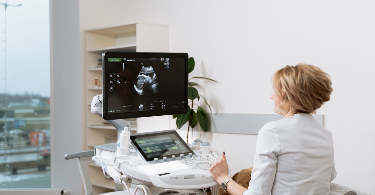
Ultrasound may be utilized initially and typically demonstrates a solid and cystic mass with variable blood flow. However, ultrasound lacks the detailed anatomical mapping required for comprehensive surgical planning.
10. Tissue Biopsy
Clinical examination and imaging cannot provide a definitive cellular diagnosis. A tissue biopsy is an absolute requirement. Surgeons generally perform a core needle biopsy to safely extract a cylinder of tissue from the solid components of the mass.
During histological evaluation, the pathologist utilizes specific immunohistochemical stains. The tumor cells commonly express specific markers such as desmin, CD99, and epithelial membrane antigen, while testing negative for vascular endothelial markers like CD31, proving the blood spaces are not true blood vessels.
The final diagnostic step involves fluorescence in situ hybridization to confirm the presence of the EWSR1 or FUS gene rearrangement. This molecular confirmation finalizes the pathology report and guides the multidisciplinary treatment team.
11. Surgical Resection
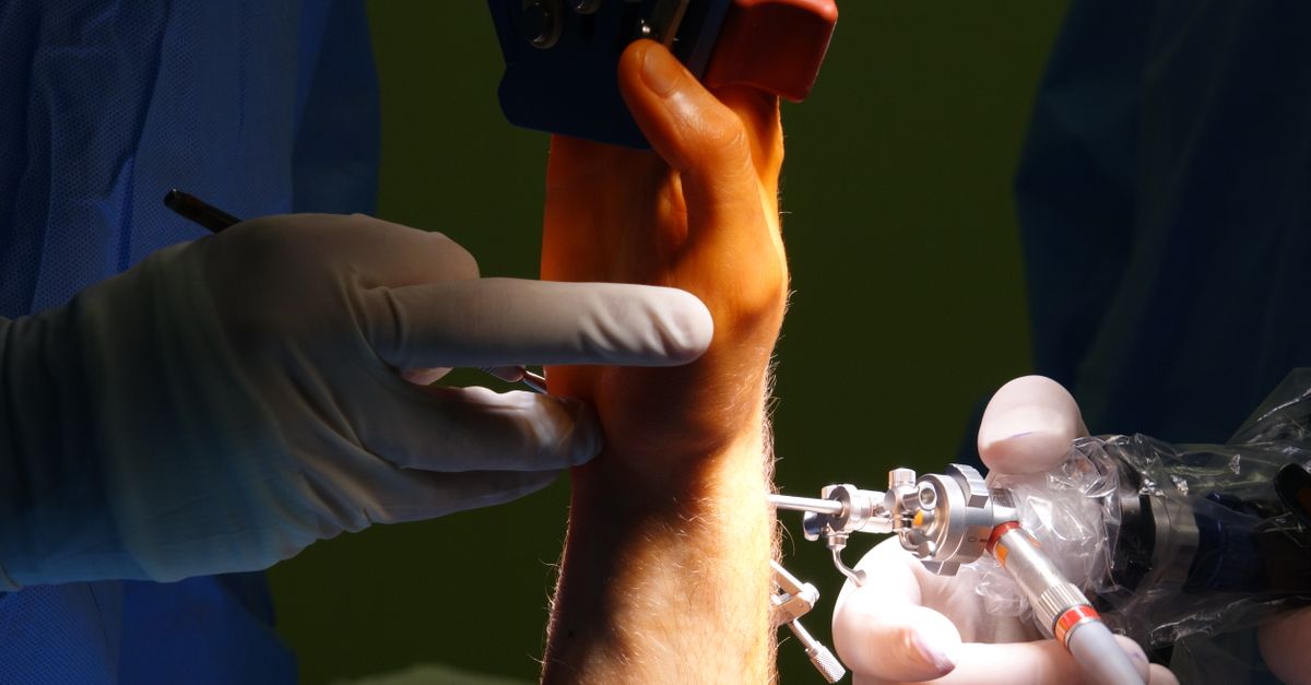
Complete surgical excision is the primary, definitive, and most successful treatment for this condition. The overarching surgical objective is to remove the entire tumor mass intact, achieving clear, negative microscopic margins to prevent any abnormal cells from remaining in the local tissue bed.
Because the tumor is generally well-circumscribed by its fibrous capsule, surgical removal is often straightforward when located in superficial subcutaneous tissues. The surgeon meticulously dissects around the capsule, removing a small margin of surrounding normal fat or muscle tissue to ensure complete eradication.
If the tumor is located deep within a limb and is intimately involved with critical structures, the surgical approach requires meticulous care to preserve the patient’s neurological and mechanical function while still satisfying the oncological requirement for clear margins.
12. Role of Lymph Node Dissection
Unlike most soft tissue tumors, angiomatoid fibrous histiocytoma possesses a recognized, albeit low, potential to spread to regional lymph nodes. This occurs in a small minority of cases, typically estimated at less than five percent.
Due to this specific biological behavior, the preoperative clinical examination and imaging must include a thorough assessment of the lymph node basins corresponding to the tumor location. If a lymph node appears abnormally enlarged or metabolically active on imaging, a biopsy of the node is performed.
Routine, preventive surgical removal of lymph nodes is generally not recommended unless there is confirmed microscopic evidence of tumor spread. Confirmed lymph node involvement necessitates a formal regional lymph node dissection performed by a surgical oncologist.
13. Systemic Medical Management
Systemic therapies, including traditional cytotoxic chemotherapy, are not routinely utilized in the standard management of this disease. The tumor is categorized as a low-grade neoplasm, and its cells do not divide rapidly enough to be vulnerable to standard chemotherapeutic protocols.
Medical management is focused almost entirely on the localized surgical intervention. In the highly exceptional event that the disease presents with distant metastatic spread or becomes surgically inoperable, patients are evaluated at specialized sarcoma centers for potential inclusion in clinical trials exploring novel targeted therapies aimed at the specific EWSR1 gene fusions.
For the vast majority of patients, localized surgical excision provides a complete and permanent cure without the need for systemic medications.
14. Post-Treatment Surveillance
The overall clinical prognosis for individuals diagnosed with this condition is excellent following complete surgical removal. The primary clinical focus remains on vigilant surveillance to detect any potential local recurrence or regional lymph node involvement.
The post-treatment protocol typically involves regular physical examinations of the surgical site and the regional lymph nodes. Periodic imaging, such as localized MRI scans, is scheduled frequently during the first few years and gradually spaced out as the statistical risk of recurrence diminishes.
Adherence to the recommended surveillance schedule is critical. Early detection of a local recurrence or lymph node metastasis allows for prompt salvage surgery, which remains highly effective in controlling the disease.
15. Rehabilitation and Recovery
Recovering from the surgical excision involves both physical healing and adapting to any temporary functional limitations. The recovery timeframe depends entirely on the anatomical location and depth of the surgery. Operations on the limbs may require a brief period of physical therapy to restore full joint mobility and muscle strength.
For patients who experienced systemic symptoms such as fever or anemia prior to surgery, the postoperative period brings rapid relief. Energy levels and blood counts typically return to normal within a few weeks as the source of the inflammatory cytokines has been removed.
Establishing a supportive dialogue with the healthcare team helps alleviate any lingering anxiety regarding the diagnosis. Understanding the low-grade nature of the disease provides immense psychological relief during the long-term recovery phase.
16. When to See a Doctor
It is advisable to schedule a medical evaluation if you or your child develop a new, unexplained lump anywhere on the body, particularly on the arms or legs. While most soft tissue swellings are harmless benign cysts, a persistent mass that slowly enlarges over several weeks warrants professional assessment.
Seek prompt medical attention if a localized lump is accompanied by systemic symptoms such as unexplainable fever, drenching night sweats, or chronic fatigue. Early clinical evaluation and appropriate diagnostic imaging are the most effective ways to secure an accurate diagnosis and achieve the best possible medical outcome.
17. Frequently Asked Questions FAQ
1. Is angiomatoid fibrous histiocytoma a deadly cancer?
No, it is classified as a low-grade tumor. It is highly curable with surgery and very rarely poses a threat to life or spreads to internal organs.
2. Why did my doctor think it was a swollen lymph node?
The tumor naturally attracts a massive cluster of immune cells around its border, making it look and feel exactly like a swollen, inflamed lymph node during initial physical exams and on some scans.
3. Can this tumor be treated without surgery?
No. There are no medications or radiation protocols proven to cure this condition. Complete surgical removal is the only definitive treatment.
4. Will I need to have my lymph nodes removed?
Lymph nodes are only removed if a biopsy proves the tumor has spread to them. Preventive removal is not recommended for most patients.
5. Do the systemic symptoms mean the disease has spread?
No. Symptoms like fever and anemia are caused by inflammatory chemicals released by the localized tumor. These symptoms resolve entirely once the primary mass is surgically removed.
18. Bibliography
Disclaimer: The content is for informational purposes only and does not replace medical advice. Always consult your doctor for personalized treatment.