1. Introduction
Early signs of osteoporosis in your hips and lower back include a gradual, unexplainable loss of height, the development of a stooped posture, and a dull, continuous ache in the lower spine. You should seek an orthopedic or endocrinological evaluation if you experience sudden, sharp back pain after a minor movement like bending or lifting, which strongly suggests a vertebral compression fracture. Because bone density loss occurs silently over many years, recognizing these structural shifts is vital for initiating medical therapies that strengthen the skeletal matrix and prevent debilitating hip fractures.
2. Bone Remodeling and Skeletal Integrity
The human skeleton is not a static, dead structure; it is living tissue that undergoes a continuous, lifelong process known as bone remodeling. This process relies on the synchronized action of two specialized types of bone cells.
Osteoclasts are responsible for bone resorption; they break down and remove old, damaged bone tissue. Osteoblasts are responsible for bone formation; they lay down new, healthy bone matrix to replace what was removed. In a healthy adult, this cycle is perfectly balanced, maintaining optimal bone mass and structural strength. This delicate equilibrium is heavily regulated by hormones, particularly estrogen and testosterone, as well as mechanical stress from physical activity.
3. The Pathophysiology of Osteoporosis
Osteoporosis develops when the delicate balance of bone remodeling becomes deeply skewed. The activity of the osteoclasts begins to outpace the activity of the osteoblasts.
More bone is broken down than is rebuilt. Consequently, the bones lose their vital mineral content, specifically calcium and phosphorus, and their internal microarchitecture degenerates. The dense, honeycomb-like structure inside the bone becomes porous, with the spaces between the bone struts growing significantly larger. This pathological loss of density leaves the bone fragile, brittle, and highly susceptible to fracturing under forces that a healthy bone would easily withstand.
4. The Silent Nature of Bone Density Loss
The most dangerous aspect of osteoporosis is its profoundly silent progression. The actual loss of bone mineral density does not stimulate any pain receptors within the skeletal system.
A patient can lose a substantial percentage of their bone mass over a decade without feeling a single symptom. There are no warning aches or warning stiffness associated directly with the thinning of the bone cortex. Therefore, clinical signs only become apparent when the microarchitecture has become so weak that it begins to fail mechanically under the normal forces of gravity and daily movement.
5. Loss of Height and Vertebral Compression
One of the most reliable, observable early signs of advanced bone density loss is a progressive reduction in overall height. This is most frequently noticed during a routine medical examination or when a patient realizes their clothing no longer fits as it used to.
This height loss is not a result of poor posture alone; it is caused by silent vertebral compression fractures. The vertebrae in the lower and middle back consist largely of spongy trabecular bone, which is highly vulnerable to osteoporosis. The weakened vertebrae simply collapse or crush under the weight of the upper body, resulting in a permanent loss of vertical height.
6. Postural Changes and Kyphosis
As multiple vertebrae in the spine suffer anterior wedge compression fractures, the anatomical alignment of the spinal column is permanently altered. The front of the vertebrae collapses while the back remains relatively intact.
This forces the spine to curve forward, creating a pronounced, stooped posture. In the upper back, this severe forward curvature is clinically termed kyphosis, commonly referred to as a dowager’s hump. This postural deformity not only causes chronic muscular strain but also compresses the chest and abdominal cavities, leading to secondary respiratory difficulties and severe gastrointestinal discomfort.
7. Unexplained Lower Back Aches
While the thinning of the bone itself is painless, the mechanical failure it causes is not. Patients often experience a continuous, dull, deep ache in the lower back that worsens with prolonged standing or walking.
This pain frequently originates from the severe strain placed on the paraspinal muscles and ligaments. Because the structural integrity of the spinal column is compromised, the surrounding soft tissues must work exceptionally hard to keep the body upright. The chronic fatigue and spasm of these supporting muscles generate a constant, nagging lower back pain that does not resolve with standard rest or over-the-counter analgesics.
8. The Fragility Fracture: The First Overt Sign
For many individuals, the absolute first clinical sign that they possess severe osteoporosis is experiencing a fragility fracture. A fragility fracture is defined as a broken bone resulting from a low-energy trauma that would not normally injure a healthy skeleton.
This could occur from a simple fall from standing height, slipping on a rug, or even bumping into a piece of heavy furniture. The most common locations for these devastating fragility fractures are the wrist, the proximal humerus near the shoulder, the vertebral bodies of the spine, and critically, the neck of the femur in the hip joint.
9. Hip Joint Vulnerability and Groin Pain
The hip joint bears the entire mechanical load of the upper body during walking and standing. The neck of the femur, connecting the ball of the joint to the main thigh bone, is particularly susceptible to osteoporotic thinning.
Before a complete fracture occurs, a patient may develop microscopic stress fractures in the femoral neck. These stress fractures present as a deep, vague, aching pain located primarily in the groin or deep within the buttock. This pain typically worsens with weight-bearing activities and improves with rest. Ignoring this localized joint pain is exceedingly dangerous, as a stress fracture can quickly propagate into a complete, displaced hip fracture.
10. Decreased Mobility and Functional Decline
The combination of chronic lower back pain, altered spinal mechanics, and the fear of falling leads to a substantial decline in daily mobility. Patients unconsciously alter their gait, taking shorter, shuffling steps to maintain balance.
They may begin to heavily rely on furniture or mobility aids to navigate their own homes. This reduction in physical activity creates a vicious, self-perpetuating cycle. Because bone requires the mechanical stress of weight-bearing exercise to stimulate osteoblast activity, a sedentary lifestyle accelerates the rate of bone loss, making the skeleton even weaker and further increasing the risk of a catastrophic fall.
11. Differentiating Osteoporosis from Osteoarthritis
These two common skeletal conditions are frequently confused but represent entirely different pathological processes.
| Clinical Feature | Osteoporosis (Bone Density Loss) | Osteoarthritis (Joint Degeneration) |
|---|---|---|
| Primary Pathology | Bones become porous and brittle. | Cartilage wearing away in the joints. |
| Primary Symptom | Silent until a fracture occurs or height is lost. | Pain, stiffness, and joint swelling. |
| Most Common Sites | Spine, hips, wrists. | Knees, hips, hands, lower back joints. |
| Systemic Nature | Affects the entire skeletal system. | Localized to specific heavily used joints. |
12. Recognizing Risk Factors and Hormonal Influences
Because early symptoms are virtually nonexistent, identifying clinical risk factors is the primary diagnostic strategy. Postmenopausal women are at the highest risk due to the sudden, precipitous drop in circulating estrogen, a hormone essential for inhibiting osteoclast activity.
Other profound risk factors include a slender body frame, advanced age, a family history of hip fractures, and chronic tobacco or alcohol use. Furthermore, long-term treatment with systemic corticosteroids, frequently prescribed for asthma or autoimmune diseases, heavily suppresses osteoblast function and dramatically accelerates the rate of bone mineral density loss.
13. Warning Signs Requiring Urgent Orthopedic Assessment
While chronic posture changes warrant a scheduled evaluation, certain events require immediate medical attention. Urgent orthopedic intervention is mandatory if a patient experiences sudden, agonizing back pain immediately after lifting a light object, coughing forcefully, or stepping off a curb.
This presentation is highly indicative of an acute vertebral compression fracture. Additionally, if an older adult falls and subsequently cannot bear any weight on one leg, or if the leg appears shortened and externally rotated, it almost certainly indicates a complete fracture of the hip, requiring emergency surgical fixation.
14. Dual-Energy X-Ray Absorptiometry Bone Scans
The definitive diagnostic tool for evaluating osteoporosis is the Dual-Energy X-Ray Absorptiometry scan. This rapid, non-invasive imaging test measures the exact bone mineral density, typically focusing on the lumbar spine and the hip.
The machine calculates a T-score, which compares the patient’s bone density to the optimal peak bone density of a healthy thirty-year-old adult of the same sex. A T-score of negative two point five or lower confirms the clinical diagnosis of osteoporosis. Regular scanning provides objective data to monitor the rate of bone loss over time and assess the effectiveness of prescribed pharmacological therapies.
15. Pharmacological Bone Resorption Inhibitors
The management of osteoporosis involves aggressive lifestyle modifications, including adequate calcium and vitamin D supplementation and weight-bearing exercise. However, when the risk of fracture is high, pharmacological intervention is necessary.
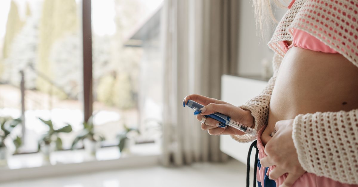
The most frequently prescribed medications are bisphosphonates. These drugs bind directly to the bone matrix and inhibit the action of the osteoclasts, severely slowing the rate of bone breakdown. This allows the osteoblasts time to rebuild the microarchitecture, increasing bone density and significantly reducing the statistical risk of future vertebral and hip fractures. For more severe cases, anabolic agents that directly stimulate new bone formation are administered via daily injections.
16. Frequently Asked Questions (FAQ)
1. Can drinking milk cure my osteoporosis?
While dietary calcium is absolutely necessary to provide the raw materials for bone building, calcium alone cannot stop the overactive osteoclasts from breaking the bone down. Medical therapy is required to alter the biological remodeling cycle.
2. Is it safe to lift weights if I have thin bones?
Supervised, low-impact resistance training is actually highly recommended. The mechanical tension created by muscles pulling on the bones actively stimulates the osteoblasts to lay down new bone tissue, improving density and strength.
3. Will a routine X-ray show that I have osteoporosis?
A standard X-ray will only show bone loss after roughly thirty to forty percent of the bone mass has already been destroyed, or if a fracture is already present. A specialized DEXA scan is required for early, accurate detection.
4. Does osteoporosis cause the bones to ache constantly?
No, the thinning of the bone itself is completely painless. If you have chronic bone pain, it is due to a fracture, severe muscle strain compensating for a weak spine, or a different medical condition entirely.
5. Are men at risk for developing osteoporosis?
Yes, while it is more common in women due to menopause, men can and do develop severe osteoporosis, particularly as testosterone levels decline in older age, or if they have taken corticosteroid medications for chronic illnesses.
17. Bibliography
Disclaimer: The content is for informational purposes only and does not replace medical advice. Always consult your doctor for personalized treatment.