
1. Introduction
A glial tumor, broadly referred to as a glioma, is a primary central nervous system neoplasm that originates from the supportive glial cells of the brain and spinal cord. These tumors represent the most prevalent group of primary brain tumors in adults. Unlike metastatic cancers that travel to the brain from other organs, glial tumors begin within the delicate neural architecture, growing and infiltrating the surrounding healthy brain tissue.
Patients facing this diagnosis often experience a range of neurological symptoms depending on the tumor’s precise location, including persistent headaches, unprovoked seizures, or subtle changes in personality and motor function. Because these tumors grow within the rigid confines of the skull, even small neoplasms can cause substantial neurological disruption by increasing pressure on vital brain structures.
Clinical management involves a highly specialized neuro-oncological team. Treatment strategies are completely dependent on the specific cell type, the genetic profile of the tumor, and its grade of malignancy. The approach routinely involves a combination of maximal safe surgical resection, precision radiation therapy, and targeted systemic chemotherapy to control tumor growth and preserve the patient’s neurological function and quality of life.
2. Cellular Origins and Pathophysiology
The brain is composed of specialized neurons that transmit electrical signals and a vast network of supportive cells known as neuroglia, or simply glial cells. Glial cells are responsible for maintaining the chemical environment, providing structural support, and insulating the neurons. When these supportive cells undergo genetic mutations that cause them to divide uncontrollably, a glial tumor is formed.
There are three primary types of glial cells: astrocytes, oligodendrocytes, and ependymal cells. Tumors can arise from any of these lineages. Astrocytes are star-shaped cells that regulate the blood-brain barrier and nutrient supply. Oligodendrocytes produce myelin, the fatty insulating sheath that wraps around nerve fibers to speed up electrical conduction. Ependymal cells line the ventricles, the fluid-filled cavities within the brain, and assist in producing cerebrospinal fluid.
The pathophysiology of a glioma is characterized by its infiltrative nature. Unlike benign tumors that have a distinct, smooth border, most glial tumors send microscopic tentacle-like extensions deep into the adjacent healthy brain tissue. This infiltration makes complete surgical removal practically impossible without causing severe, irreversible neurological damage.
3. Astrocytic Tumors
Astrocytomas are the most frequent type of glial tumor. They arise from astrocytes and can occur anywhere in the brain or spinal cord, though they are most commonly found in the cerebrum in adults and the cerebellum in children. These tumors span a wide spectrum of aggressiveness, from slow-growing, low-grade lesions to highly malignant, rapidly fatal tumors.
Low-grade astrocytomas grow slowly and may remain stable for many years. However, they carry an inherent biological tendency to mutate further and progress into higher-grade, more aggressive tumors over time.
The most malignant and aggressive form of astrocytoma is glioblastoma multiforme. This grade four tumor grows extraordinarily fast, creates its own chaotic blood supply, and features areas of central cellular necrosis. It requires immediate, aggressive intervention and remains one of the most challenging tumors to treat in modern oncology.
4. Oligodendroglial Tumors
Oligodendrogliomas arise from the myelin-producing cells of the central nervous system. They are typically found in the cerebral hemispheres, particularly in the frontal and temporal lobes. These tumors are uniquely prone to forming tiny calcium deposits, called calcifications, which are often visible on initial CT scans.
Biologically, oligodendrogliomas often exhibit a more favorable prognosis and a slower growth rate compared to astrocytomas of the same grade. A critical factor in their management is specific genetic testing. The presence of a combined loss of genetic material on chromosomes 1p and 19q, termed a 1p/19q codeletion, is a defining hallmark.
Tumors possessing this specific codeletion are remarkably responsive to radiation and specific chemotherapy regimens. Identifying this genetic signature is a mandatory step in neuro-oncology, as it directly dictates the treatment protocol and provides a significantly better long-term survival outlook for the patient.
5. Ependymal Tumors
Ependymomas develop from the ependymal cells lining the brain’s ventricular system and the central canal of the spinal cord. Because they grow within or immediately adjacent to these fluid-filled spaces, they frequently block the normal flow of cerebrospinal fluid. This blockage causes hydrocephalus, an acute accumulation of fluid that leads to a rapid and dangerous increase in intracranial pressure.
In children, ependymomas are most commonly located in the posterior fossa, the lower back compartment of the brain near the cerebellum and brainstem. In adults, they are more frequently found within the spinal cord.
Unlike astrocytomas, ependymomas tend to push adjacent brain tissue away rather than infiltrating deeply into it. This distinct growth pattern often makes them more amenable to complete surgical resection, provided they are not attached to critical vascular structures or deep brainstem nuclei.
6. Grading and Aggressiveness
The World Health Organization utilizes a strict grading system from Grade 1 to Grade 4 to classify the biological aggressiveness and anticipated clinical behavior of glial tumors.
| Tumor Grade | Characteristics and Clinical Behavior |
|---|---|
| Grade 1 | Slow-growing, distinct borders, usually benign. Often curable with complete surgical removal. (e.g., Pilocytic Astrocytoma). |
| Grade 2 | Slow-growing but infiltrative. Tendency to recur and progress to higher grades over time. |
| Grade 3 | Malignant, fast-growing, with actively dividing cells. Requires aggressive adjuvant treatment. (e.g., Anaplastic Astrocytoma). |
| Grade 4 | Highly malignant, aggressive, rapid growth, extensive blood vessel formation, and tissue necrosis. |
The grade is determined by a neuropathologist who examines the biopsied tissue under a microscope, looking for signs of rapid cellular division, abnormal blood vessel growth, and dead tissue.
7. Neurological Symptoms
The symptoms of a glial tumor are entirely dependent on its precise anatomical location within the brain. The brain is highly compartmentalized, with different regions controlling specific functions. As the tumor grows, it disrupts the local electrical pathways and destroys local neurons.
A tumor in the frontal lobe often causes profound changes in personality, diminished executive function, poor judgment, and weakness on one side of the body. If the tumor is located in the temporal lobe, the patient may experience significant memory loss, difficulty understanding language, or auditory hallucinations.
Parietal lobe tumors frequently result in sensory disturbances, such as numbness, tingling, or an inability to recognize objects by touch. Tumors situated in the occipital lobe primarily cause visual field defects, while those in the cerebellum lead to severe coordination issues, poor balance, and an unsteady gait.
8. Intracranial Pressure Signs
Regardless of location, all growing brain tumors add volume to the restricted space inside the skull. This leads to generalized symptoms of elevated intracranial pressure. The most common symptom is a new, persistent headache. Tumor-related headaches are often worst in the morning upon waking and may be exacerbated by coughing, sneezing, or bending over.
Nausea and unexplained vomiting, particularly occurring in the morning without associated gastrointestinal illness, are critical warning signs of increased pressure compressing the brainstem vomit centers.
Unprovoked seizures are the presenting symptom in a substantial percentage of patients, particularly those with low-grade, slow-growing cortical tumors. The tumor tissue acts as a physical irritant to the surrounding cerebral cortex, triggering abnormal electrical discharges that manifest as focal or generalized convulsions.
9. Diagnostic Imaging
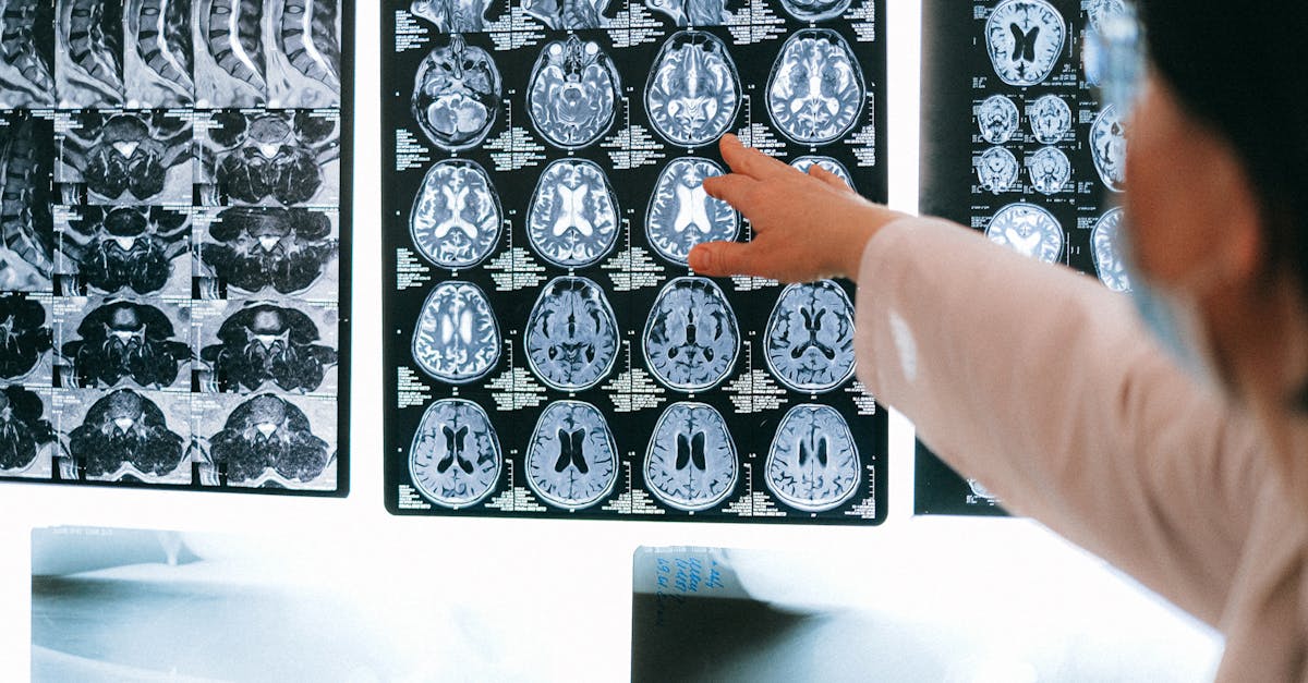
When a brain tumor is suspected based on neurological symptoms, advanced diagnostic imaging is immediately required. Magnetic resonance imaging, universally referred to as MRI, is the gold standard for evaluating brain anatomy. An MRI provides exceptionally detailed images of the brain’s soft tissues, far superior to a CT scan.
To visualize the tumor comprehensively, a contrast agent containing gadolinium is injected into the patient’s vein during the scan. Malignant glial tumors, particularly Grade 3 and Grade 4 lesions, have abnormal, leaky blood vessels. The contrast dye leaks out of these vessels, causing the tumor to enhance brightly on the MRI, indicating high-grade aggressive behavior.
Advanced MRI techniques, such as functional MRI and diffusion tensor imaging, are utilized for surgical planning. These specialized scans map the exact locations of critical motor and speech centers and trace the major nerve fiber tracts, allowing the neurosurgeon to plan a safe trajectory that avoids paralyzing the patient.
10. Biopsy and Molecular Profiling
While imaging provides crucial anatomical information, a definitive diagnosis requires analyzing the actual tumor tissue. This is achieved through a surgical biopsy. If the tumor is safely accessible, the biopsy is usually performed concurrently with an open surgical resection. If the tumor is deep or located in a critical area, a minimally invasive stereotactic needle biopsy is performed.
The tissue is examined by a neuropathologist to determine the exact cell type and the World Health Organization grade. Crucially, modern neuro-oncology relies heavily on molecular and genetic profiling of the tumor tissue.
Testing for the IDH gene mutation and MGMT promoter methylation status is standard protocol. These molecular markers provide vital prognostic information. Tumors with an IDH mutation generally have a significantly better prognosis, while MGMT promoter methylation indicates that the tumor will likely respond favorably to specific chemotherapy agents.
11. Surgical Resection Strategies
The primary therapeutic intervention for most glial tumors is maximal safe surgical resection. The surgical objective is to remove as much of the tumor mass as possible without causing new, permanent neurological deficits. This procedure is called a craniotomy.
Because glial tumors infiltrate normal brain tissue, defining the exact border during surgery is exceptionally difficult. Neurosurgeons employ advanced technologies, such as intraoperative MRI and fluorescence-guided surgery, where the patient ingests a specific dye before surgery that causes the tumor cells to glow under a special blue light in the operating room.
For tumors located near speech or motor centers, awake craniotomy techniques are frequently utilized. The patient is awakened during the critical part of the operation and asked to perform specific tasks, such as speaking or moving their hands. This allows the surgeon to map the brain in real-time, ensuring that the vital functional areas remain completely intact during the tumor removal.
12. Radiation Therapy
Following surgery, or as a primary treatment for inoperable tumors, radiation therapy is utilized to destroy any remaining microscopic tumor cells. Standard external beam radiation delivers high-energy X-rays precisely targeted at the tumor bed and a margin of surrounding brain tissue where infiltrating cells likely reside.
The treatment is typically fractionated, meaning the total radiation dose is divided into small daily treatments administered over several weeks. This approach maximizes the damage to the rapidly dividing cancer cells while allowing the healthy, slower-dividing brain cells time to repair themselves between sessions.
In certain specific cases, stereotactic radiosurgery may be employed. This technique delivers a single, highly concentrated dose of radiation to a very precise, small target. It is generally reserved for small, well-defined recurrences rather than the primary treatment of large, diffuse gliomas.
13. Systemic Chemotherapy
Chemotherapy utilizes powerful systemic drugs to halt cellular division and induce tumor cell death. The blood-brain barrier naturally prevents most circulating toxins from entering the central nervous system, which historically made treating brain tumors with chemotherapy challenging.
Temozolomide is an oral chemotherapy agent uniquely designed to cross the blood-brain barrier effectively. It is the global standard of care for high-grade gliomas, routinely administered concurrently with daily radiation therapy and continued for several months thereafter as a maintenance regimen.
The effectiveness of temozolomide is heavily influenced by the tumor’s genetic profile. Tumors exhibiting MGMT promoter methylation are unable to repair the DNA damage caused by the chemotherapy, making the drug exceptionally effective. Conversely, unmethylated tumors are highly resistant to this standard treatment.
14. Frequently Asked Questions
1. Are glial tumors considered brain cancer?
Yes, most glial tumors, especially Grade 3 and Grade 4, are malignant brain cancers. Grade 1 and some Grade 2 tumors are considered benign or low-grade but still require serious medical management.
2. What causes a glial tumor to form?
The exact cause remains unknown. There are very few known risk factors, aside from prior high-dose therapeutic radiation to the head and certain rare inherited genetic syndromes.
3. Can a glial tumor be completely cured?
Grade 1 tumors can often be cured with complete surgical removal. However, Grade 2, 3, and 4 tumors are infiltrative and almost always recur, making them manageable but generally not entirely curable.
4. Is surgery always required for a brain tumor?
Surgery is usually the first and most critical step to remove the mass and obtain tissue for a definitive diagnosis. If the tumor is in a highly dangerous location, a needle biopsy followed by radiation may be preferred.
5. What is the difference between an MRI and a CT scan for a brain tumor?
An MRI uses powerful magnets to create exceptionally detailed images of the brain’s soft tissues and is far superior to a CT scan for accurately defining the size, exact location, and characteristics of a brain tumor.
15. Bibliography
Disclaimer: The content is for informational purposes only and does not replace medical advice. Always consult your doctor for personalized treatment.
