
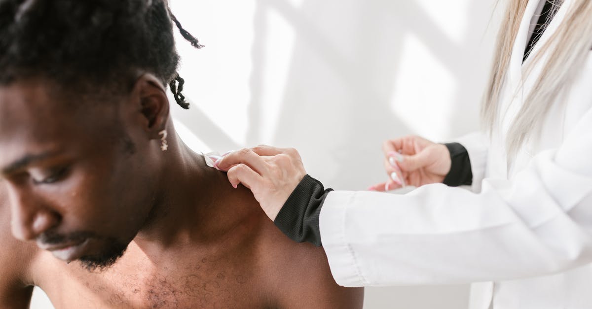
1. Introduction to Acromioclavicular Joint Pain
Pain of the acromioclavicular joint is primarily driven by traumatic ligamentous sprains, repetitive mechanical wear, or progressive osteoarthritis at the superior aspect of the shoulder. Effective management relies on resting the joint, modulating inflammation, and progressively restoring structural stability through targeted rehabilitation. This specific anatomical region is highly vulnerable to both acute injury and chronic degeneration due to its vital role in shoulder mechanics.
Patients experiencing pathology in this joint typically report localized discomfort at the very top of the shoulder, precisely where the collarbone meets the shoulder blade. The pain often intensifies during overhead activities, heavy lifting, or when reaching across the body. The mechanical nature of the pain means that it directly correlates with specific movements, serving as a reliable clinical diagnostic clue.
Proper clinical assessment is required to distinguish structural problems in the acromioclavicular joint from issues originating in the adjacent rotator cuff or cervical spine. A clear understanding of the exact pathological mechanism guides the clinician in formulating a successful therapeutic intervention, whether through conservative physical therapy or targeted surgical stabilization.
2. Anatomy of the Acromioclavicular Joint
The acromioclavicular joint is a small but functionally critical articulation located at the highest point of the shoulder complex. It forms the direct structural connection between the distal end of the clavicle and the acromion process of the scapula. This joint essentially links the entire upper extremity to the axial skeleton of the chest wall.
A specialized fibrocartilaginous disc resides within the joint space, serving as a crucial shock absorber. This articular disc mitigates the compressive forces generated during arm movement. The integrity of the joint is maintained by the acromioclavicular ligaments, which control horizontal stability, and the robust coracoclavicular ligaments, which provide essential vertical stability and prevent upward displacement of the clavicle.
The acromioclavicular joint functions primarily to facilitate the complex rotational movements of the scapula necessary for full overhead elevation of the arm. Because the joint surfaces are small and the mechanical forces transmitted through them are substantial, the articular cartilage is highly susceptible to mechanical wear, microscopic trauma, and early degenerative changes.
3. Acute Traumatic Separations
Traumatic disruption of the joint, commonly referred to as a shoulder separation, is a frequent cause of acute, severe superior shoulder pain. This injury typically occurs as a result of a direct forceful impact to the point of the shoulder, often sustained during contact sports like rugby or football, or from a high-velocity fall directly onto the lateral aspect of the joint.
The downward force of the impact drives the acromion process inferiorly while the clavicle remains relatively stationary. This shear force stretches or tears the stabilizing ligaments. Clinical classification of these injuries ranges from Grade I, involving a mild sprain of the acromioclavicular ligaments without joint displacement, to Grade VI, representing catastrophic disruption of all ligamentous support with severe displacement.
Patients present with immediate sharp pain, localized soft tissue swelling, and distinct tenderness over the joint line. In more severe grades, the unanchored distal clavicle displaces upward, creating a visible bony prominence or “step-off” deformity beneath the skin. Clinical evaluation dictates the management pathway based strictly on the severity of the ligamentous disruption.
4. Osteoarthritis of the Acromioclavicular Joint
Progressive osteoarthritis is the most prevalent chronic condition affecting the acromioclavicular joint, especially in individuals over the age of forty. Decades of continuous joint loading, overhead motion, and microtrauma gradually erode the protective fibrocartilaginous disc and the articular cartilage lining the bone ends.
As the cartilage deteriorates, the underlying subchondral bone is exposed, leading to increased mechanical friction and localized inflammation. The body attempts to stabilize the failing joint by forming irregular bony outgrowths known as osteophytes. These bone spurs alter joint mechanics and can cause secondary impingement upon the underlying rotator cuff tendons during overhead reaching.
Patients with acromioclavicular osteoarthritis experience a deep, aching pain at the top of the shoulder that worsens with activity and improves with rest. The pain may occasionally radiate into the base of the neck or down the anterior aspect of the arm. Morning stiffness and a palpable clicking or grinding sensation during arm movement are common clinical features.
5. Distal Clavicular Osteolysis
Distal clavicular osteolysis, frequently termed “weightlifter’s shoulder,” is a unique pathological process characterized by the localized resorption and cystic degeneration of bone at the distal end of the clavicle. This condition is overwhelmingly associated with repetitive, heavy microtrauma, most notably from resistance training exercises such as the bench press or overhead military press.
The exact pathophysiology involves repetitive microfractures in the subchondral bone of the clavicle. When the rate of repetitive injury exceeds the body’s natural bone remodeling capacity, an inflammatory response ensues, leading to osteoclastic hyperactivation. This results in the active dissolving of the bone matrix at the joint margin.
Patients present with an insidious onset of dull, aching pain localized directly over the acromioclavicular joint. The pain is severely exacerbated by specific weight-bearing exercises and cross-body adduction movements. Early recognition and immediate cessation of the offending activities are necessary to halt the destructive osteolytic process and allow the bone matrix to recover.
6. Biomechanical Stress and Poor Posture
Biomechanical dysfunction within the entire shoulder girdle significantly alters the load distribution across the acromioclavicular joint. The scapula must move dynamically and fluidly along the thoracic wall to maintain optimal joint alignment during arm elevation. This coordinated movement is known as scapulohumeral rhythm.
When muscular imbalances exist, such as tightness in the pectoral muscles combined with weakness in the lower trapezius and serratus anterior, the scapula rests in an abnormal, protracted position. This forward-tilted posture places constant, non-physiological stress directly on the superior aspect of the shoulder, accelerating mechanical wear on the acromioclavicular joint.
Correcting these underlying postural deficits is a fundamental component of managing non-traumatic joint pain. Physical therapy interventions aim to restore normal resting posture and dynamic scapular control, thereby reducing the compressive forces transmitted through the acromioclavicular joint during daily activities.
7. Inflammatory Arthritis
While degenerative osteoarthritis is common, systemic inflammatory arthropathies can also aggressively target the acromioclavicular joint. Conditions such as rheumatoid arthritis, ankylosing spondylitis, or gout involve a systemic immune or metabolic process that triggers severe inflammation within the synovial lining of the joint capsule.
In rheumatoid arthritis, the hyperactive immune system attacks the synovium, causing it to thicken and produce excess fluid. This profound inflammatory state leads to rapid destruction of the articular cartilage and subchondral bone, resulting in severe pain, warmth, and joint swelling. The pain is typically worse in the morning and is accompanied by prolonged joint stiffness.
Diagnosing inflammatory arthritis requires a comprehensive clinical evaluation, including serological testing for specific autoimmune markers and acute phase reactants. Management necessitates the use of systemic disease-modifying medications to control the aberrant immune response and preserve long-term joint integrity.
8. Differentiating Rotator Cuff Pathology
A critical challenge in evaluating superior shoulder pain is distinguishing acromioclavicular joint pathology from conditions affecting the underlying rotator cuff. Because these anatomical structures are in close proximity, inflammation in one area frequently mimics or exacerbates problems in the other.
Rotator cuff tendinopathy typically presents with pain located slightly lower on the lateral aspect of the shoulder, often radiating down the side of the upper arm. Unlike isolated acromioclavicular pain, rotator cuff issues are notoriously associated with significant night pain that disrupts sleep, particularly when the patient rolls onto the affected shoulder.
A careful physical examination utilizing specific provocative tests is required to isolate the anatomical source of the symptoms. While the cross-body adduction test targets the acromioclavicular joint, tests like the empty can test or external rotation resistance testing are highly specific for identifying primary rotator cuff tears or tendinopathy.
9. Referred Pain from the Cervical Spine
Pain perceived in the region of the acromioclavicular joint may actually originate from the cervical spine. Pathology affecting the lower cervical nerve roots, particularly C4 and C5, can refer pain directly to the superior aspect of the shoulder and the trapezius muscle ridge.
This referred pain phenomenon occurs because the sensory nerve pathways from the neck and the shoulder converge within the spinal cord. Pain of cervical origin is often described as burning or electrical and may be accompanied by a sensation of numbness or tingling that extends down the arm.
Clinical differentiation requires examining the patient’s neck. If cervical spine movements, such as looking upward or tilting the head toward the painful shoulder, reproduce the symptoms at the acromioclavicular joint, a cervical etiology must be strongly suspected and investigated with appropriate neurological testing.
10. Clinical Presentation and Provocative Testing
The physical examination of the acromioclavicular joint is precise and heavily reliant on specific clinical maneuvers. The clinician begins by inspecting for any visible asymmetry, localized swelling, or a prominent distal clavicle indicative of a prior separation. Direct palpation of the joint line usually elicits pinpoint tenderness in affected individuals.
| Clinical Test | Mechanism and Diagnostic Goal |
|---|---|
| Cross-Body Adduction Test | The patient elevates the arm and pulls it across the chest. This compresses the AC joint, producing distinct localized pain. |
| Active Compression Test (O’Brien) | Evaluates for both AC joint pathology and labral tears by applying downward resistance to the internally rotated arm. |
| Paxinos Sign | The examiner applies direct pressure squeezing the clavicle and acromion together to reproduce joint line pain. |
| AC Joint Injection Test | A diagnostic injection of local anesthetic directly into the joint space. Immediate relief of pain confirms the AC joint as the pain generator. |
The combination of a positive cross-body adduction test and pinpoint joint tenderness strongly correlates with primary acromioclavicular pathology.
11. Diagnostic Imaging Modalities
Radiographic evaluation begins with standard plain X-rays of the shoulder. A specialized view, known as the Zanca view, is specifically utilized to visualize the acromioclavicular joint without overlapping bony shadows. X-rays are excellent for identifying acute fractures, clavicular displacement in separations, and the hallmark signs of chronic osteoarthritis such as joint space narrowing and bone spurs.
When soft tissue injury is suspected, or when the diagnosis remains unclear after standard radiography, magnetic resonance imaging provides unparalleled detail. Magnetic resonance imaging precisely evaluates the integrity of the coracoclavicular ligaments, detects subtle bone marrow edema indicative of distal clavicular osteolysis, and rules out concurrent rotator cuff tears.
Diagnostic ultrasound is an increasingly valuable tool in the clinical setting. It allows for real-time dynamic assessment of joint stability under stress and can be used to accurately guide therapeutic injections directly into the narrow acromioclavicular joint space.
12. Conservative Management Strategies
The initial therapeutic approach for most conditions affecting the acromioclavicular joint relies on non-operative measures. Activity modification is the absolute cornerstone of treatment. Patients must temporarily cease heavy lifting, overhead reaching, and any resistance exercises that compress the joint, allowing the localized inflammatory cascade to subside.
Pharmacological management includes the use of oral nonsteroidal anti-inflammatory drugs or topical analgesic gels to control pain and reduce soft tissue swelling. Applying ice packs to the superior shoulder for short intervals multiple times a day provides significant symptomatic relief during acute inflammatory flares.
For patients who experience severe, recalcitrant pain that fails to respond to oral medications, an intra-articular injection of a corticosteroid provides potent, localized anti-inflammatory effects. This intervention often breaks the cycle of chronic pain, providing a crucial window of relief that allows the patient to effectively participate in rehabilitative therapy.
13. Physical Therapy and Rehabilitation
Structured physical therapy is essential for optimizing shoulder biomechanics and ensuring long-term recovery. The rehabilitation protocol focuses on restoring proper scapular kinematics and addressing the underlying muscular imbalances that contribute to joint stress.
Early therapy emphasizes stretching tight anterior structures, particularly the pectoralis minor, to prevent the scapula from tilting forward. Subsequent phases focus on strengthening the dynamic stabilizers of the shoulder girdle, primarily the lower trapezius, rhomboids, and serratus anterior. These muscles act synergistically to retract and depress the scapula, unloading the acromioclavicular joint during arm elevation.
Patients recovering from acute joint separations follow a specialized, progressive protocol that respects the biological healing time of the ligaments. Premature return to heavy loading before adequate fibrous tissue formation has occurred significantly increases the risk of chronic joint instability and subsequent painful osteoarthritis.
14. Surgical Interventions
Surgical management is considered when rigorous conservative measures fail to provide adequate pain relief, or when acute trauma results in a severe, high-grade joint separation that severely impairs limb function. The specific surgical procedure is tailored to the underlying pathology.
For chronic, debilitating osteoarthritis or osteolysis that remains painful despite injections and therapy, a distal clavicle excision, known as the Mumford procedure, is highly effective. In this surgery, a few millimeters of bone are resected from the end of the clavicle, preventing the arthritic bone surfaces from grinding against each other.
In cases of severe acute traumatic separation, surgical stabilization is required to realign the joint and reconstruct the torn coracoclavicular ligaments. Modern surgical techniques utilize specialized synthetic grafts or robust suture-button devices to anatomically reduce the clavicle and restore physiological stability to the shoulder complex.
15. Frequently Asked Questions (FAQ)
1. Can an AC joint separation heal on its own?
Low-grade separations involving mild ligament stretching typically heal well with a period of rest and use of a sling. The ligaments form strong scar tissue, though a slight permanent bump may remain. High-grade complete tears often require surgical repair.
2. Why does the top of my shoulder hurt when I bench press?
The bench press maneuver places immense compressive stress directly across the acromioclavicular joint. This repetitive high-load compression is a primary cause of distal clavicular osteolysis, leading to localized inflammation and bone resorption.
3. Is surgery always required for arthritis in this joint?
No. The vast majority of acromioclavicular osteoarthritis cases are managed successfully with activity modification, anti-inflammatory medications, and localized corticosteroid injections. Surgery is strictly reserved for severe cases unresponsive to conservative care.
4. Can poor sleeping position aggravate AC joint pain?
Yes. Sleeping directly on the affected shoulder or with the arm extended forcefully across the chest places continuous mechanical compression on the joint, frequently exacerbating the underlying pain and inflammation.
5. How long does a corticosteroid injection last for AC joint pain?
The duration of relief varies significantly among patients. It can provide profound pain relief lasting anywhere from a few weeks to several months. The injection reduces acute inflammation but does not reverse structural degenerative changes.
Bibliography
Disclaimer: The content is for informational purposes only and does not replace medical advice. Always consult your doctor for personalized treatment.
