1. Introduction
A fetal disorder resulting from a breech delivery occurs when the fetus presents buttocks or feet first, leading to mechanical and physiological complications during childbirth. In a standard vertex presentation, the fetal head acts as a natural dilating wedge, safely paving the way for the rest of the body to navigate the maternal pelvis. When this orientation is reversed, the dynamics of labor shift profoundly, increasing the risk of mechanical obstruction and prolonged fetal stress.
Navigating a breech presentation requires precise obstetrical planning and continuous fetal monitoring. The physiological challenges arise because the largest and least compressible part of the fetus, the head, is delivered last. This reversed sequence leaves the fetal head vulnerable to entrapment behind the maternal pelvic bones or an incompletely dilated cervix.
Clinical management centers on identifying the breech position well before the onset of active labor. Physicians carefully evaluate maternal pelvic capacity and fetal size to determine the safest mode of delivery. Modern obstetrical protocols strictly limit vaginal breech deliveries to select cases, prioritizing surgical delivery to prevent structural and neurological injuries to the newborn.
2. Understanding Breech Presentation
A breech presentation signifies an abnormal longitudinal lie where the fetal pelvis engages the maternal pelvic inlet. This orientation occurs in a small percentage of full-term pregnancies. Throughout early gestation, the fetus changes position frequently within the amniotic fluid. As the pregnancy approaches term, the fetus typically settles into a head-down position due to the shape of the uterine cavity and the relative weight of the fetal head.
Certain maternal and fetal factors predispose a pregnancy to a persistent breech presentation. Uterine anomalies, such as a septate or bicornuate uterus, physically restrict the ability of the fetus to rotate. Additionally, multiple gestations limit the available space for spontaneous turning. Variations in amniotic fluid volume, including both significant excess and marked deficiency, also impede normal fetal rotation.
Fetal conditions can contribute to an abnormal presentation. A fetus with neuromuscular impairments or structural anomalies may lack the ability to execute the natural kicking movements required to achieve a cephalic presentation. Identifying the underlying cause of the breech position assists clinicians in anticipating potential neonatal complications upon delivery.
3. Biomechanics of Breech Delivery
The mechanical forces involved in a vaginal breech delivery differ substantially from those of a standard vertex birth. When the fetal buttocks enter the birth canal, they do not apply uniform, firm pressure against the cervix. This lack of uniform pressure often results in slower, less efficient cervical dilation, leading to a prolonged labor process.
As the fetal body descends, the chest and shoulders must navigate the maternal pelvis. The arms of the fetus can occasionally become extended above the head rather than remaining folded across the chest. Extended arms create a broader mechanical profile, causing the descent to arrest and requiring complex obstetrical maneuvers to safely sweep the arms downward.
The final and most critical phase of the delivery involves the fetal head. Because the softer, smaller lower body has already passed through the cervix, the cervix may begin to contract before the rigid fetal skull can pass. This sequence forces the delivering physician to execute precise rotational maneuvers to extract the head safely without applying dangerous traction to the fetal neck.
4. Risks of Head Entrapment
Head entrapment is a severe, life-threatening complication unique to breech deliveries, particularly involving preterm infants. In a premature fetus, the circumference of the head is significantly larger than the circumference of the abdomen. The small lower body can slip through a partially dilated cervix, but the larger head becomes firmly trapped behind the cervical ring.
When the head is trapped, the umbilical cord is compressed between the fetal skull and the maternal pelvis. This compression instantly halts the flow of oxygenated blood to the fetus. The delivering physician has a very narrow window to relieve this compression. Interventions include administering uterine relaxants to soften the cervix or performing surgical incisions on the cervix to widen the opening.
The mechanical forces required to extract an entrapped head can inflict profound structural damage. The delicate cervical spine of the fetus is subjected to severe stretching, elevating the risk of spinal cord trauma. Consequently, vaginal breech delivery is generally contraindicated for premature infants to eliminate the risk of this profound mechanical complication.
5. Umbilical Cord Prolapse
Umbilical cord prolapse represents another critical emergency frequently associated with breech presentations, specifically the footling breech variety. In a footling breech, one or both feet point downward toward the cervix. Because the feet and legs do not completely fill the maternal pelvis, empty space remains around the fetal presenting parts.
When the amniotic sac ruptures, the flow of amniotic fluid can easily wash the umbilical cord down past the fetal feet and into the vaginal canal. Once the cord drops into the birth canal, the descending body of the fetus compresses it against the rigid pelvic bones. This complete occlusion of the cord halts fetal oxygenation instantly.
Diagnosing a cord prolapse requires immediate clinical recognition. The fetal heart rate monitor will display profound, sustained decelerations. The obstetrical team must immediately elevate the presenting part of the fetus to relieve the pressure on the cord and transfer the mother to the operating room for an emergent surgical delivery.
6. Brachial Plexus and Nerve Injuries
The extraction of a breech fetus frequently requires the physician to apply manual traction and rotation to the fetal body. This manipulation places substantial stress on the delicate neuromuscular structures of the neonate. The brachial plexus, a network of nerves originating in the neck and controlling the arm, is particularly vulnerable to stretching injuries during these maneuvers.
If the physician must apply lateral traction to the fetal body to deliver an entrapped shoulder or an extended arm, the nerve roots can become bruised or stretched. This trauma manifests clinically as Erb palsy or Klumpke palsy, depending on which specific nerve roots are affected. The infant will present with an asymmetric arm posture and decreased spontaneous movement on the injured side.
While many of these nerve injuries resolve spontaneously with gentle physical therapy, severe traction can lead to the complete avulsion of the nerve roots from the spinal cord. This specific trauma causes permanent paralysis of the affected limb and requires specialized microsurgical intervention to restore even limited functionality.
7. Skeletal Trauma During Extraction
Skeletal fractures are a recognized complication of the mechanical forces exerted during a difficult breech extraction. The fetal clavicle, or collarbone, is the most frequently injured bone. Fractures often occur as the physician attempts to sweep down extended fetal arms or rotate the shoulders through a narrow maternal pelvis.
Fractures of the long bones, such as the femur or humerus, can also occur. The physician must grasp the fetal pelvis or lower limbs to guide the body downward. If the bones are fragile or if significant force is required to overcome a mechanical arrest, the bones can fracture. These injuries present postnatally with localized swelling, pain upon movement, and decreased spontaneous use of the affected limb.
Fortunately, the regenerative capacity of a newborn skeleton is robust. Most clavicular and long bone fractures heal rapidly without long-term structural deformity when properly immobilized. The pediatric team will confirm the fracture with a radiograph and provide supportive care to manage neonatal discomfort during the healing process.
8. Hypoxic-Ischemic Encephalopathy
The culmination of mechanical delays, cord compression, and head entrapment significantly elevates the risk of prolonged fetal hypoxia. When the brain is deprived of adequate oxygen and blood flow for a sustained period, the fetus is at risk of developing hypoxic-ischemic encephalopathy. This is a severe neurological condition that can lead to permanent developmental deficits.
The lack of oxygen forces the fetal cells to switch to anaerobic metabolism, which generates substantial amounts of lactic acid. This metabolic acidosis depresses the central nervous system and impairs the function of vital organs, including the fetal heart and kidneys. Infants born after a prolonged hypoxic event often require extensive resuscitation in the delivery room.
The clinical presentation of this encephalopathy includes abnormal neonatal reflexes, poor muscle tone, and the potential for neonatal seizures. Modern neonatal intensive care units utilize therapeutic hypothermia, cooling the infant body temperature for several days, to slow cellular metabolism and limit the extent of the permanent brain injury.
9. External Cephalic Version
To prevent the complications of a breech delivery, obstetricians frequently attempt an external cephalic version before the onset of active labor. This procedure involves the physician applying firm, targeted pressure to the maternal abdomen to manually rotate the fetus from a breech position into a head-down, cephalic orientation.
This intervention is typically offered around thirty-seven weeks of gestation. By this time, the fetus is mature, but adequate amniotic fluid generally remains to facilitate rotation. The procedure is performed under continuous fetal monitoring, usually within a facility equipped for emergency surgical delivery should the fetal heart rate drop unexpectedly.
While an external cephalic version is successful in a significant percentage of cases, it carries intrinsic risks. The manual manipulation can occasionally cause the placenta to detach prematurely or entangle the umbilical cord. If the procedure is successful, the mother can proceed with a standard, lower-risk vaginal delivery.
10. Identifying Breech Presentation Prior to Labor
Accurate and timely diagnosis of fetal presentation is a cornerstone of safe obstetrical care. During routine prenatal visits in the third trimester, the physician performs specific abdominal palpations known as Leopold maneuvers. These maneuvers allow the clinician to identify the location of the fetal head, back, and pelvis.
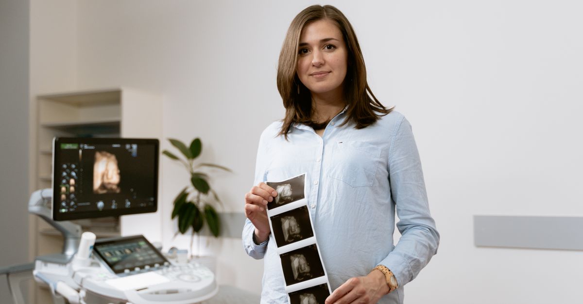
If physical examination suggests a non-cephalic presentation, a standard ultrasound is utilized to confirm the exact position. The ultrasound provides detailed information regarding the type of breech presentation, the estimated fetal weight, the location of the placenta, and the volume of amniotic fluid.
Confirming the breech status prior to the onset of labor is vital. It provides the medical team and the expectant parents ample time to discuss the risks, evaluate the criteria for a safe delivery plan, and schedule an elective surgical delivery if a vaginal trial is deemed unsafe.
11. Criteria for a Safe Vaginal Breech Trial
In specific, rigorously selected cases, a vaginal breech delivery may be attempted. The criteria for this trial of labor are strict to minimize fetal trauma. The fetus must be presenting as a frank breech, where the buttocks are down and the legs are extended straight up toward the fetal head, as this position provides the best dilating wedge.
The estimated fetal weight must fall within a specific, safe range; the fetus cannot be macrosomic, nor can it be significantly premature. The maternal pelvis must be clinically evaluated and deemed adequately spacious. Furthermore, the fetal head must be tucked forward, in a flexed position. An extended fetal head presents a larger diameter and dramatically increases the risk of entrapment.
Finally, the hospital must have continuous fetal monitoring capabilities, an experienced obstetrician skilled in complex breech extraction maneuvers, and an anesthesiologist readily available. If labor fails to progress rapidly and smoothly, the trial is abandoned immediately in favor of a surgical delivery.
12. Structured Data: Types of Breech Presentations
Understanding the specific orientation dictates the medical risks and the delivery strategy.
| Presentation Type | Anatomical Position | Clinical Implications |
|---|---|---|
| Frank Breech | Buttocks present first, legs extended upward | Safest for vaginal trial, forms the best dilating wedge |
| Complete Breech | Buttocks present first, hips and knees flexed | Moderate risk, feet may slip into the birth canal |
| Incomplete Breech | One or both hips extended | Higher risk of cord prolapse and uneven cervical dilation |
| Footling Breech | One or both feet present first | Highest risk for cord prolapse, vaginal delivery contraindicated |
13. The Role of Planned Cesarean Section
Due to the profound risks of structural trauma and neurological compromise, planned surgical delivery is the definitive standard of care for the vast majority of breech presentations in modern obstetrics. A scheduled cesarean section completely bypasses the mechanical bottleneck of the maternal pelvis, preventing head entrapment and cord prolapse.
The procedure is typically scheduled at thirty-nine weeks of gestation to ensure fetal lung maturity while preempting the onset of spontaneous labor. Regional anesthesia allows the mother to remain awake, and the surgeon carefully extracts the fetus through an abdominal incision.
While surgical delivery provides significant safety for the breech fetus, it introduces surgical risks for the mother, including infection, hemorrhage, and an increased risk of complications in subsequent pregnancies. The decision requires a detailed clinical discussion balancing maternal surgical risks against the profound safety benefits for the neonate.
14. Neonatal Resuscitation Readiness
Whenever a breech delivery is planned or occurs unexpectedly, the delivery room must be fully equipped with specialized neonatal resuscitation personnel. The obstetrical team anticipates that a breech infant may experience a transient period of hypoxia during the final stages of extraction, resulting in depressed respiratory drive at birth.
The neonatal team prepares equipment for immediate positive pressure ventilation, continuous oxygen saturation monitoring, and advanced airway management. The immediate goal is to establish regular, spontaneous breathing and stabilize the neonatal heart rate.
Clear communication between the delivering obstetrician and the pediatric team is essential. Information regarding the duration of the labor, the presence of meconium, and any difficulties encountered during the extraction of the fetal head guides the intensity and specific protocols of the neonatal resuscitation effort.
15. Postpartum Neonatal Assessment
Following stabilization, the newborn requires a meticulous physical examination to identify any occult birth trauma resulting from the breech extraction. The pediatrician will carefully palpate the clavicles and long bones to detect crepitus, which indicates a fracture.
The infant is observed for symmetrical movement of all extremities to rule out nerve injuries or brachial plexus trauma. The physician will also examine the hips. Breech infants face a statistically elevated risk of developmental dysplasia of the hip, a condition where the hip joint is unstable or dislocated due to the abnormal positioning in utero.
An ultrasound of the neonatal hips is routinely recommended for all infants born in a breech presentation, typically performed several weeks after birth. Early detection of hip dysplasia allows for simple, non-invasive bracing treatments that ensure normal joint development.
16. Long-Term Neurological Follow-Up
Infants who experience significant mechanical difficulty or prolonged hypoxia during a breech delivery require structured, long-term neurodevelopmental monitoring. The subtle effects of transient oxygen deprivation or significant cranial compression may not become apparent until the child reaches early developmental milestones.
Pediatricians track the acquisition of motor skills, speech development, and cognitive progress. If the infant suffered a recognized hypoxic-ischemic event or required advanced resuscitation, they are referred to specialized early intervention programs.
These programs provide physical therapy, occupational therapy, and developmental support. Consistent, proactive monitoring ensures that any neurological deficits or learning challenges are identified early, providing the child with the therapies necessary to maximize their functional and educational potential.
17. When to Seek Medical Attention
Expectant mothers should attend all scheduled prenatal visits to allow the clinician to monitor fetal presentation. If a woman reaches the late third trimester and experiences a sudden change in fetal movement patterns or suspects the fetus has flipped, a prompt ultrasound evaluation is necessary.
If a mother with a known breech presentation goes into spontaneous labor or experiences a rupture of membranes, she must proceed immediately to the hospital. Prompt medical attention is critical because the risk of umbilical cord prolapse is substantial once the amniotic fluid is released.
Following delivery, parents should contact their pediatrician if they notice the newborn favoring one arm, crying continuously when a specific limb is touched, or demonstrating an asymmetrical facial expression. These signs suggest a potential birth trauma requiring prompt orthopedic or neurological evaluation.
18. Frequently Asked Questions (FAQ)
1. Can a breech baby flip to a normal position right before labor?
Yes, some fetuses will spontaneously rotate to a head-down position even in the final weeks of pregnancy. However, the probability of spontaneous rotation decreases significantly as the fetus grows larger and space becomes limited.
2. Are certain exercises effective at turning a breech baby?
While various postural exercises and alternative therapies are often discussed, clinical evidence proving their effectiveness is limited. You should always consult your obstetrician before attempting any physical maneuvers to alter fetal position.
3. Why is footling breech considered the most dangerous?
When the feet point downward, there is significant empty space in the pelvis. If your water breaks, the umbilical cord can easily wash down past the feet, becoming trapped and cutting off the oxygen supply to the baby.
4. Will a planned surgical delivery guarantee the baby has no injuries?
While a cesarean section eliminates the risks of navigating the birth canal, the infant can still have hip instability related to their posture in the womb. The surgical extraction is significantly safer but carries small inherent surgical risks.
5. How long does a broken collarbone take to heal in a newborn?
Newborn bones possess remarkable regenerative capacity. A fractured clavicle typically heals completely within a few weeks without any specific casting, leaving no permanent structural deformity.
19. Bibliography
Disclaimer: The content is for informational purposes only and does not replace medical advice. Always consult your doctor for personalized treatment.