
1. Introduction
Carcinoma in situ represents a critical diagnostic category in modern oncology, denoting a pre-invasive stage of cellular malignancy. The designation “not otherwise specified” indicates that while the cells exhibit clear malignant features and remain confined to their original tissue layer, the exact histological subtype or specific cellular architecture cannot be definitively categorized into a more precise sub-classification. This diagnosis serves as a vital early warning, identifying a localized accumulation of abnormal cells that possess the potential to progress into a fully invasive and dangerous cancer if left untreated.
Clinical management centers on the definitive, localized eradication of the abnormal cellular clone. Because the neoplastic cells have not yet breached the fundamental tissue barriers required for systemic spread, the condition is considered entirely curable. A multidisciplinary approach is required to ensure precise anatomical mapping, complete surgical excision, and the implementation of appropriate long-term surveillance protocols to protect the patient from future oncological risks.
2. Cellular Pathophysiology
The development of any carcinoma in situ is driven by a localized breakdown in normal cellular regulation. Epithelial cells, which line the internal and external surfaces of the body, undergo continuous replication. When these cells accumulate specific genetic mutations due to environmental carcinogens or inherited genetic anomalies, they begin to proliferate at an abnormal rate.
Under microscopic evaluation, these cells exhibit profound morphological abnormalities known as severe dysplasia. The cells are frequently enlarged, possess dark and irregularly shaped nuclei, and demonstrate an accelerated rate of mitotic division. Despite these malignant visual characteristics, the fundamental biological mechanism driving systemic invasion remains inactive at this stage, trapping the cells within their native anatomical compartment.
3. The Basement Membrane Barrier
The defining characteristic that separates carcinoma in situ from an invasive malignancy is the absolute preservation of the basement membrane. The basement membrane is a thin, specialized layer of extracellular matrix that provides structural support to epithelial tissues. It acts as a strict physical boundary, separating the surface epithelium from the deeper stromal connective tissue, which contains the blood vessels and lymphatic channels.
In carcinoma in situ, the rapidly dividing neoplastic cells completely replace the normal epithelial layer but fail to penetrate this underlying membrane. Because they cannot access the circulatory or lymphatic systems, these cells are biologically incapable of metastasizing to distant organs. The primary goal of medical intervention is to remove these cells before they acquire the genetic mutations required to dissolve this protective barrier.
4. The “Not Otherwise Specified” Designation
The addition of the “not otherwise specified” label reflects specific limitations encountered during the pathological evaluation of the tissue sample. In many organ systems, carcinoma in situ is divided into distinct architectural subtypes, such as ductal or lobular in the breast, or squamous versus glandular in other mucous membranes.
When a pathologist assigns a not otherwise specified diagnosis, it means the biopsied cells lack the distinct structural formations necessary for subtyping, or the provided tissue sample was too small or fragmented to allow for a more precise classification. While this ambiguity requires careful consideration, it does not alter the fundamental pre-invasive nature of the disease or the immediate requirement for localized therapeutic intervention.
5. Clinical Presentation
Because the abnormal cells are strictly confined to the superficial epithelial layer, carcinoma in situ generally does not produce profound systemic symptoms such as unintended weight loss, severe pain, or fatigue. The clinical presentation is dictated entirely by the anatomical location of the lesion.
Cutaneous or mucosal lesions may present as persistent, localized discolorations, scaly patches, or areas of thickened tissue. In glandular organs, the proliferation of cells within microscopic ducts may remain entirely asymptomatic. The absence of a distinct, palpable mass or systemic illness underscores the insidious nature of pre-invasive disease and highlights the critical necessity of structured medical screening programs.
6. The Role of Population Screening
The vast majority of asymptomatic carcinoma in situ diagnoses are discovered through routine, population-based medical screening protocols. These programs are specifically designed to detect cellular abnormalities at the pre-invasive stage.
For example, routine cytological examinations of the cervix identify severe dysplasia before it breaches the basement membrane. Similarly, screening mammography is optimized to detect the microscopic calcium deposits frequently left behind by dying cells within an in situ breast lesion. Regular dermatological skin checks serve an identical preventative function, allowing clinicians to identify and remove pre-cancerous epidermal changes early.
7. Diagnostic Imaging Modalities
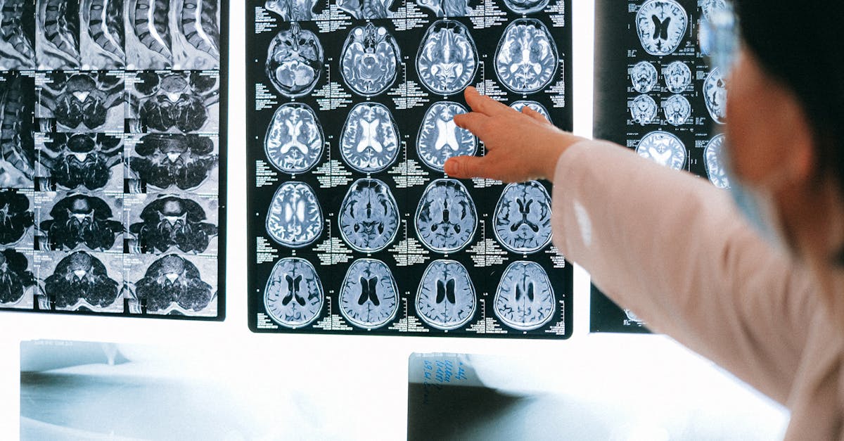
When a screening test yields a suspicious result, advanced radiological imaging is often required to define the full extent of the abnormality. Standard imaging techniques are tailored to the specific organ system involved.
In solid organs, ultrasonography or computed tomography can help delineate areas of architectural distortion or abnormal tissue enhancement. Magnetic resonance imaging is frequently utilized for its superior soft-tissue contrast, accurately mapping the boundaries of the pre-invasive disease and ensuring that no occult, fully invasive lesions are hiding in the adjacent tissue parenchyma prior to surgical planning.
8. Tissue Procurement and Biopsy
A definitive diagnosis of carcinoma in situ relies exclusively on rigorous microscopic tissue evaluation. Procuring an adequate and representative tissue sample is critical. A minimally invasive core needle biopsy or a localized excisional biopsy provides the pathologist with a comprehensive view of the tissue architecture.
The pathologist must carefully examine the entire perimeter of the lesion to ensure the basement membrane remains completely intact throughout the sample. This meticulous review is necessary to definitively rule out the presence of occult microinvasion, which would instantly escalate the diagnosis to an invasive carcinoma and profoundly alter the treatment algorithm.
9. Immunohistochemical Staining
To overcome the ambiguity of a not otherwise specified diagnosis, pathology laboratories frequently employ advanced immunohistochemical staining. This technique utilizes specific antibodies that bind to unique proteins present within the tumor cells.
By observing which proteins are expressed, pathologists can often trace the cellular lineage back to a specific cell type, confirming the epithelial origin of the lesion and occasionally resolving the not otherwise specified designation into a precise, targeted diagnosis. This molecular profiling is essential for refining the long-term therapeutic strategy.
10. Differential Diagnosis
Expert pathological review is essential to distinguish severe pre-invasive dysplasia from benign conditions and true invasion.
| Diagnostic Stage | Pathological and Clinical Characteristics |
|---|---|
| Low-Grade Dysplasia | Mild cellular abnormalities; frequently resolves spontaneously; often managed with active surveillance. |
| Carcinoma In Situ, NOS | Severe cellular abnormalities occupying the full epithelial layer; intact basement membrane; requires definitive localized treatment. |
| Invasive Carcinoma | Malignant cells have actively breached the basement membrane; capable of metastasis; requires aggressive multimodal therapy. |
11. Localized Ablation Therapies
For specific mucosal or cutaneous lesions where the abnormal cells are strictly superficial, localized ablation therapies are frequently employed. Techniques such as cryotherapy, which freezes the tissue, or targeted laser ablation physically destroy the abnormal cellular clone without requiring deep surgical incisions.
These minimally invasive techniques are profoundly effective for treating in situ disease in anatomically sensitive areas, offering excellent cure rates while promoting rapid recovery and preserving the maximum amount of surrounding healthy tissue.
12. Surgical Excision Protocols
For larger lesions, or those located within complex glandular organs, surgical excision remains the absolute standard of care. The surgical objective is the complete, localized removal of the in situ neoplasm with clear, negative microscopic margins of healthy surrounding tissue.
Achieving clear margins guarantees that no abnormal cells remain to instigate a local recurrence or progress to an invasive cancer. The surgeon carefully plans the resection based on preoperative imaging, prioritizing the complete extirpation of the abnormal tissue while maintaining optimal organ function and aesthetic form.
13. Adjuvant Radiation Therapy
Depending on the specific organ system involved and the anatomical extent of the in situ lesion, adjuvant radiation therapy may be incorporated into the treatment plan following surgical excision.
Targeted high-energy ionizing radiation is applied to the surgical bed to eradicate any microscopic, residual neoplastic cells that may persist near the surgical boundaries. This localized intervention significantly minimizes the statistical probability of a regional recurrence, ensuring durable local disease control.
14. Systemic Therapy Considerations
Because carcinoma in situ represents a strictly localized, non-invasive condition, traditional systemic cytotoxic chemotherapy is completely unwarranted. Chemotherapy is designed to target rapidly dividing cells circulating throughout the entire body, a biological scenario absent in pre-invasive disease.
However, targeted chemoprevention strategies may be utilized. If the pre-invasive cells express specific hormone receptors, oral endocrine therapies may be prescribed for several years. These medications suppress systemic hormonal pathways, significantly reducing the risk of a new, independent pre-invasive or invasive lesion developing in the future.
15. The Importance of Active Surveillance
The successful eradication of an in situ neoplasm transitions the patient into a critical phase of active, long-term surveillance. The initial diagnosis confirms that the patient’s specific epithelial tissue possesses an elevated baseline susceptibility to neoplastic transformation.
The clinical team establishes a structured monitoring protocol involving frequent localized physical examinations, specialized radiological imaging, and routine cytological screenings. This vigilant approach ensures that any new pre-cancerous changes are detected and managed promptly.
16. Long-Term Clinical Prognosis
The clinical prognosis for individuals diagnosed with carcinoma in situ, not otherwise specified, is exceptionally outstanding. Because the condition fundamentally lacks the biological mechanisms required for metastasis, the disease-specific survival rate approaches one hundred percent following appropriate, localized definitive treatment. The primary clinical focus remains on diligent adherence to surgical recommendations and lifelong surveillance protocols, providing profound reassurance to the patient.
17. Frequently Asked Questions (FAQ)
1. Is carcinoma in situ considered a true cancer?
It is technically classified as a pre-cancer or a non-invasive cancer. The cells look exactly like cancer cells under a microscope, but they have not acquired the ability to break through the tissue barriers and spread to other organs.
2. If it cannot spread, why is surgery necessary?
If left untreated, the abnormal cells will continue to mutate. Eventually, they will develop the ability to destroy the tissue barrier and become a dangerous, invasive cancer. Surgery is required to stop this inevitable progression.
3. Why could the pathologist not give a more specific name to my condition?
The “not otherwise specified” label means the biopsy sample lacked the distinct architectural shapes needed to identify the exact subtype. However, this does not change the fact that the cells are pre-invasive and require complete removal.
4. Will I lose my hair from chemotherapy for this condition?
No. Because carcinoma in situ is strictly localized and cannot spread through the bloodstream, traditional systemic chemotherapy is entirely unnecessary and completely avoided.
18. Bibliography
Disclaimer: The content is for informational purposes only and does not replace medical advice. Always consult your doctor for personalized treatment.

