1. Introduction
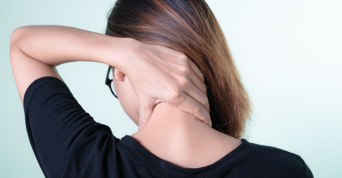
Pain in the cervical spine, commonly referred to as neck pain, is a highly prevalent musculoskeletal disorder that profoundly impacts functional mobility and quality of life. The cervical spine is a biomechanically complex structure engineered to support the weight of the human head while providing a remarkable degree of multi-axial flexibility. This intricate balance of mobility and stability renders the cervical region highly susceptible to acute traumatic injuries, postural fatigue, and chronic degenerative changes.
The clinical presentation of cervical spine pain is highly variable. A patient may experience an isolated, self-limiting muscular ache, or they may present with severe, radiating neuropathic pain accompanied by sensory and motor deficits. Effective medical management demands a precise differential diagnosis, distinguishing between benign mechanical neck pain and serious neurological compromise, such as cervical radiculopathy or myelopathy. Identifying the exact anatomical pain generator allows for the implementation of targeted pharmacological, physical, and interventional therapies.
2. Anatomy of the Cervical Spine
The cervical spine comprises seven distinct vertebrae, designated C1 through C7. The upper two vertebrae, the atlas (C1) and the axis (C2), are uniquely structured to facilitate the rotation and nodding movements of the head. The remaining subaxial vertebrae (C3-C7) are separated by intervertebral discs, which act as fibrocartilaginous shock absorbers.
The stability of this osseous column is maintained by a complex array of ligaments and paraspinal muscles. Posteriorly, the vertebrae articulate via the facet (zygapophyseal) joints, which guide and limit the range of motion. The cervical spine houses the spinal cord within its central canal, while the cervical nerve roots exit laterally through the intervertebral foramina to innervate the shoulders, arms, and hands. Pathology in any of these structural elements can generate significant localized or referred pain.
3. Muscular and Ligamentous Strain
The most frequent cause of acute cervical spine pain is muscular and ligamentous strain. The muscles of the posterior neck, particularly the trapezius, levator scapulae, and the deep cervical erector spinae, are constantly engaged to maintain the upright posture of the head.
Strains occur when these tissues are overloaded by acute mechanical forces or prolonged, poor ergonomic posturing—frequently observed in individuals engaging in extended computer use (“text neck”). The patient experiences localized, dull, aching pain, muscle spasms, and significant stiffness that restricts the range of motion. The pain is mechanical in nature, worsening with active movement and improving with rest.
4. Cervical Spondylosis (Osteoarthritis)
Cervical spondylosis is the clinical term for age-related degenerative osteoarthritis of the cervical spine. As individuals age, the intervertebral discs undergo desiccation (loss of hydration), leading to a reduction in disc height. This disc space narrowing alters the biomechanics of the cervical spine, shifting an increased load onto the posterior facet joints.
The facet joints respond to this abnormal stress by developing subchondral sclerosis and hypertrophic bone spurs known as osteophytes. Patients with cervical spondylosis present with a chronic, gradual onset of neck pain and stiffness. The pain often radiates to the base of the skull or the tops of the shoulders and is characteristically worse in the morning, gradually improving as the joints mobilize throughout the day.
5. Cervical Radiculopathy
Cervical radiculopathy occurs when a spinal nerve root is compressed or inflamed as it exits the intervertebral foramen. This compression is most frequently caused by an acute herniation of an intervertebral disc (the soft nucleus pulposus extrudes through the annulus fibrosus) or by chronic impingement from bony osteophytes.
The resulting pain is neuropathic. Patients report a severe, sharp, burning, or electrical pain that originates in the neck and radiates down the arm, following a specific dermatomal distribution. Neurological deficits are common, including numbness, tingling, and diminished reflexes. A patient may present for a clinical evaluation of unexplained arm numbness, where the primary underlying pathology is discovered to reside entirely within the cervical spine.
6. Cervical Myelopathy
Cervical myelopathy represents a severe, progressive condition where the spinal cord itself is compressed within the central spinal canal. This central stenosis is typically the end result of advanced cervical spondylosis, a massive central disc herniation, or the ossification of the posterior longitudinal ligament.
Unlike radiculopathy, which affects the peripheral nerves of the arm, myelopathy affects the central nervous system tracts. Neck pain may be mild or entirely absent. The clinical hallmarks include a loss of fine motor skills in the hands (clumsiness, dropping objects), an unsteady, spastic gait, and generalized weakness. If left untreated, cervical myelopathy can lead to permanent spinal cord damage and severe disability.
7. Whiplash-Associated Disorders
Whiplash is an acceleration-deceleration injury mechanism commonly resulting from rear-end motor vehicle collisions. The rapid, forceful hyperextension followed by hyperflexion of the cervical spine causes widespread micro-trauma to the ligaments, muscles, facet joints, and intervertebral discs.
Patients with whiplash-associated disorders frequently experience a delayed onset of symptoms, with severe neck pain and stiffness developing 24 to 48 hours post-injury. The pain is often accompanied by tension-type headaches, dizziness, and cognitive difficulties, such as poor concentration. The recovery trajectory for whiplash is highly variable, with a subset of patients developing chronic, intractable neck pain.
8. Facet Joint Arthropathy
The facet joints are paired synovial joints located on the posterior aspect of each vertebral level. These joints are richly innervated by the medial branches of the dorsal rami. Degeneration or traumatic injury to the facet joints results in facet joint arthropathy, a prominent source of localized neck pain.
Facet joint pain is typically described as a deep, aching discomfort that does not radiate past the shoulder. The pain is characteristically exacerbated by extending the neck (looking upward) and rotating the head toward the affected side, as these movements mechanically compress the inflamed joint surfaces.
9. Differentiating Cervical Pathologies
Accurate clinical differentiation guides the diagnostic workup. The following table contrasts the major cervical spine pathologies:
| Condition | Pain Quality and Location | Associated Neurological Symptoms |
|---|---|---|
| Muscular Strain | Dull ache, stiffness in posterior neck and shoulders. | None. |
| Cervical Spondylosis | Chronic ache, grinding sensation, worse in the morning. | None, unless nerve compression is present. |
| Cervical Radiculopathy | Sharp, burning pain radiating down one arm. | Numbness, tingling, weakness in specific fingers/arm muscles. |
| Cervical Myelopathy | Variable neck pain; often painless. | Clumsy hands, unsteady gait, hyperreflexia. |
10. Clinical Evaluation and Neurological Testing
A rigorous physical and neurological examination is essential. The clinician inspects the neck for deformity and assesses the active range of motion. Palpation identifies localized muscular trigger points or tenderness over the spinous processes.
Neurological testing involves a systematic evaluation of motor strength, sensory perception, and deep tendon reflexes in the upper extremities. Specific provocative tests are utilized. The Spurling test (neck extension and side-bending with applied downward pressure) is highly specific for reproducing the radiating pain of cervical radiculopathy. The Hoffman sign and assessment for hyperreflexia are used to screen for upper motor neuron lesions indicative of cervical myelopathy.
11. Advanced Diagnostic Imaging
For patients with red flag symptoms, persistent neurological deficits, or pain refractory to conservative care, advanced diagnostic imaging is required. Plain radiographs provide a baseline evaluation of bone alignment, loss of disc height, and the presence of osteophytes.
Magnetic Resonance Imaging (MRI) is the gold standard for evaluating cervical soft tissues. It provides excellent visualization of the intervertebral discs, nerve roots, and the spinal cord, definitively identifying herniations, stenosis, and spinal cord signal changes. Computed Tomography (CT) scans offer superior bony detail, often utilized when evaluating complex fractures or detailed osteophyte anatomy prior to surgery.
12. Pharmacological Management
Pharmacological therapy targets inflammation and specific pain pathways. Nonsteroidal anti-inflammatory drugs (NSAIDs) are the first line of defense for acute muscular strains and inflammatory facet joint pain. Muscle relaxants (such as cyclobenzaprine) are prescribed for short-term use to alleviate severe muscular spasms.
For patients suffering from the neuropathic pain of cervical radiculopathy, standard analgesics are often ineffective. Adjuvant medications, specifically gabapentinoids (gabapentin or pregabalin) or tricyclic antidepressants, are utilized to modulate nerve excitability and reduce the sharp, burning neuropathic sensations.
13. Physical Therapy and Ergonomic Modification
Physical therapy is a cornerstone of conservative cervical spine management. Therapists employ manual techniques, including joint mobilization and soft tissue release, to restore normal segmental mobility. A targeted exercise program focuses on strengthening the deep cervical flexors and stabilizing the scapular musculature, thereby reducing the mechanical load on the cervical spine.
Ergonomic modifications are crucial for preventing recurrence. Patients are educated on maintaining a neutral cervical posture during computer work and utilizing supportive pillows during sleep to maintain the natural lordotic curve of the neck.
14. Interventional Pain Procedures
When oral medications and physical therapy provide insufficient relief, interventional procedures are considered. Cervical epidural steroid injections are highly effective for managing severe radiculopathy. A potent corticosteroid is injected under fluoroscopic guidance directly into the epidural space, bathing the inflamed nerve root and rapidly suppressing the inflammatory cascade.
For chronic facet joint arthropathy, medial branch blocks are utilized diagnostically. A local anesthetic is injected over the nerves supplying the facet joint. If the patient experiences significant pain relief, a radiofrequency ablation can be performed, which uses thermal energy to temporarily deactivate the sensory nerve, providing long-term pain relief.
15. Indications for Surgical Intervention
Surgical intervention is indicated when conservative measures fail, when the patient experiences intractable, excruciating pain, or when there is evidence of progressive neurological deficit. The presence of cervical myelopathy requires urgent surgical evaluation to decompress the spinal cord and halt neurological decline.
The most common surgical procedure for cervical disc herniation is the Anterior Cervical Discectomy and Fusion (ACDF). The surgeon removes the offending disc from the front of the neck, relieving pressure on the spinal cord or nerve roots, and fuses the adjacent vertebrae together to restore stability. Alternatively, artificial disc replacement maintains motion at the operated level in carefully selected patients.
16. Frequently Asked Questions (FAQ)
1. What is causing the sharp pain shooting down my arm?
Sharp, electrical pain radiating down the arm is a classic symptom of cervical radiculopathy. This occurs when a nerve root in your neck is pinched or inflamed, usually by a herniated disc or a bone spur.
2. Is it bad to crack my neck to relieve pain?
Self-manipulation or forceful cracking of the neck is strongly discouraged. It can overstretch the ligaments, leading to joint instability, and in rare cases, violent manipulation can damage the major blood vessels supplying the brain.
3. What does “text neck” mean?
“Text neck” is a modern clinical term for the muscle strain and pain caused by looking down at electronic devices for long periods. The forward head posture exponentially increases the mechanical weight your neck muscles must support, leading to chronic spasms and pain.
4. How do I know if my neck pain is serious?
You should seek immediate medical evaluation if your neck pain is accompanied by numbness or weakness in your arms or hands, difficulty maintaining your balance while walking, or loss of bowel or bladder control. These are signs of spinal cord compression.
5. Will a neck brace help my pain?
Soft cervical collars are rarely recommended for more than a few days following an acute injury like whiplash. Wearing a brace long-term causes the neck muscles to rapidly weaken and atrophy, which ultimately makes the pain and instability worse.
17. Bibliography
Disclaimer: The content is for informational purposes only and does not replace medical advice. Always consult your doctor for personalized treatment.