1. Introduction to Closed Skull Vault Fractures
A closed fracture of the vault of the skull accompanied by an intracranial hemorrhage represents a severe, complex traumatic brain injury. The cranial vault, or calvaria, is the rigid bony dome that houses and protects the brain. When high-impact biomechanical forces fracture this bone without tearing the overlying scalp—a “closed” fracture—the structural integrity of the cranium is compromised without external bleeding.
While the fracture itself represents significant skeletal trauma, the true medical emergency lies in the associated internal injury. The same kinetic energy that cracked the skull is transmitted directly into the cranial cavity, tearing the delicate blood vessels that run along the inner surface of the bone or through the surface of the brain itself.
This results in an acute intracranial hemorrhage, where blood rapidly accumulates within the fixed volume of the skull. This expanding mass of blood compresses the underlying brain tissue, leading to an exponential rise in intracranial pressure. Prompt recognition, immediate neuroimaging, and rapid neurosurgical intervention are critical to evacuate the hematoma, relieve the crushing pressure on the brain, and prevent permanent neurological devastation or death.
2. Anatomy of the Cranial Vault
To understand the mechanics of this injury, one must review the structure of the cranial vault and its relationship to the underlying neurovascular structures. The vault is composed of several large, flat bones—predominantly the frontal, parietal, temporal, and occipital bones—interlocked by fibrous joints called sutures.
These bones have a unique structure: two dense plates of compact bone separated by a layer of spongy bone (diploë). This architecture is designed to absorb and dissipate significant impact forces. The inner surface of the cranial vault is not smooth; it contains distinct grooves that perfectly house the meningeal arteries.
The most critical of these is the middle meningeal artery, which runs through a deep groove on the inner surface of the temporal and parietal bones. Directly beneath the bone lie the meninges—the dura mater, arachnoid mater, and pia mater—which encase the brain. The intimate physical relationship between the bone, the meningeal arteries, and the dural venous sinuses means that any fracture of the vault puts these major blood vessels at immediate risk of laceration.
3. Biomechanics of a Closed Fracture
A closed fracture of the cranial vault requires significant blunt force trauma. Motor vehicle collisions, falls from significant heights, and high-energy sports impacts are the primary mechanisms. The term “closed” explicitly means that the scalp remains intact; there is no open wound exposing the bone to the external environment, reducing the immediate risk of central nervous system infection but masking the severity of the internal injury.
Fractures of the vault are generally categorized as linear or depressed. A linear fracture is a single crack in the bone without any displacement of the bone fragments. While the bone maintains its anatomical position, the crack can traverse directly across the path of a major artery.
A depressed fracture occurs when the blunt force is concentrated over a smaller area, driving a fragment of the fractured skull inward. In a depressed fracture, the sharp edges of the displaced bone fragment can directly lacerate the underlying dura mater and physically slice into the cerebral cortex, causing immediate internal bleeding and severe, localized brain tissue destruction.
4. Pathophysiology of Epidural Hemorrhage
The most classic and rapidly fatal intracranial hemorrhage associated with a skull vault fracture is the epidural hematoma. This specific type of bleed occurs almost exclusively in conjunction with a fracture, typically a linear fracture involving the temporal or parietal bone (the side of the head).
When the fracture line crosses the groove housing the middle meningeal artery, the sharp bone edges tear the artery wide open. Because this is an arterial bleed, blood escapes under high systemic pressure. The blood aggressively forces its way into the potential space between the inner table of the skull and the tough, adherent dura mater.
This creates a rapidly expanding, lens-shaped (biconvex) pocket of blood. The high-pressure arterial bleeding quickly strips the dura away from the bone, compressing the underlying brain tissue. An epidural hematoma is a profound neurosurgical emergency; because it is arterial, the volume expands rapidly, driving up intracranial pressure and leading to brainstem herniation in a matter of hours if not surgically evacuated.
5. Pathophysiology of Subdural Hemorrhage
A closed skull fracture can also result in a subdural hematoma, though the mechanism is distinctly different. A subdural hemorrhage involves bleeding into the space between the dura mater and the delicate arachnoid mater.
This type of bleed is primarily venous in origin. The brain is connected to the large dural venous sinuses by fragile “bridging veins.” During a high-impact head injury, the skull abruptly stops moving, but the gelatinous brain continues to shift rapidly within the cranial fluid (acceleration-deceleration injury). This violent shifting physically tears these delicate bridging veins.
The torn veins bleed slowly into the subdural space, creating a crescent-shaped hematoma that spreads diffusely over the hemisphere of the brain. While venous bleeding is under lower pressure than arterial bleeding, an acute subdural hematoma accompanied by a skull fracture and severe underlying brain contusion (bruising) causes massive brain swelling and carries a notoriously high mortality rate.
6. Coup and Contrecoup Brain Injuries
The kinetic energy associated with a closed skull fracture rarely limits its damage to a single location. The brain undergoes significant internal trauma known as coup and contrecoup injuries.
The “coup” injury represents the immediate contusion (bruising) and microscopic hemorrhage that occurs in the brain tissue directly beneath the site of the skull impact. The fractured bone or the sheer force of the blow directly damages the localized neural tissue and cortical blood vessels.
The “contrecoup” injury occurs on the exact opposite side of the brain. As the head recoils from the impact, the brain sloshes violently backward, striking the hard, jagged internal surface of the opposite side of the skull. This results in severe bruising and intraparenchymal hemorrhage (bleeding inside the brain tissue itself) far away from the actual fracture site, compounding the overall neurological deficit and brain swelling.
7. Clinical Presentation and the “Lucid Interval”
The clinical presentation of a patient with a closed skull fracture and intracranial hemorrhage depends heavily on the specific type of bleed. A hallmark presentation of an epidural hematoma involves a phenomenon known as the “lucid interval.”
Following the initial blunt trauma, the patient may experience a brief loss of consciousness due to the concussive force. They then awaken and may appear completely lucid, speaking and acting normally. However, this is deceptive. During this lucid interval, the torn meningeal artery is actively pumping blood into the epidural space.
As the hematoma expands and reaches a critical volume, the intracranial pressure spikes. The patient’s condition rapidly deteriorates. They quickly develop a severe headache, projectile vomiting, and acute confusion, rapidly sinking into a deep coma. A classic physical sign at this late stage is anisocoria—one pupil becomes massively dilated and unreactive to light, signaling that the expanding blood clot is crushing the brainstem and the oculomotor nerve.
8. Assessing Altered Consciousness: The GCS
Emergency medical responders and physicians rely on a standardized clinical tool to objectively measure the severity of a traumatic brain injury: the Glasgow Coma Scale (GCS). This scale assesses the patient’s neurological function based on three distinct parameters: eye-opening response, verbal response, and motor response.
The GCS score ranges from 3 (deep coma or death) to 15 (fully awake and oriented). A patient who can open their eyes spontaneously, converse coherently, and obey motor commands scores a 15. A patient who does not open their eyes to pain, makes incomprehensible sounds, and exhibits abnormal posturing scores significantly lower.
A GCS score of 8 or less defines a severe traumatic brain injury. In the context of a closed skull fracture, a rapidly declining GCS score—for example, a patient who drops from a 14 to an 8 within thirty minutes—is the most ominous clinical indicator of an actively expanding intracranial hemorrhage requiring immediate surgical intervention.
9. Diagnostic Imaging Protocols
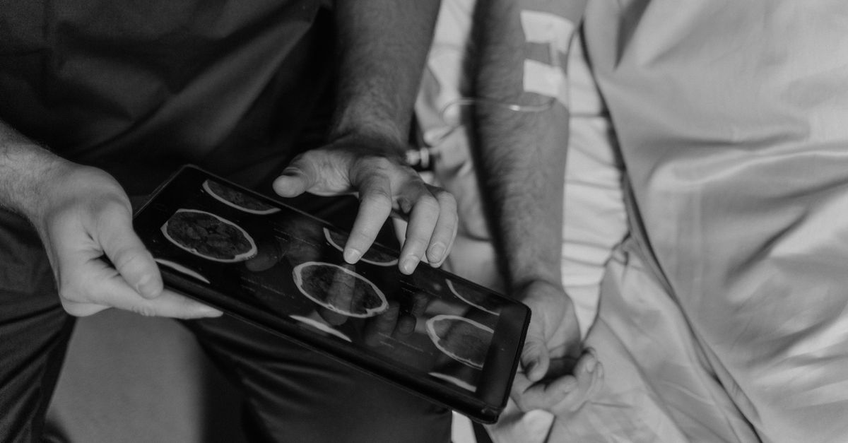
Upon arrival at a trauma center, rapid and definitive imaging is the absolute priority. A non-contrast Computed Tomography (CT) scan of the head is the gold standard diagnostic modality. It is incredibly fast and highly sensitive for detecting both skull fractures and acute blood.
The CT scan provides a detailed map of the cranial vault, clearly identifying linear or depressed fractures. More importantly, it immediately visualizes the acute intracranial hemorrhage. An epidural hematoma appears as a bright white, lens-shaped (biconvex) mass pushing inward against the brain. A subdural hematoma appears as a bright white, crescent-shaped mass hugging the curve of the skull.
The CT scan also provides critical information regarding the secondary effects of the bleed. The radiologist measures the “midline shift”—how many millimeters the hematoma has physically pushed the brain structures off their normal central axis. A significant midline shift is a direct indicator of critically high intracranial pressure and imminent brain herniation.
10. Comparing Traumatic Hemorrhages
Understanding the differences between the two most common fracture-associated hemorrhages dictates the surgical urgency. The table below highlights these key clinical distinctions.
| Feature | Epidural Hematoma (EDH) | Subdural Hematoma (SDH) |
|---|---|---|
| Source of Bleeding | Arterial (Middle meningeal artery) | Venous (Bridging veins) |
| Location | Between skull and dura mater | Between dura and arachnoid mater |
| CT Scan Appearance | Biconvex (lens-shaped) | Crescent-shaped |
| Classic Presentation | “Lucid interval” followed by rapid coma | Gradual decline, severe underlying brain injury |
11. Initial Trauma Resuscitation
The initial medical management of a patient with a severe head injury follows strict Advanced Trauma Life Support (ATLS) protocols. Because the brain requires a continuous supply of oxygen and glucose to survive, systemic stabilization is as critical as addressing the brain bleed itself.
Airway protection is the foremost priority. If the patient has a GCS of 8 or less, or is actively seizing, they must be intubated. An endotracheal tube is placed, and mechanical ventilation is initiated to prevent hypoxia and control blood carbon dioxide levels, as elevated carbon dioxide causes cerebral blood vessels to dilate, exacerbating brain swelling.
Intravenous fluids are administered to maintain adequate systemic blood pressure. If the blood pressure drops (hypotension), the injured brain will not receive enough blood flow (cerebral perfusion), leading to secondary ischemic brain damage. If the patient is on blood thinners, rapid reversal agents must be administered immediately to restore the blood’s clotting ability.
12. Neurosurgical Intervention and Craniotomy
If the CT scan reveals a significant epidural or subdural hematoma causing a midline shift or severe neurological deficit, emergency neurosurgery is strictly indicated. The goal of surgery is to evacuate the blood clot, control the active bleeding, and relieve the lethal pressure on the brainstem.
The neurosurgeon performs a craniotomy in the operating room. A large flap of the fractured skull is temporarily removed. If the fracture is depressed, the sharp bone fragments that have been driven into the brain must be carefully elevated and removed.
Once the skull is open, the pressurized hematoma is meticulously suctioned away. The surgeon identifies the torn meningeal artery or bleeding bridging vein and seals it using electrocautery to achieve hemostasis. The skull flap is then replaced and secured with small titanium plates. In cases of massive brain swelling, the surgeon may perform a decompressive craniectomy, leaving the bone flap off and placing it in a tissue bank to allow the swollen brain room to expand outward rather than crushing inward.
13. Medical Management of Intracranial Pressure
For patients whose hemorrhage is not large enough to require immediate surgery, or for patients recovering post-operatively, the primary focus in the neurocritical care unit is the aggressive medical management of intracranial pressure (ICP).
An ICP monitor, a small fiber-optic catheter, may be drilled through the skull and placed directly into the brain tissue to provide continuous, real-time pressure readings. If the pressure begins to rise dangerously, the medical team initiates hyperosmolar therapy.
Intravenous mannitol or hypertonic saline solutions are administered. These highly concentrated fluids use osmosis to pull excess water directly out of the swollen, uninjured brain tissue and into the bloodstream, physically shrinking the brain and relieving the pressure within the cranial vault. Deep sedation and targeted temperature management (mild hypothermia) may also be used to reduce the brain’s metabolic demand.
14. Post-Concussive Syndrome and Complications
Even after successful surgical evacuation and stabilization, patients recovering from a closed skull fracture with hemorrhage face a high risk of significant, long-term complications. Post-traumatic seizures are a common occurrence, as the bruised cerebral cortex forms epileptogenic scar tissue. Anticonvulsant medications are routinely prescribed for several months post-injury.
Patients frequently suffer from severe post-concussive syndrome. This manifests as chronic headaches, profound fatigue, dizziness, severe light and sound sensitivity, and persistent sleep disturbances.
Cognitive and psychiatric complications are profound. The sheer force of the trauma often causes diffuse axonal injury—the microscopic shearing of the neural connections throughout the brain. This results in significant memory deficits, emotional lability, profound depression, and profound changes in personality and executive functioning, profoundly impacting the patient’s quality of life.
15. Long-Term Neurological Rehabilitation
Recovery from a severe traumatic brain injury is a marathon, not a sprint. The brain tissue that was directly crushed by the initial impact or starved of oxygen by the hematoma cannot regenerate. However, through the process of neuroplasticity, the brain can slowly reorganize itself to bypass the damaged areas.
Intensive, multidisciplinary neurorehabilitation must begin as soon as the patient awakens. Physical therapists focus on overcoming paralysis, restoring balance, and relearning how to walk. Occupational therapists assist the patient in regaining fine motor skills and adapting to their deficits to perform daily activities like dressing and eating independently.
Speech-language pathologists play a critical role in treating aphasia (language deficits) and dysphagia (swallowing difficulties). Cognitive rehabilitation is equally vital, focusing on memory strategies, problem-solving skills, and emotional regulation to help the patient and their family navigate the profound psychological impacts of a severe brain injury.
16. When to Seek Emergency Medical Attention
Any significant impact to the head requires immediate medical evaluation, as a closed skull fracture presents no outward signs of bone damage. The symptoms of a life-threatening internal hemorrhage can develop minutes to hours after the initial blow.
Immediate emergency transport via ambulance is required if the individual experiences any loss of consciousness, even if they wake up quickly and seem fine. A worsening, severe headache, repeated vomiting, or acute confusion and agitation are major red flags of rising intracranial pressure.
Furthermore, if the patient develops new physical signs such as unequal pupil sizes, clear fluid draining from the nose or ears (cerebrospinal fluid leak), or new weakness on one side of their body, these indicate severe, acute neurological compromise that demands immediate trauma center evaluation.
17. Frequently Asked Questions (FAQ)
1. Can a closed skull fracture heal on its own?
Yes, a simple, linear closed skull fracture that does not have an associated brain bleed and is not pushed inward (depressed) will heal on its own over several months, similar to a broken arm. However, the patient must still be monitored closely for internal bleeding.
2. Why is the “lucid interval” so dangerous?
It provides a false sense of security. The patient wakes up after a concussion and feels relatively okay, so they may refuse medical care or go to sleep. Meanwhile, the torn artery is rapidly filling the skull with blood, leading to sudden coma and death shortly after.
3. Does wearing a helmet prevent a brain bleed?
Helmets are highly effective at preventing severe skull fractures by absorbing and distributing the impact force. While they cannot prevent all brain movement inside the skull (and thus cannot prevent all concussions), they drastically reduce the likelihood of the skull cracking and tearing the meningeal arteries.
4. Will someone fully recover from a severe brain bleed?
Recovery is highly variable and depends entirely on how much brain tissue was damaged before the surgery relieved the pressure. While many people make remarkable recoveries through extensive rehabilitation, some degree of permanent physical, cognitive, or emotional change is common after a severe traumatic brain injury.
18. Bibliography
Disclaimer: The content is for informational purposes only and does not replace medical advice. Always consult your doctor for personalized treatment.