1. Introduction
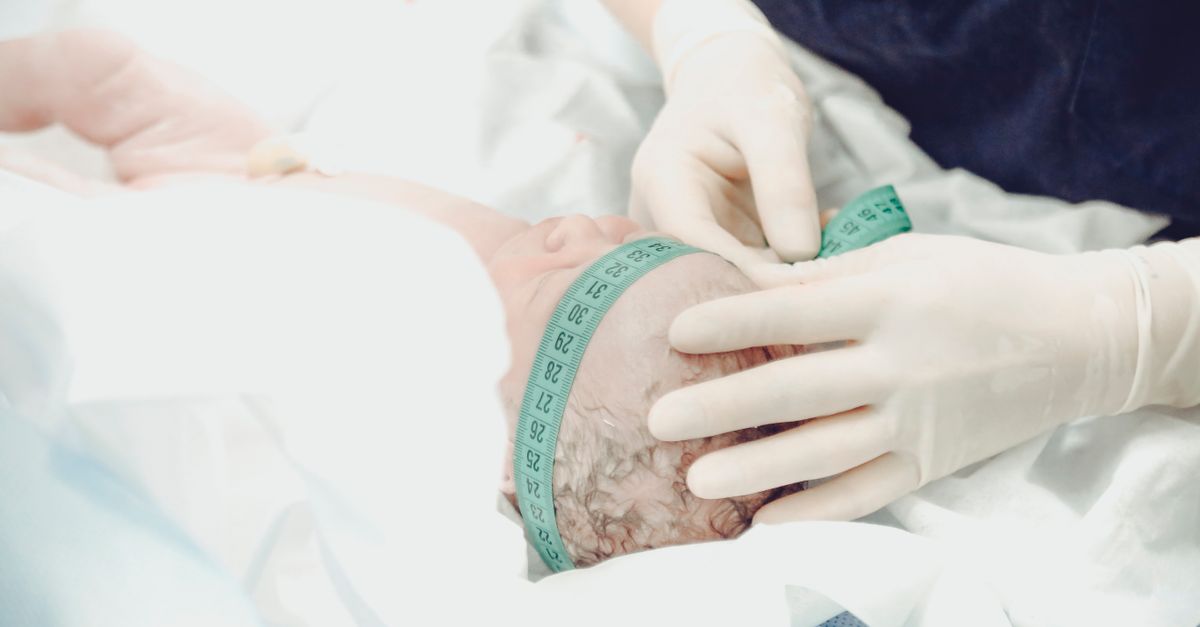
Fetal birth trauma refers to structural damage or physical injury sustained by an infant during the mechanical process of labor and delivery. The journey through the maternal pelvis is a rigorous biomechanical event. The fetus is subjected to immense compressive forces from powerful uterine contractions and intense rotational friction against the bony architecture of the birth canal. While the infant body is uniquely designed to withstand significant pressure, the limits of this biological resilience can be exceeded under specific obstetrical conditions, resulting in unintended physical trauma.
The incidence of severe birth trauma has decreased dramatically in modern obstetrics due to the widespread implementation of advanced fetal monitoring, precise ultrasound sizing, and the liberal use of the cesarean section for high-risk deliveries. However, unexpected mechanical arrests and emergency vaginal deliveries still occur, rendering birth injuries an ongoing and critical clinical concern. Injuries range from minor, self-resolving soft tissue bruises to profound neurological damage and skeletal fractures requiring specialized surgical intervention.
The clinical management of a newborn who has experienced a traumatic birth requires an immediate, meticulous physical examination. The neonatal medical team must rapidly identify the extent of the structural damage, implement targeted neuroprotective or orthopedic therapies, and provide clear, empathetic communication to the distressed parents regarding the anticipated recovery timeline and the long-term prognosis of their child.
2. The Biomechanics of Delivery
To comprehend how birth trauma occurs, one must understand the biomechanics of a normal vaginal delivery. The fetal head, which is typically the largest and least compressible part of the body, serves as a battering ram, dilating the cervix and navigating the rigid maternal pelvis. To fit through this tight passage, the skull bones of the fetus are not fused; they overlap in a process called molding.
Following the emergence of the head, the fetal shoulders must rotate and collapse slightly inward to pass under the maternal pubic bone. The neck acts as a flexible pivot point, allowing the head to extend and rotate as the body follows. These cardinal movements of labor require perfect anatomical alignment and smooth execution.
Trauma occurs when this intricate mechanical dance is interrupted. If the driving force of the uterine contractions pushes the fetus against an unyielding bony obstruction, or if the delivering physician must apply external traction, twisting, or leveraging forces to extract an impacted infant, the mechanical stress is transferred directly to the fragile skeletal and neurological structures of the newborn.
3. Major Risk Factors for Injury
Certain maternal and fetal characteristics significantly elevate the statistical probability of a traumatic delivery. Fetal macrosomia, defined as a birth weight exceeding four thousand grams, is the single most critical risk factor. A large infant possesses a significantly wider shoulder diameter, drastically increasing the likelihood of the shoulders becoming physically wedged behind the maternal pubic bone, a terrifying emergency known as shoulder dystocia.
Maternal factors include a small or abnormally shaped pelvis, which creates a narrow physical bottleneck, and maternal obesity, which can obscure the clinical assessment of fetal size and pelvic capacity. Abnormal fetal presentations are also highly dangerous. A breech presentation, where the buttocks or feet enter the pelvis first, places the head and the delicate cervical spine at massive risk of entrapment and severe traction injury during the final stages of extraction.
The use of operative vaginal instruments is inherently linked to an increased risk of specific traumas. When a labor stalls and the fetus exhibits signs of acute distress, the physician may utilize an obstetrical vacuum extractor or metal forceps applied to the fetal head. While these tools are frequently life-saving, they exert intense, localized mechanical force that can tear blood vessels and fracture delicate bones if not applied with absolute precision.
4. Cranial and Scalp Injuries
The head absorbs the brunt of the mechanical forces during a vaginal delivery, making cranial injuries the most frequently observed form of birth trauma. Caput succedaneum is a minor, highly common condition characterized by generalized, boggy swelling of the scalp. It is caused by prolonged pressure against the dilating cervix, disrupting local venous return. It crosses the suture lines of the skull and resolves harmlessly within a few days.
A more significant cranial injury is a cephalohematoma. This involves a collection of blood pooling beneath the periosteum, the tough fibrous membrane covering the skull bone. Because the bleeding is trapped under this tightly adhered membrane, the swelling forms a firm, distinct lump that strictly does not cross the bony suture lines. Cephalohematomas are frequently associated with the use of vacuum extractors and forceps.
The most severe, life-threatening cranial injury is a subgaleal hemorrhage. In this condition, the emissary veins connecting the scalp to the brain tear, allowing massive quantities of blood to hemorrhage into the loose space beneath the scalp aponeurosis. This space spans the entire head, and a newborn can rapidly bleed out into this compartment, leading to profound hypovolemic shock and sudden cardiovascular collapse.
5. Brachial Plexus Nerve Injuries
Brachial plexus injuries are devastating neurological traumas that occur when the network of thick nerve fibers supplying the arm and hand is severely stretched or completely torn. This trauma is almost exclusively associated with shoulder dystocia, where the physician must apply downward lateral traction to the fetal head to physically dislodge the impacted shoulder from behind the maternal pubic bone.
The most common presentation is Erb palsy, which involves injury to the upper nerves of the plexus. The infant arm hangs limply at their side, internally rotated, with the wrist flexed, assuming a classic “waiter’s tip” posture. The infant cannot actively lift the arm or bend the elbow, although finger movement is often preserved.
A more severe, rarer injury is Klumpke palsy, involving the lower nerves of the plexus. This results in the paralysis of the hand and wrist muscles, presenting as a clawed hand with absent grasp reflexes. If the traction force is severe enough, the nerve roots can be entirely avulsed, or torn directly out of the spinal cord, causing permanent, irreversible paralysis of the entire upper extremity.
6. Skeletal Fractures
The forceful mechanical compression of delivery can fracture the fragile bones of the newborn. A clavicle, or collarbone, fracture is the most frequently encountered skeletal injury. It often occurs spontaneously as the wide fetal shoulders are compressed while passing through a narrow pelvis, or it may be intentionally fractured by the physician as a last-resort maneuver to rapidly resolve a severe shoulder dystocia and save the infant from asphyxiation.
Infants with a fractured clavicle typically exhibit decreased movement of the arm on the affected side, a clinical sign termed pseudoparalysis. The physician may feel a distinct clicking sensation, known as crepitus, or a firm bony lump over the collarbone indicating a healing callus.
Fractures of the long bones, specifically the humerus in the upper arm or the femur in the thigh, are much less common but occur most frequently during complex breech extractions. These fractures present with obvious deformity, profound local swelling, and severe pain when the limb is manipulated during diaper changes or routine medical examinations.
7. Facial Nerve and Soft Tissue Injuries
The facial nerve, which controls the muscles of facial expression, exits the skull just beneath the earlobe, making it highly vulnerable to localized pressure. Facial nerve palsy typically occurs when the head is pressed intensely against the rigid maternal sacrum during a prolonged labor, or it can be a direct complication of the rigid blades of obstetrical forceps compressing the nerve.
The diagnosis is highly visual and becomes apparent when the infant cries. The face appears distinctly asymmetrical; the affected side remains smooth and motionless, the eyelid may not close completely, and the mouth is drawn over to the healthy, functional side. While visually distressing for the parents, this injury is usually the result of temporary nerve bruising rather than permanent severing.
Soft tissue injuries are extremely common and include significant facial bruising, petechiae (tiny pinpoint hemorrhages), and superficial lacerations. Petechiae are frequently seen on the face and neck of infants who experienced a rapid, explosive delivery or a tight nuchal cord wrapped around the neck. Lacerations, occasionally requiring minor suturing, can occur inadvertently during the use of a scalpel during a cesarean section.
8. Identifying Internal Hemorrhage
While external injuries are immediately apparent, the mechanical forces of delivery can cause insidious, life-threatening internal trauma. Rupture of the liver or the spleen is a rare but catastrophic complication, typically associated with extreme macrosomia, breech deliveries, or aggressive cardiopulmonary resuscitation applied immediately after birth.
These solid organs are highly vascular, and a rupture leads to massive, concealed internal hemorrhage into the abdominal cavity. The infant will not exhibit external bruising but will present with signs of profound, unexplained cardiovascular shock: a racing heart rate, extremely pale skin, weak pulses, and a distinctly distended, tight abdomen.
Intracranial hemorrhage, bleeding deep within the brain tissues or the surrounding fluid spaces, can occur due to the extreme molding and shearing forces applied to the fetal skull. Symptoms of internal brain bleeding include a high-pitched, abnormal cry, profound lethargy, poor feeding reflexes, and the sudden onset of focal or generalized neurological seizures.
9. Comprehensive Clinical Assessment
The immediate management of a newborn following a difficult or instrumental delivery begins with a highly focused physical examination. The neonatal physician systematically palpates the entire skull, meticulously differentiating between the harmless boggy swelling of a caput and the dangerous fluid wave of a subgaleal hemorrhage.
The physician carefully evaluates the symmetry of movement in all four limbs. They will elicit standard neonatal reflexes, particularly the Moro, or startle reflex. In a healthy infant, both arms flare outward symmetrically. If one arm fails to move, it provides an immediate clinical clue pointing toward a fractured clavicle or a brachial plexus nerve injury.
The eyes and facial movements are assessed during crying to rule out facial nerve palsy, and the abdomen is palpated gently to ensure it is soft and non-distended. Vitals signs are monitored continuously, with a specific focus on maintaining stable blood pressure and heart rate, ensuring no hidden internal bleeding is destabilizing the cardiovascular system.
10. Structured Data: Differentiating Cranial Swelling
Accurately diagnosing the type of head swelling dictates whether the infant requires observation or immediate life-saving blood transfusions.
| Condition | Anatomical Location of Swelling | Clinical Characteristics |
|---|---|---|
| Caput Succedaneum | Superficial, above the periosteum | Soft, boggy, crosses skull suture lines, resolves in days |
| Cephalohematoma | Trapped beneath the periosteum | Firm, distinct lump, never crosses suture lines, takes weeks to resolve |
| Subgaleal Hemorrhage | Beneath the scalp aponeurosis (entire head) | Massive, fluid-like swelling, ears pushed outward, causes severe blood loss |
11. Diagnostic Imaging and Laboratory Protocols
When physical trauma is suspected, specific radiological imaging is rapidly deployed. For infants presenting with decreased arm movement, a plain radiograph of the chest and upper extremities is the standard initial test. The X-ray rapidly confirms the presence and exact location of a clavicle or humerus fracture, allowing the team to stabilize the bone.
If the physician suspects an internal brain injury or an intracranial hemorrhage based on abnormal neurological signs or seizures, a cranial ultrasound is frequently the first imaging modality utilized. It is a rapid, radiation-free bedside test performed through the soft fontanelle of the skull. For more detailed imaging, a magnetic resonance imaging scan of the brain is the gold standard for defining the extent of the brain tissue damage.
Laboratory testing is essential for monitoring blood loss. Serial complete blood counts are drawn to track the hemoglobin and hematocrit levels. A rapid drop in these values confirms active, ongoing internal bleeding, heavily guiding the decision to initiate emergency blood transfusions or surgical exploration.
12. Management of Cranial Trauma
The vast majority of superficial cranial traumas, including caput succedaneum and cephalohematomas, require no specific medical intervention. The body will naturally reabsorb the pooled fluids and blood over a period of weeks. However, infants with significant cephalohematomas are at a high risk of developing severe jaundice as the trapped red blood cells break down, frequently requiring phototherapy.
In stark contrast, a subgaleal hemorrhage is an extreme medical emergency managed in the neonatal intensive care unit. The primary intervention is aggressive volume resuscitation. The infant frequently requires the rapid, massive transfusion of cross-matched blood and coagulation factors to replace the blood lost into the scalp space and prevent fatal hypovolemic shock.
If the infant suffers a severe depressed skull fracture, where the bone is pushed deeply inward against the brain tissue resembling a dented ping-pong ball, a pediatric neurosurgeon is consulted immediately. While some minor dents elevate spontaneously with time, deep compressions require surgical elevation to prevent localized brain damage and future seizure disorders.
13. Rehabilitation for Brachial Plexus Injuries
The management of a brachial plexus nerve injury is a long-term, multidisciplinary process. In the first few days of life, the affected arm is simply positioned carefully across the abdomen of the infant to prevent further stretching or mechanical tension on the damaged nerve roots.
Following the initial acute phase, rigorous physical therapy and occupational therapy are the cornerstones of treatment. The therapist instructs the parents on specific, gentle range-of-motion exercises. The goal is to keep the joints of the shoulder, elbow, and wrist perfectly supple and prevent severe, permanent muscle contractures while waiting for the injured nerves to heal and regenerate.
The majority of these nerve injuries involve stretching rather than tearing, and the infant will slowly regain spontaneous movement over the first three to six months of life. However, if there is absolutely no clinical improvement by three to six months, specialized microsurgical nerve grafting by a pediatric orthopedic surgeon is required to restore functional connectivity to the arm.
14. Orthopedic Interventions
Skeletal fractures sustained during delivery are surprisingly robust and heal remarkably fast due to the aggressive bone-forming capacity of a newborn. A fractured clavicle generally requires no active casting or surgical setting. The primary intervention involves simply pinning the sleeve of the infant to their shirt across their chest.
This simple immobilization stabilizes the arm, prevents sharp pain during movement, and allows the collarbone to heal naturally. A large, firm lump of calcium, known as a callus, will form over the fracture site within a few weeks and will gradually remodel and disappear entirely over the first year of life.
Fractures of the long bones, such as the humerus or femur, require slightly more intervention. A pediatric orthopedic specialist will frequently apply a soft, customized splint or a specialized harness to keep the limb properly aligned. Due to the immense regenerative capacity of newborn bones, these fractures heal perfectly, leaving no long-term structural deformity or limb length discrepancy.
15. Pain Management and Supportive Care
Providing adequate pain relief is a fundamental component of managing any infant with a birth injury. Newborns experience profound pain, and untreated pain destabilizes their heart rate, breathing patterns, and feeding routines. For minor injuries, simple interventions like swaddling, skin-to-skin contact, and the provision of a pacifier dipped in sweet sucrose solution are highly effective at blunting the pain response.
For distinct skeletal fractures or severe soft tissue trauma, scheduled doses of infant acetaminophen are utilized. Stronger, intravenous opioid pain medications are strictly reserved for infants suffering from massive trauma or those recovering from major reconstructive surgeries in the intensive care unit.
Supportive care also extends heavily to the parents. Witnessing a newborn suffer a traumatic injury is psychologically devastating. The medical team must provide continuous, transparent updates regarding the medical plan, facilitating bonding between the parents and the infant despite the presence of splints, medical equipment, or the environment of the intensive care unit.
16. Long-Term Prognosis
The long-term prognosis for an infant who has suffered a birth injury depends entirely on the specific anatomical structure damaged. Soft tissue injuries, simple skull fractures, and clavicle fractures boast an excellent prognosis. They heal rapidly and completely, leaving absolutely no residual physical or developmental consequences as the child grows.
The prognosis for facial nerve palsy is also generally highly favorable. Because the nerve is usually only bruised by pressure, full symmetrical facial movement typically returns completely within a few weeks to a few months.
The prognosis for severe brachial plexus injuries or intracranial hemorrhage is much more guarded. While many nerve injuries recover fully, severe avulsions can result in permanent, lifelong weakness, stunted growth of the affected arm, and reduced fine motor control. Infants who suffer significant bleeding in the brain require years of neurological monitoring, as they face a high risk of developing cerebral palsy, learning disabilities, and epilepsy.
17. Preventative Obstetrical Strategies
The primary medical focus remains entirely on the prevention of these traumatic injuries. Modern obstetrical protocols heavily utilize serial ultrasound imaging to carefully track fetal growth trajectories, specifically looking for signs of severe macrosomia. If an infant is estimated to weigh significantly over the safe anatomical limits, an elective cesarean section is strongly recommended to completely avoid the mechanical dangers of the birth canal.
During active labor, continuous electronic fetal monitoring is utilized. If the fetal heart rate indicates that the infant is not tolerating the mechanical forces of the contractions, the trial of labor is swiftly aborted. Furthermore, physicians are trained in highly specific, rapid physical maneuvers to resolve a shoulder dystocia without applying dangerous downward traction on the delicate fetal neck.
The use of operative vaginal instruments, like forceps or vacuums, is now highly regulated and restricted. These tools are only applied when specific, strict clinical criteria are met, and the delivering physician possesses extensive, specialized training in their precise mechanical application, minimizing the risk of devastating cranial trauma.
18. Frequently Asked Questions (FAQ)
1. Will my baby have brain damage from the swelling on their head?
The vast majority of head swelling (caput or cephalohematoma) is entirely on the outside of the skull bone and does not affect the brain tissue underneath. It looks concerning but is generally completely harmless to brain development.
2. Did the doctor break my baby collarbone on purpose?
Sometimes, yes. In a terrifying emergency where the baby shoulders are completely stuck and the baby cannot breathe (shoulder dystocia), the doctor may intentionally fracture the collarbone to instantly collapse the shoulder width and save the life of the infant.
3. Will the broken collarbone heal crooked?
No. Newborn bones heal exceptionally fast and remodel themselves perfectly as the baby grows. Within a few months, it is usually impossible to tell on an X-ray that the bone was ever broken.
4. Why is my baby arm completely limp?
This is likely a brachial plexus nerve injury caused by the neck stretching during delivery. Most of these injuries are bruises to the nerve and will slowly heal with physical therapy, though severe tears require surgery.
5. How long will the facial drooping last?
Facial nerve palsy caused by pressure during birth usually resolves completely on its own within a few weeks. The doctor will monitor the baby to ensure the eye is protected and stays moist until the eyelid can close fully again.
19. Bibliography
Disclaimer: The content is for informational purposes only and does not replace medical advice. Always consult your doctor for personalized treatment.