
1. Introduction to Focal Epilepsy
Focal epilepsy is a complex neurological disorder characterized by recurrent, unprovoked seizures that originate within a specific, localized network of neurons in one hemisphere of the brain. Historically referred to as partial epilepsy, this condition stands in direct contrast to generalized epilepsy, where the abnormal electrical discharges simultaneously involve extensive networks across both cerebral hemispheres from the absolute onset.
The clinical presentation of a focal seizure is highly diverse and entirely dictated by the precise anatomical location of the hyperactive neuronal network. Depending on the brain region involved, a patient may experience sudden motor spasms in a single limb, profound sensory hallucinations, intense emotional shifts, or sudden alterations in conscious awareness. Because these symptoms can be subtle or mimic psychiatric conditions, accurate diagnosis requires meticulous clinical observation and advanced neurological testing.
Management of focal epilepsy has advanced significantly. The primary goal of therapy is the complete cessation of seizures with minimal pharmacological side effects. Through the use of targeted antiseizure medications, advanced neuroimaging to identify structural brain lesions, and potential surgical interventions for drug-resistant cases, modern neurology offers robust strategies to help patients achieve long-term seizure freedom and maintain an excellent quality of life.
2. Brain Anatomy and Cortical Networks
To comprehend the diverse symptoms of focal epilepsy, it is essential to understand the functional mapping of the human cerebral cortex. The brain is divided into two distinct hemispheres, each containing four primary lobes: the frontal, parietal, temporal, and occipital lobes. Each of these lobes is highly specialized to process specific neurological functions.
The frontal lobe is the command center for voluntary motor execution, expressive language, and complex executive functions like decision-making. The parietal lobe processes somatosensory information, including touch, temperature, and spatial awareness. The temporal lobe is critical for auditory processing, memory consolidation, and emotional regulation. The occipital lobe is exclusively dedicated to visual processing.
In focal epilepsy, a microscopic cluster of neurons within one of these specific lobes becomes unstable. When this cluster misfires, it disrupts the normal function of that specific cortical area. If the electrical storm remains confined to that small area, the symptoms remain highly localized. However, the brain is densely interconnected by tracts of white matter. If the abnormal electrical discharge travels along these tracts, the seizure can spread to adjacent lobes or even cross the corpus callosum to involve the entire brain.
3. Pathophysiology of Epileptogenesis
The biological transition of a normal neuronal network into a hyperactive, seizure-generating focus is a process known as epileptogenesis. In a healthy brain, there is a strict, elegant balance between excitatory neurotransmitters, primarily glutamate, which stimulate neurons to fire, and inhibitory neurotransmitters, primarily Gamma-Aminobutyric Acid (GABA), which suppress neuronal firing.
A focal seizure occurs when this delicate balance collapses in a localized area. A cluster of neurons suddenly begins to fire in rapid, synchronous bursts, a phenomenon termed a paroxysmal depolarizing shift. This localized hyperactivity overwhelms the surrounding inhibitory networks designed to contain it.
The underlying cause of this instability is often a structural defect in the brain tissue. A microscopic scar, a malformed blood vessel, or an area of abnormal cellular development acts as a constant irritant. The neurons surrounding this structural lesion undergo maladaptive physical and chemical changes, altering their ion channels and receptor densities, permanently lowering their threshold for initiating a massive, synchronized electrical discharge.
4. Classification of Focal Seizures
The International League Against Epilepsy continually refines the classification of seizures to ensure precise clinical communication. Focal seizures are primarily categorized based on whether the patient’s conscious awareness remains fully intact during the electrical event, and whether the seizure involves physical movement.
A “focal aware seizure,” previously known as a simple partial seizure, occurs when the patient remains completely conscious, alert, and able to interact with their environment throughout the episode, despite experiencing intense, involuntary neurological symptoms. The patient remembers the event clearly.
A “focal impaired awareness seizure,” historically termed a complex partial seizure, occurs when the abnormal electrical activity spreads to the deeper structures of the brain responsible for maintaining consciousness, such as the temporal lobes or the reticular activating system. The patient may appear awake, with their eyes open, but they are entirely disconnected from their environment, unresponsive to commands, and typically have complete amnesia regarding the event once the seizure concludes.
5. Auras and Sensory Symptoms
An aura is technically a focal aware seizure that produces purely subjective sensory, emotional, or cognitive symptoms without any observable physical signs. For many patients, the aura serves as an invaluable, albeit brief, warning sign that a more severe seizure is imminent, allowing them to sit down or seek safety.
The specific sensation of an aura depends entirely on the anatomical location of the seizure focus. If the seizure originates in the occipital lobe, the patient may experience sudden visual hallucinations, such as flashing lights or geometric shapes. A parietal lobe focus often generates intense paresthesias—a sensation of tingling, numbness, or electric shocks traveling down one side of the body.
Temporal lobe auras are exceptionally complex. Patients frequently report an intense, sudden feeling of déjà vu, profound unexplained fear or euphoria, or sudden olfactory hallucinations, such as the distinct, overpowering smell of burning rubber or sulfur, despite no actual odor being present in the environment.
6. Motor Manifestations and Automatisms
When a focal seizure involves the motor cortex in the frontal lobe, the clinical presentation is dramatically visible. The patient may exhibit clonic movements, which are rhythmic, forceful jerking motions of a single limb, such as the right arm or the left side of the face. A classic presentation is the “Jacksonian march,” where the jerking movement starts in a small area, like the thumb, and progressively spreads up the arm as the electrical discharge moves sequentially across the motor cortex map.
In focal impaired awareness seizures, particularly those originating in the temporal or frontal lobes, patients frequently display automatisms. Automatisms are repetitive, purposeless, and involuntary physical movements performed while the patient is completely unconscious of their actions.
Oral automatisms include continuous lip-smacking, chewing, or swallowing motions. Manual automatisms often involve the patient aimlessly picking at their clothing, fumbling with objects, or repeatedly rubbing their hands together. Recognizing these specific movements is crucial for the neurologist, as they strongly point toward a temporal lobe origin for the epilepsy.
7. The Postictal State
The termination of a focal seizure is rarely immediate. As the massive electrical storm within the brain finally exhausts the metabolic energy of the neurons, the seizure stops, and the patient enters the postictal state. This is a crucial physiological recovery period during which the affected brain networks are profoundly suppressed and attempting to restore their normal chemical balance.
Following a focal aware seizure, the postictal state may be brief or virtually unnoticeable. However, following a focal impaired awareness seizure, the postictal period can last from several minutes to several hours. During this time, the patient is typically profoundly confused, disoriented, and deeply lethargic.
A unique phenomenon known as Todd’s paralysis can occur during the postictal state following a focal motor seizure. The patient may experience severe, temporary weakness or complete paralysis in the specific limb that was convulsing during the seizure. This localized paralysis can persist for hours to days and is often clinically mistaken for an acute stroke, requiring careful diagnostic differentiation.
8. Structural Causes and Brain Lesions
Unlike generalized epilepsy, which often has a strong, diffuse genetic basis, focal epilepsy is predominantly a symptomatic disorder, meaning it is the direct consequence of a specific, identifiable structural lesion within the brain parenchyma. Identifying this lesion is a primary goal of the neurological evaluation.
Mesial temporal sclerosis is the single most common structural cause of focal epilepsy in adults. This condition involves severe scarring and loss of neurons deep within the hippocampus, a critical structure in the temporal lobe. Head trauma from severe concussions or traumatic brain injuries can leave areas of cortical scarring (gliosis) that become highly epileptogenic years after the initial injury.
Other structural etiologies include cerebrovascular accidents (strokes), where the boundary zone of dead tissue becomes electrically unstable; slow-growing, benign brain tumors such as gangliogliomas; and vascular malformations like cavernous hemangiomas, which occasionally leak microscopic amounts of blood, severely irritating the surrounding cortical neurons.
9. Diagnostic Evaluation with Electroencephalography
The definitive diagnostic tool for evaluating any seizure disorder is the Electroencephalogram (EEG). This non-invasive test involves placing highly sensitive electrodes across the patient’s scalp to record the continuous, spontaneous electrical activity of the cerebral cortex.
In a patient with focal epilepsy, the neurologist analyzes the EEG tracing for interictal epileptiform discharges—brief, sharp spikes or waves of abnormal electricity that occur between actual clinical seizures. The specific geographical location of these spikes on the scalp allows the neurologist to map the exact brain lobe where the hyperactive network resides.
Often, a routine twenty-minute EEG will appear completely normal because the brain is not actively misfiring at that precise moment. In these cases, the patient is admitted to an epilepsy monitoring unit for continuous Video-EEG telemetry. The patient is monitored continuously for several days while their antiseizure medications are slowly reduced, intending to capture and record the electrical and physical manifestations of an actual seizure in a controlled, safe environment.
10. Neuroimaging in Epilepsy
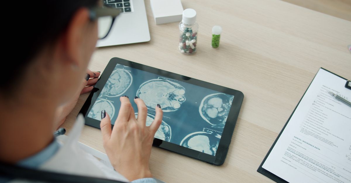
While the EEG reveals the electrical pathology, advanced neuroimaging is strictly required to visualize the structural pathology causing the focal epilepsy. Magnetic Resonance Imaging (MRI) is the absolute gold standard imaging modality. A routine MRI is insufficient; an epilepsy-protocol MRI utilizes ultra-high-resolution, thin-slice sequences designed specifically to detect microscopic cortical malformations or subtle hippocampal scarring.
If the MRI identifies a clear structural lesion that perfectly correlates with the electrical location found on the EEG, the physician can establish a definitive, highly specific diagnosis, which is critical if surgical intervention is considered.
| Diagnostic Modality | Primary Purpose | Typical Finding in Focal Epilepsy |
|---|---|---|
| Routine EEG | Measure electrical brain activity. | Localized sharp spikes in a specific brain lobe. |
| Video-EEG Monitoring | Correlate physical symptoms with brain waves. | Captures the exact electrical onset of the clinical seizure. |
| Epilepsy-Protocol MRI | Identify structural brain defects. | Visualizes tumors, vascular lesions, or hippocampal scarring. |
11. Pharmacological Management
The cornerstone of treatment for focal epilepsy is the daily administration of antiseizure medications. These powerful pharmacological agents do not cure the underlying brain lesion; rather, they alter the chemical environment of the brain to suppress abnormal electrical firing and prevent a localized seizure from initiating or spreading.
Medications function through various mechanisms. Some, like carbamazepine and lamotrigine, physically block voltage-gated sodium channels on the neuron’s surface, preventing the cell from firing rapid, repetitive electrical impulses. Others, like levetiracetam, bind to specific synaptic vesicle proteins to modulate neurotransmitter release.
Selecting the appropriate medication requires significant clinical expertise. The neurologist must consider the specific type of focal seizure, the patient’s age, potential side effects (such as lethargy, dizziness, or cognitive slowing), and any potential interactions with other medications the patient is currently taking. The goal is always monotherapy—controlling the seizures completely using a single medication at the lowest effective dose.
12. Drug-Resistant Epilepsy and Surgical Interventions
While many patients achieve excellent seizure control with medication, approximately one-third of individuals will develop drug-resistant focal epilepsy, defined as the failure of two appropriately chosen and tolerated antiseizure medications to achieve seizure freedom. For these patients, surgical intervention offers a highly effective, potentially curative option.
The most common and successful surgical procedure is a focal cortical resection. If comprehensive testing confirms that all the seizures originate from a single, specific location in the brain, and that removing this small area of tissue will not cause significant neurological deficits (such as a loss of speech or motor control), a neurosurgeon will physically remove the epileptogenic focus.
For patients with temporal lobe epilepsy caused by mesial temporal sclerosis, the surgical removal of the scarred hippocampus results in complete seizure freedom for a substantial majority of patients, dramatically transforming their quality of life.
13. Neuromodulation Devices
When a patient has drug-resistant focal epilepsy but is not a candidate for resective surgery—often because the seizure focus is located in a critical, irreplaceable language or motor area of the brain—neuromodulation devices offer a valuable therapeutic alternative.
Vagus Nerve Stimulation involves surgically implanting a small device, similar to a cardiac pacemaker, under the skin of the chest. It sends regular, mild electrical pulses up the vagus nerve in the neck directly to the brain, which desynchronizes the cortical networks and reduces the frequency and severity of seizures.
Responsive Neurostimulation is a more advanced, targeted therapy. A small computer is implanted in the skull, with tiny electrodes placed directly over the seizure focus. The device continuously monitors the brain’s electrical activity. When it detects the specific electrical signature of a seizure beginning, it immediately delivers a microscopic, imperceptible electrical counter-shock to the tissue, instantly terminating the seizure before the patient even experiences a symptom.
14. Lifestyle Management and Safety
Comprehensive care for focal epilepsy extends beyond medication and surgery; strict lifestyle management is critical for reducing seizure frequency. Sleep deprivation is one of the most potent triggers for focal seizures. Patients must prioritize rigorous sleep hygiene, ensuring they achieve sufficient, uninterrupted rest every night.
Managing psychological stress, minimizing alcohol consumption, and strictly avoiding illicit drugs are also vital, as these factors significantly lower the brain’s seizure threshold. Furthermore, patients must adhere flawlessly to their medication schedule. Missing even a single dose of an antiseizure medication causes a sudden drop in blood levels, drastically increasing the risk of a breakthrough seizure.
Safety considerations are paramount. Patients with focal impaired awareness seizures must adhere to legal driving restrictions mandated by their local jurisdictions until they have achieved a documented period of complete seizure freedom. Precautions should be taken around open water, heights, and heavy machinery to prevent severe traumatic injury during a sudden loss of awareness.
15. Frequently Asked Questions
1. What is the difference between a focal seizure and a grand mal seizure?
A focal seizure starts in one small, specific area of the brain, causing localized symptoms. A grand mal (generalized tonic-clonic) seizure involves massive electrical firing across the entire brain simultaneously, causing total unconsciousness and full-body convulsions.
2. Can a focal seizure turn into a full-body seizure?
Yes. If the abnormal electricity from a focal seizure is strong enough, it can travel along the brain’s pathways and spread to the rest of the brain, evolving into a generalized tonic-clonic seizure.
3. Why do I smell something burning before I have a seizure?
If the seizure originates in the temporal lobe, which processes smell and memory, the misfiring neurons can create a powerful olfactory hallucination. This is called an aura, and it is actually the very beginning of the seizure.
4. Will I have to take seizure medication for the rest of my life?
It depends entirely on the cause. If the epilepsy is caused by a permanent structural scar in the brain, long-term medication is usually required. If you remain completely seizure-free for several years, your neurologist may discuss a very slow, controlled trial of tapering off the medication.
5. Why is the doctor asking about repetitive lip-smacking?
Lip-smacking, chewing, or aimlessly picking at clothes are called automatisms. They are involuntary movements that frequently occur during focal seizures originating in the temporal lobe. Identifying them helps the neurologist pinpoint where the seizure starts.
6. Is brain surgery a safe option for focal epilepsy?
For patients whose seizures cannot be controlled by medication, epilepsy surgery is a highly refined and safe option. Advanced imaging and mapping ensure that the surgeon only removes the damaged tissue causing the seizures while strictly preserving normal brain function.
16. Bibliography
Disclaimer: The content is for informational purposes only and does not replace medical advice. Always consult your doctor for personalized treatment.
