1. Introduction
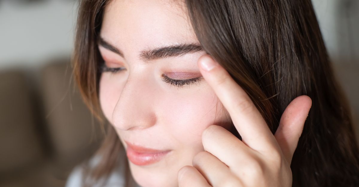
Having dark circles under the eyes of genetic origin is completely normal and represents a benign anatomical variation rather than a medical pathology. This condition, clinically known as periorbital hyperpigmentation, frequently runs in families and is caused by a combination of concentrated melanin deposits and naturally thin eyelid skin that reveals underlying vascular beds. Recognizing the genetic basis of these dark circles is essential for distinguishing them from lifestyle-induced or pathological changes.
The skin surrounding the eyes is uniquely structured compared to the rest of the face. It is exceptionally delicate and lacks a robust layer of subcutaneous fat. Consequently, the anatomical features beneath the surface play a prominent role in the visible appearance of the lower eyelid. When genetic factors dictate specific pigment distribution or structural bone contours, dark circles emerge as a permanent facial characteristic.
Understanding that this pigmentation is an inherited trait helps individuals manage their expectations regarding cosmetic treatments. While topical creams claim to erase dark shadows, genuine genetic periorbital hyperpigmentation requires a deeper comprehension of dermal anatomy and targeted clinical interventions for effective modification.
2. Anatomy of the Periorbital Region
The periorbital region encompasses the tissues immediately surrounding the eye. The epidermis in this specific area is the thinnest on the entire human body, measuring roughly a half millimeter in thickness. Beneath this delicate outer layer lies a complex network of tiny blood vessels and capillaries.
Because the skin acts as a semi-transparent window, the underlying vascular structures naturally cast a dark, bluish, or purplish hue visible on the surface. This anatomical reality guarantees that the under-eye area will always appear slightly darker than the thicker skin of the cheeks or forehead.
Individuals inherit the baseline thickness of their skin from their parents. Those who genetically inherit exceptionally thin periorbital skin will invariably exhibit more pronounced dark circles simply due to the enhanced visibility of their normal venous network.
3. Melanin and Hyperpigmentation
Melanin is the biological pigment responsible for giving skin, hair, and eyes their color. Melanocytes are the specialized cells located in the deeper layers of the epidermis that produce this pigment.
In cases of genetic periorbital hyperpigmentation, the melanocytes in the skin around the eyes are either more numerous or produce an excess of melanin compared to adjacent facial skin. This localized overproduction creates a distinct brownish or olive-colored band under the lower lash line.
This specific pattern of pigment deposition is particularly prevalent in populations with Mediterranean, Middle Eastern, and South Asian ancestry. It is a dominant genetic trait, meaning it frequently passes directly from one generation to the next, becoming a defining familial characteristic.
4. The Tear Trough Deformity
Genetics do not only dictate skin color and thickness; they also dictate bone structure. The tear trough is the natural anatomical depression running from the inner corner of the eye down toward the upper cheek.
Some individuals inherit a prominent, deeply recessed tear trough due to the specific shape of their orbital bone and the natural distribution of their facial fat pads. A deep tear trough creates a physical shadow under ambient lighting.
This shadow is frequently mistaken for skin pigmentation. No amount of skin lightening will resolve a dark circle caused by a structural shadow. Differentiating between true pigment and a structural tear trough deformity is a critical step in clinical aesthetic assessments.
5. Vascular Contributions
The orbicularis oculi muscle is a circular muscle that controls the closing of the eyelids. This muscle lies immediately beneath the thin periorbital skin and is richly supplied with deep red and purple blood vessels.
Genetics determine the proximity of this muscle to the surface of the skin. If the muscle sits superficially, the dark purplish hue of the muscle tissue and its vascular network bleeds through the translucent skin, mimicking the appearance of a bruise.
Furthermore, sluggish venous drainage in this region, which can also be an inherited trait, allows deoxygenated blood to pool slightly in the periorbital capillaries, amplifying the dark, shadowy appearance.
6. Differentiating Genetics from Lifestyle Factors
While genetics establish the baseline appearance of the under-eye area, lifestyle factors frequently exacerbate the condition. A lack of restorative sleep causes systemic peripheral vasodilation.
When blood vessels dilate to compensate for fatigue, the vessels under the eyes expand, making the genetic dark circles appear significantly darker and more prominent. Similarly, systemic dehydration causes the skin to lose its natural turgor, sinking inward and deepening structural shadows.
Patients often report that their dark circles worsen during periods of stress or illness. It is crucial to understand that while lifestyle factors modify the intensity of the pigmentation, the foundational presence of the dark circles remains anchored in the individual’s DNA.
7. The Role of Atopic Dermatitis and Allergies
Genetic predispositions extend to the immune system. Individuals with a family history of atopic dermatitis, eczema, or severe seasonal allergies frequently develop dark circles through a secondary mechanism known as post-inflammatory hyperpigmentation.
Chronic allergies cause intense itching. The repetitive mechanical trauma of rubbing and scratching the delicate periorbital skin induces localized inflammation. The body responds to this inflammation by stimulating melanocytes to produce excess pigment.
This condition, colloquially termed allergic shiners, presents as a dark, thickened band of skin under the eyes. Managing the underlying allergic response is mandatory to prevent the continual deposition of new melanin in these genetically susceptible individuals.
8. Aging and Collagen Loss
The natural aging process acts as an amplifier for genetic dark circles. As the body ages, the production of collagen and elastin fibers within the dermis steadily declines.
This loss of structural proteins causes the already thin periorbital skin to become even more translucent and fragile. Furthermore, the supportive fat pads in the upper cheek slowly descend with gravity, unmasking the orbital rim and deepening the tear trough shadow.
Individuals who possessed mild genetic dark circles in their youth often find the condition becomes far more pronounced as they transition through their forties and fifties, driven entirely by these predictable, age-related anatomical shifts.
9. Data: Types of Dark Circles
| Category | Primary Cause | Visual Characteristics |
|---|---|---|
| Pigmented (Melanin) | Genetic melanin overproduction | Brown or olive-colored, uniform band |
| Vascular | Thin skin over blood vessels | Blue, pink, or purplish hue |
| Structural | Deep bone structure or fat loss | Shadows that change with lighting angles |
| Mixed | Combination of the above factors | Complex hues and structural depressions |
10. Dermatological Assessment
A precise clinical assessment by a dermatologist determines the exact etiology of the dark circles. The physician performs a simple manual stretch test to differentiate the underlying causes.
If the skin under the eye is gently stretched and the dark color dissipates or turns slightly lighter, the darkness is primarily vascular; the stretching physically empties the blood vessels.
If the color remains a constant, dark brown regardless of how the skin is stretched, the issue is strictly pigment-related. If the darkness disappears completely when a light is shone directly onto the area from the front, it proves the dark circle is a structural shadow rather than actual skin coloration.
11. Topical Treatments and Limitations
The commercial market is flooded with eye creams promising to eradicate dark circles. However, topical treatments possess significant clinical limitations when addressing deeply ingrained genetic traits.
Ingredients such as hydroquinone, kojic acid, and vitamin C act as tyrosinase inhibitors, which slow down the production of new melanin. While these can slightly fade pigmented dark circles, they require months of diligent application and cannot alter the baseline genetic instruction of the melanocytes.
Topical caffeine is frequently utilized for vascular dark circles. Caffeine acts as a temporary vasoconstrictor, shrinking the underlying blood vessels and slightly reducing the purplish hue, but the effect lasts only a few hours and provides no structural change.
12. Clinical Interventions for Pigmentation
For patients seeking definitive modification of genetic hyperpigmentation, dermatologists employ advanced in-office procedures. Chemical peels utilizing trichloroacetic acid or glycolic acid carefully exfoliate the top layers of the epidermis, drawing the excess melanin out of the skin.
Q-switched lasers and intense pulsed light therapies specifically target and shatter the melanin pigment deposits within the dermis without damaging the surrounding delicate tissue.
These energy-based devices offer profound improvements for purely pigmented dark circles, but they require precise calibration by a skilled clinician to avoid paradoxical worsening of the pigmentation, a risk notably higher in patients with darker skin tones.
13. Addressing Structural Shadows
If the genetic dark circles are driven by a tear trough deformity or a lack of orbital fat, topical creams and lasers are entirely ineffective. The treatment must address the structural deficit by replacing the lost volume.
Dermal fillers, typically composed of hyaluronic acid, are injected directly into the deep tissue overlying the orbital bone. This physically lifts the skin up and away from the underlying vascular muscle and smooths out the anatomical depression.
By eliminating the physical valley under the eye, the ambient light no longer casts a shadow, instantly resolving the appearance of the dark circle. This procedure provides immediate results but requires maintenance injections every nine to twelve months.
14. Surgical Considerations
In cases where genetics have dictated large, bulging fat pads under the eyes alongside severe skin laxity, nonsurgical interventions may prove inadequate. These bulging fat pads cast significant shadows downward onto the cheeks.
A lower blepharoplasty is a surgical procedure performed by an oculoplastic surgeon. The surgeon makes a microscopic incision inside the lower eyelid to remove or reposition the excess orbital fat.
This surgery permanently modifies the inherited anatomical contours of the periorbital region, creating a smooth transition from the eye to the cheek and permanently eliminating the structural shadows that mimicked dark pigmentation.
15. The Psychological Impact
While medically benign, genetic dark circles carry a substantial psychological weight. Cultural beauty standards frequently equate dark circles with exhaustion, illness, or premature aging.
Individuals with this genetic trait frequently face unsolicited comments inquiring if they are tired or feeling unwell, even when they are fully rested and vibrant. This continuous external validation of a perceived flaw can lead to significant social anxiety and a reliance on heavy cosmetic concealers.
Counseling patients involves validating their aesthetic concerns while firmly reinforcing the physiological reality that their appearance is a normal expression of their healthy DNA, completely unrelated to chronic fatigue or systemic pathology.
16. When to Seek Medical Care
Genetic dark circles remain stable over time. A medical evaluation is necessary if the periorbital region undergoes a sudden, rapid change in appearance.
If dark circles appear abruptly, are highly asymmetrical, or are accompanied by severe swelling, profound redness, or localized pain, an acute inflammatory process, infection, or trauma is likely responsible.
Furthermore, if the sudden darkening of the skin is accompanied by systemic symptoms such as profound weight loss, severe lethargy, or joint pain, a comprehensive medical workup is required to rule out underlying autoimmune or endocrine disorders.
17. Frequently Asked Questions FAQ
1. Can drinking more water cure my genetic dark circles?
No. While severe dehydration causes the skin to sink and exacerbates structural shadows, optimal hydration simply returns the skin to its baseline state. Water consumption cannot alter the inherited melanin deposits or the anatomical thinness of your eyelid skin.
2. Are cucumber slices actually effective for dark circles?
Cucumber slices provide a cooling effect that causes temporary vasoconstriction, slightly reducing vascular swelling. They also offer mild hydration to the epidermis. However, they provide only fleeting, superficial relief and do not alter the genetic causes of hyperpigmentation.
3. Will getting more sleep make genetic dark circles disappear?
Adequate sleep prevents the blood vessels under the eyes from dilating due to exhaustion, which keeps the dark circles from looking worse than their baseline. However, sleep will not erase the underlying genetic pigment or correct a structural tear trough shadow.
4. Can I use regular body bleaching creams under my eyes?
Absolutely not. The skin of the periorbital region is dangerously thin and highly sensitive. Standard bleaching agents are too harsh and will cause severe chemical burns, profound irritation, and often result in post-inflammatory hyperpigmentation, making the dark circles significantly worse.
5. At what age do genetic dark circles typically appear?
Because they are an inherited trait, genetic dark circles can be visible in early childhood. However, they typically become much more apparent during late adolescence and early adulthood as facial bone structures mature and subtle changes in skin thickness begin.
18. Bibliography
Disclaimer: The content is for informational purposes only and does not replace medical advice. Always consult your doctor for personalized treatment.