
1. Introduction to Fistulas of the Hard Palate
A fistula of the hard palate, medically termed an oronasal fistula, is an abnormal structural defect that creates a direct communication pathway between the oral cavity and the nasal cavity. In a healthy human skull, the hard palate acts as a rigid, impermeable anatomical barrier, strictly separating the mouth from the nasal passages. This separation is biologically essential for the execution of fundamental physiological functions, including the generation of precise speech, the generation of intraoral pressure for swallowing, and the prevention of food and liquids from entering the respiratory tract.
When this barrier is breached by a fistula, the resulting functional impairment is immediate and profoundly disruptive to the patient’s quality of life. The severity of the symptoms is directly correlated with the precise anatomical location and the physical dimensions of the defect. While some minute fistulas may remain relatively asymptomatic, larger defects consistently result in the distressing regurgitation of fluids through the nose and severe speech distortions.
Clinical management of a hard palate fistula is a complex endeavor requiring specialized surgical expertise. The condition demands a thorough anatomical evaluation to precisely map the defect, identify the underlying etiology, and select the appropriate reconstructive technique. Successful closure not only restores the physical barrier but also requires comprehensive post-operative rehabilitation to re-establish normal speech and swallowing mechanics.
2. Anatomy of the Hard Palate
To comprehend the pathological implications of a palatal fistula, an understanding of the local anatomy is necessary. The roof of the mouth consists of two distinct segments: the anterior hard palate and the posterior soft palate. The hard palate is a rigid, bony structure formed by the fusion of the palatine processes of the maxilla and the horizontal plates of the palatine bones.
This bony foundation is tightly covered on its inferior, oral surface by a specialized, dense mucosal layer known as the mucoperiosteum. This robust lining is designed to withstand the intense mechanical friction generated during the mastication of food. On its superior surface, the hard palate is lined with the delicate, ciliated respiratory epithelium of the nasal floor.
The structural integrity of the hard palate is vital for creating a seal during swallowing. As the tongue pushes upward against the rigid palate, it generates the negative pressure necessary to propel a food bolus backward into the pharynx. The hard palate also serves as an unyielding articulatory surface against which the tongue strikes to produce specific consonant sounds, making it indispensable for clear, intelligible speech.
3. Pathophysiology of Palatal Defects
The formation of a fistula in the hard palate represents a complete, full-thickness failure of the tissues that normally separate the oral and nasal cavities. Regardless of the underlying cause, the pathophysiology involves the necrosis, failure to fuse, or physical destruction of the bony architecture and its overlying mucosal linings.
Once an open communication tract is established, normal physiological pressures exacerbate the problem. The oral cavity routinely experiences significant positive pressure during swallowing and speech articulation. This constant positive pressure is forcefully directed upward through the path of least resistance—the fistula—into the lower-pressure environment of the nasal cavity.
This abnormal pressure dynamic prevents the delicate mucosal edges of the fistula from healing spontaneously. Furthermore, the continuous passage of saliva, food particles, and oral bacteria into the nasal passage triggers a chronic, localized inflammatory response along the margins of the defect. This chronic inflammation causes the edges of the tissue to become rigid and epithelialized, cementing the fistula as a permanent anatomical defect that cannot close without surgical intervention.
4. Congenital Causes: Cleft Palate Complications
The single most prevalent cause of a hard palate fistula is a surgical complication following the primary repair of a congenital cleft palate. A cleft palate is a birth defect where the bony plates of the roof of the mouth fail to fuse correctly during early embryonic development. Pediatric reconstructive surgeons operate to close this gap, typically within the first year of the child’s life.
Despite meticulous surgical technique, the repaired tissue is subjected to immense physical tension. If the local blood supply to the mobilized tissue flaps is compromised, or if the wound is subjected to excessive mechanical stress during the healing phase, localized tissue necrosis occurs. The surgical incision breaks down, and an oronasal fistula forms.
These post-surgical fistulas most commonly occur at the junction of the hard and soft palate or in the anterior region behind the incisors. Managing these defects is highly challenging due to the presence of dense surgical scar tissue, which severely limits the availability of pliable, healthy tissue required for a successful secondary closure attempt.
5. Acquired Causes: Trauma, Infection, and Neoplasm
While congenital complications dominate pediatric cases, hard palate fistulas in adults are almost entirely acquired due to severe external factors. Severe maxillofacial trauma, such as a gunshot wound or a high-impact facial fracture sustained in a motor vehicle accident, can physically shatter the palatal bones and tear the mucosal linings, creating massive traumatic fistulas.
Advanced infections can also destroy the palatal barrier. Tertiary syphilis, though rare in modern medicine, classically presents with gummatous lesions that aggressively erode through the hard palate. Fungal infections, such as invasive mucormycosis in severely immunocompromised patients, cause rapid, aggressive tissue necrosis that swiftly destroys the palatal vault.
Oncological factors represent a significant acquired etiology. Malignant tumors arising in the nasal cavity, the maxillary sinus, or the oral mucosa can physically erode through the bone. Furthermore, the surgical resection of these palatal tumors intentionally creates an oronasal defect to ensure clear cancer margins. Additionally, targeted radiation therapy to the head and neck can cause delayed osteoradionecrosis, where the irradiated bone loses its blood supply and crumbles away, leaving a spontaneous fistula.
6. The Impact of Illicit Substance Abuse
In contemporary clinical practice, the chronic use of intranasal cocaine is a well-documented and increasingly common etiology for severe acquired palatal fistulas. Cocaine is a potent vasoconstrictor, meaning it causes blood vessels to rapidly narrow and clamp down.
When cocaine is repeatedly snorted, the intense localized vasoconstriction severely diminishes the blood supply to the delicate tissues of the nasal septum and the floor of the nasal cavity, which sits directly atop the hard palate. Over time, this chronic ischemia results in tissue death.
The destructive process typically begins with a perforation of the nasal septum and progressively erodes downward through the hard palate bone. These toxic-induced fistulas are exceptionally difficult to manage surgically because the surrounding tissue remains chronically ischemic and unhealthy, dramatically increasing the risk of surgical failure if the patient does not achieve absolute, long-term cessation of the substance.
7. Clinical Symptoms: Speech and Swallowing
The clinical presentation of a hard palate fistula is highly characteristic and deeply disruptive to the patient’s daily functioning. The primary symptoms center around the mechanical failure of the oral-nasal barrier during the fundamental acts of eating and speaking.
Nasal regurgitation is often the most immediately distressing symptom. When the patient attempts to swallow, the pressure generated by the tongue forces liquids and food particles upward through the defect and directly into the nasal cavity. This leads to chronic nasal discharge containing food remnants, severe localized irritation, and a significantly heightened risk of aspirating the material into the lungs.
Speech is severely compromised by a condition known as hypernasality. Because the air required to articulate crisp consonants escapes upward through the fistula, the patient cannot generate sufficient intraoral pressure. Speech becomes distorted, overly nasal, and often difficult to understand, leading to profound communication barriers and social isolation.
8. Clinical Examination and Mapping
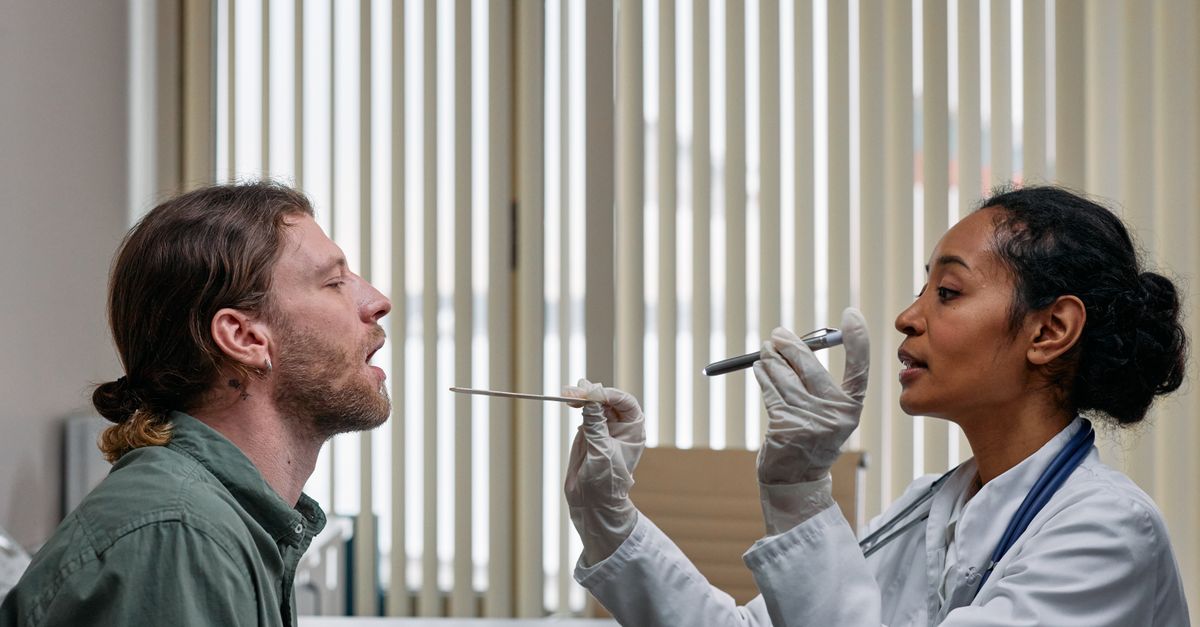
The diagnosis of a hard palate fistula is established through a meticulous physical examination of the oral cavity. Using specialized lighting and dental mirrors, the physician carefully inspects the entire expanse of the palatal vault. The exact location, size, and shape of the fistula are precisely documented.
The clinician will assess the quality of the surrounding tissue, noting the presence of heavy scarring, active inflammation, or signs of poor vascularity. To confirm that the defect communicates completely with the nasal cavity, the physician may gently pass a blunt periodontal probe through the opening or instruct the patient to forcefully exhale through the nose while holding the nostrils closed, checking for the escape of air into the mouth.
A thorough nasal examination using an endoscope is also conducted to evaluate the nasal floor, assess the integrity of the nasal septum, and determine the extent of chronic rhinosinusitis caused by the continuous influx of oral bacteria and food debris.
| Etiology Category | Common Causes | Specific Clinical Challenges |
|---|---|---|
| Congenital Complication | Cleft palate repair breakdown. | Dense scar tissue, limited tissue mobility. |
| Infectious/Inflammatory | Mucormycosis, advanced syphilis. | Requires complete eradication of underlying disease first. |
| Trauma/Oncological | Surgical tumor resection, facial fractures. | Large, complex defects requiring major reconstructive flaps. |
| Substance Abuse | Chronic intranasal cocaine use. | Poor local blood supply, high risk of surgical failure. |
9. Diagnostic Imaging
While the fistula itself is visually apparent during a clinical exam, advanced diagnostic imaging is essential for comprehensive surgical planning. A high-resolution computed tomography scan of the maxillofacial region is the gold standard imaging modality.
The scan provides a precise, three-dimensional reconstruction of the bony architecture of the hard palate. It allows the surgeon to accurately measure the extent of the bony defect, which is often significantly larger than the visible soft tissue hole. The imaging also identifies any concurrent abnormalities, such as hidden abscesses, chronic sinus disease, or the erosion of adjacent dental roots.
If the fistula is suspected to be of oncological origin, magnetic resonance imaging may be ordered in conjunction with the computed tomography scan to evaluate the extent of soft tissue tumor infiltration and determine if the disease has spread to the surrounding neurovascular structures.
10. Non-Surgical Management: Palatal Obturators
Not all patients with a hard palate fistula are immediate candidates for complex reconstructive surgery. Some may possess medical comorbidities that make general anesthesia unsafe, while others may be waiting for local tissue inflammation to subside before an operation can be attempted. In these scenarios, a palatal obturator provides a highly effective, non-surgical solution.
An obturator is a customized prosthetic device, conceptually similar to an upper dental retainer or a partial denture, constructed by a specialized prosthodontist. The device features an acrylic extension that physically plugs and covers the fistula.
When fitted correctly, the obturator creates an immediate, artificial barrier between the oral and nasal cavities. It instantly eliminates nasal regurgitation of food and dramatically improves speech clarity by preventing the escape of air. While the obturator requires daily cleaning and regular adjustments, it profoundly improves the patient’s quality of life without the need for invasive surgical intervention.
11. Principles of Surgical Closure
The definitive treatment for a hard palate fistula is surgical closure. The fundamental principle of palatal reconstruction is to achieve a tension-free, two-layer closure. The surgeon must construct a separate, distinct mucosal lining for the nasal floor and a second, overlying mucosal lining for the oral surface. This double-layer technique significantly reduces the risk of the fistula breaking open again during the healing phase.
Because the tissue surrounding the fistula is often scarred and rigid, simply pulling the edges together is guaranteed to fail due to excessive mechanical tension. Therefore, the surgeon must creatively recruit healthy, highly vascularized tissue from adjacent areas to cover the defect.
The surgical plan is dictated entirely by the size of the hole. Small fistulas can sometimes be closed using localized flaps of palatal tissue. However, larger defects require extensive tissue mobilization to ensure a robust and permanent repair.
12. Advanced Surgical Flap Techniques
For moderate to large fistulas, surgeons employ advanced flap techniques. A common approach is the use of a regional flap, such as the buccal fat pad flap. In this procedure, the surgeon accesses the dense pad of fat located in the cheek, gently mobilizes it, and pulls it over the palatal defect. The rich blood supply of the fat pad promotes rapid healing, and it eventually epithelizes to form a tough oral surface.
Another frequently utilized regional option is the facial artery musculomucosal flap, which borrows a section of tissue from the inside of the cheek and rotates it onto the palate.
For massive defects resulting from oncological resections or severe trauma, local and regional tissue is often insufficient. In these extreme cases, microvascular free tissue transfer is required. The surgeon takes a large piece of tissue—such as muscle and skin from the forearm or the thigh—and transplants it into the roof of the mouth, microscopically connecting the blood vessels of the graft to the blood vessels in the patient’s neck to ensure the tissue survives.
13. Pre-Operative Optimization and Care
Surgical success relies heavily on meticulous pre-operative optimization. The local environment of the fistula must be as healthy as possible before the operation. Patients are often required to wear an obturator for several months leading up to the surgery to block the constant influx of food and bacteria, allowing chronic inflammation at the wound edges to fully subside.
Aggressive dental hygiene is mandatory, and any actively infected teeth near the fistula must be treated or extracted prior to surgery to minimize the bacterial load. For patients with a history of substance abuse or tobacco use, strict and verifiable cessation is an absolute prerequisite, as nicotine and cocaine cause severe vasoconstriction that guarantees necrosis of the surgical flaps.
Nutritional status is evaluated and optimized to ensure the body possesses the protein and micronutrients required to support extensive tissue regeneration and wound healing.
14. Post-Operative Recovery and Rehabilitation
The immediate post-operative period following palatal reconstruction demands strict adherence to specific protocols to protect the delicate surgical repair. The newly rearranged tissue flaps are highly vulnerable to mechanical disruption. Patients are strictly placed on a clear liquid diet initially, slowly advancing to a puréed diet for several weeks to prevent any hard food particles from physically tearing the sutures.
Aggressive intraoral pressure must be avoided. Patients are instructed to sneeze with their mouth open, avoid using straws, and refrain from blowing their nose, as these actions create severe pressure spikes that can blow the repair open.
Long-term rehabilitation often requires the expertise of a speech-language pathologist. Even after a successful structural closure, patients may have developed compensatory articulation errors during the time they lived with the fistula. Targeted speech therapy helps re-train the tongue and palate to utilize the restored anatomical barrier for clear, normal speech production.
15. Frequently Asked Questions (FAQ)
1. Will a hole in the roof of the mouth heal on its own?
No. Because of the constant pressure from speaking and swallowing, and the continuous presence of saliva and bacteria, a fistula in the hard palate will not close naturally. It requires a prosthetic device or surgical repair.
2. What is the most common cause of a palatal fistula?
In children, it is a known complication that can occur after the surgical repair of a congenital cleft palate. In adults, it is often caused by trauma, surgery for oral cancer, or chronic illicit drug use.
3. Can I eat normally if I have a palatal fistula?
Without treatment, eating is very difficult because food and liquids will push up into your nose. A custom prosthetic device called an obturator can plug the hole, allowing you to eat and drink normally until surgery is possible.
4. Is the surgery to close a fistula difficult?
Yes, it is a complex and highly specialized procedure. Because the tissue on the roof of the mouth is very tight, the surgeon must use advanced techniques to move healthy tissue from the cheek or other parts of the body to cover the hole without tension.
5. Why is speech affected so severely?
Clear speech requires a solid roof of the mouth so air can be pressurized in the oral cavity to make consonant sounds. A fistula allows that air to leak out through the nose, making the voice sound highly nasal and difficult to understand.
6. Are there any restrictions after the surgery?
Yes. To protect the delicate stitches, you will be on a liquid or puréed diet for several weeks. You must also avoid using straws, blowing your nose, or sneezing with your mouth closed.
16. Bibliography
Disclaimer: The content is for informational purposes only and does not replace medical advice. Always consult your doctor for personalized treatment.
